. 2019 Oct 24;17(1):135.
doi: 10.1186/s12964-019-0451-2.

The multi-site docking protein Grb2-associated binder 1 (Gab1) enhances interleukin-6-induced MAPK-pathway activation in an SHP2-, Grb2-, and time-dependent manner

Affiliations

The multi-site docking protein Grb2-associated binder 1 (Gab1) enhances interleukin-6-induced MAPK-pathway activation in an SHP2-, Grb2-, and time-dependent manner

Hannes Bongartz et al. Cell Commun Signal. .

Abstract

Background: Cytokine-dependent activation of signalling pathways is tightly orchestrated. The spatiotemporal activation of signalling pathways dictates the specific physiological responses to cytokines. Dysregulated signalling accounts for neoplastic, developmental, and inflammatory diseases. Grb2-associated binder (Gab) family proteins are multi-site docking proteins, which expand cytokine-induced signal transduction in a spatial- and time-dependent manner by coordinating the recruitment of proteins involved in mitogen activated protein kinase (MAPK)/extracellular-signal regulated kinase (ERK) and phosphatidyl-inositol-3-kinase (PI3K) signalling. Interaction of Gab family proteins with these signalling proteins determines strength, duration and localization of active signalling cascades. However, the underlying molecular mechanisms of signal orchestration by Gab family proteins in IL-6-induced signalling are only scarcely understood.

Methods: We performed kinetic analyses of interleukin-6 (IL-6)-induced MAPK activation and analysed downstream responses. We compared signalling in wild-type cells, Gab1 knock-out cells, those reconstituted to express Gab1 mutants, and cells expressing gp130 receptors or receptor mutants.

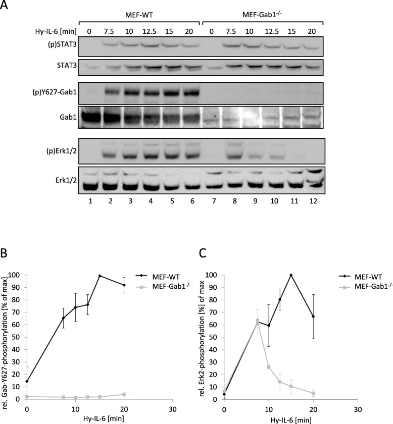
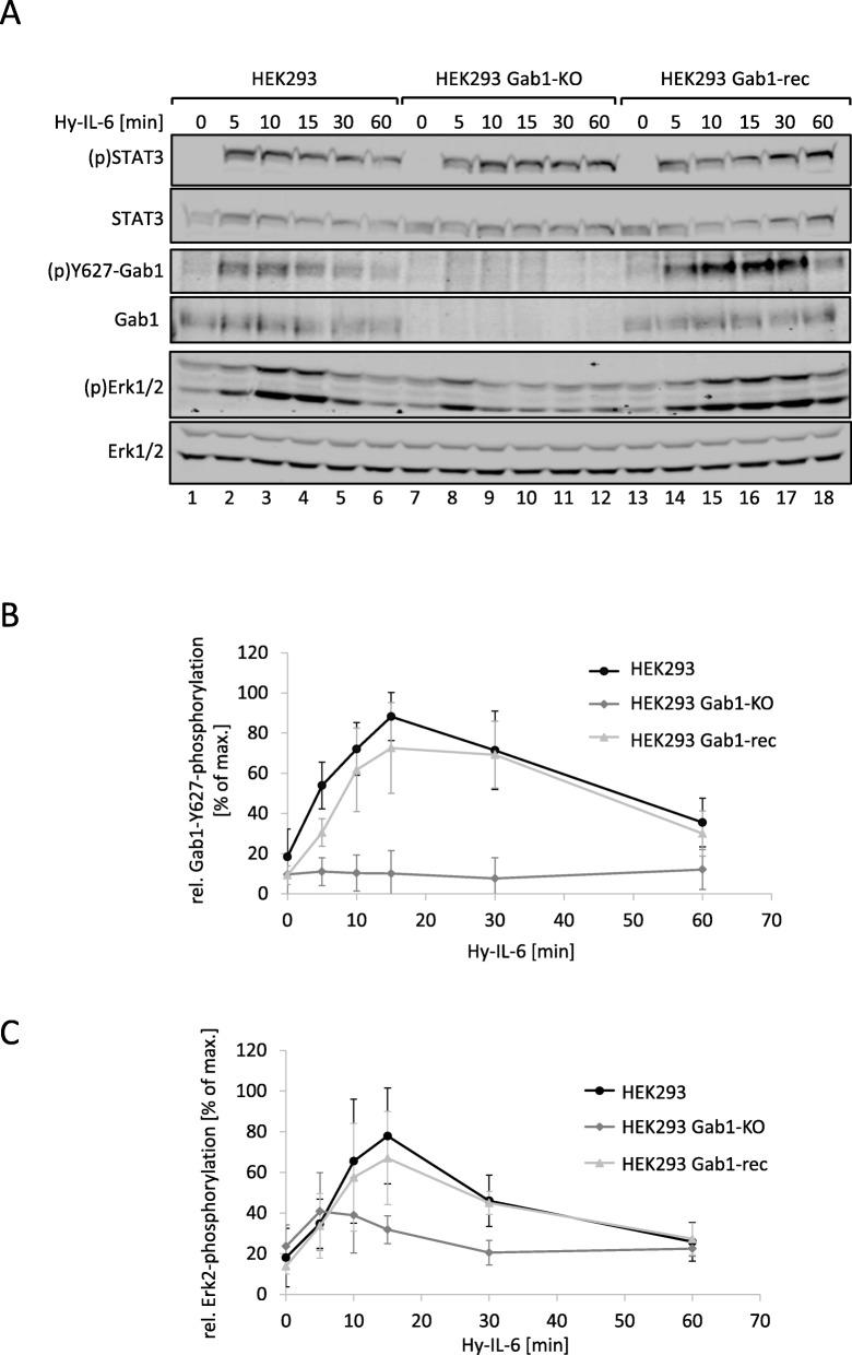
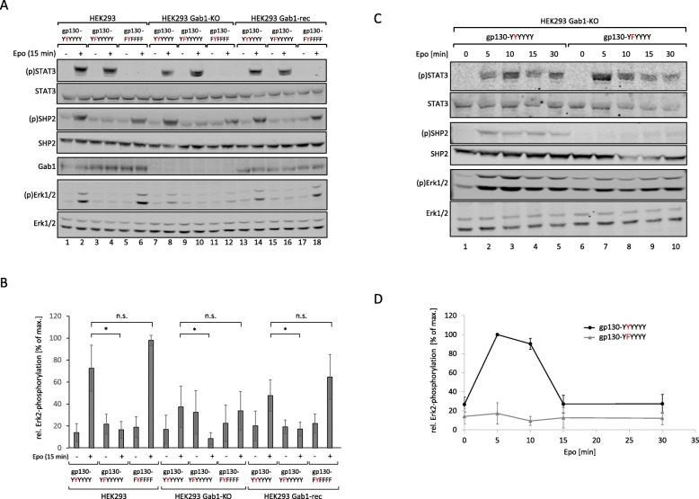
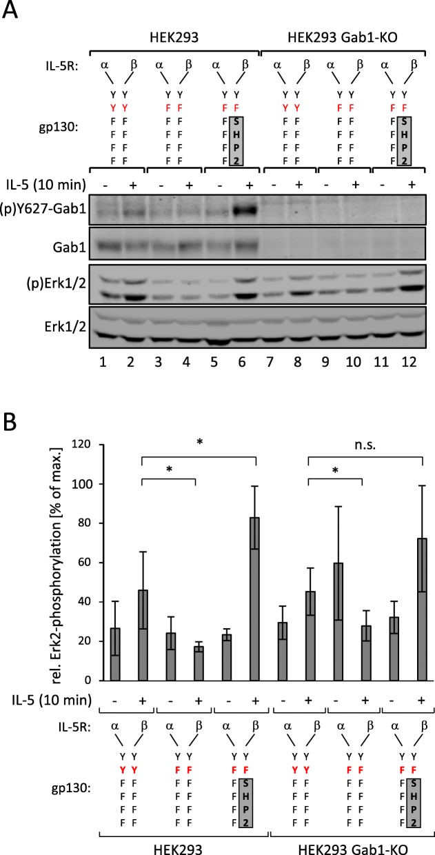
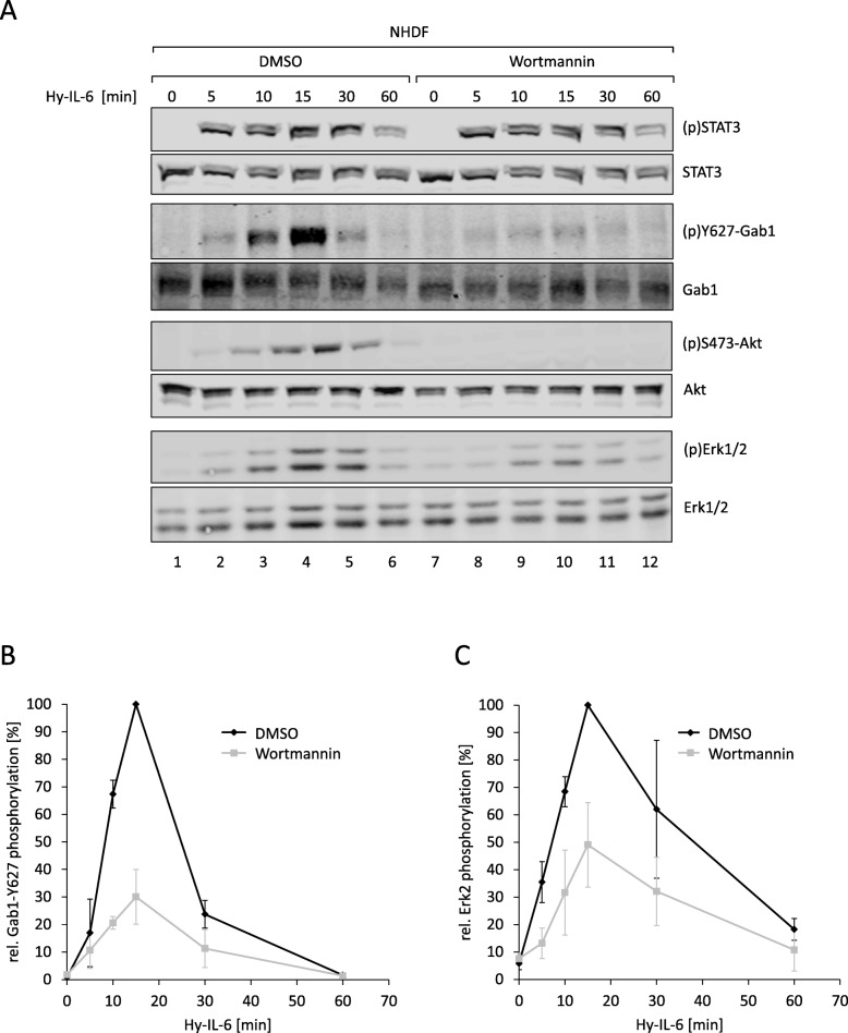
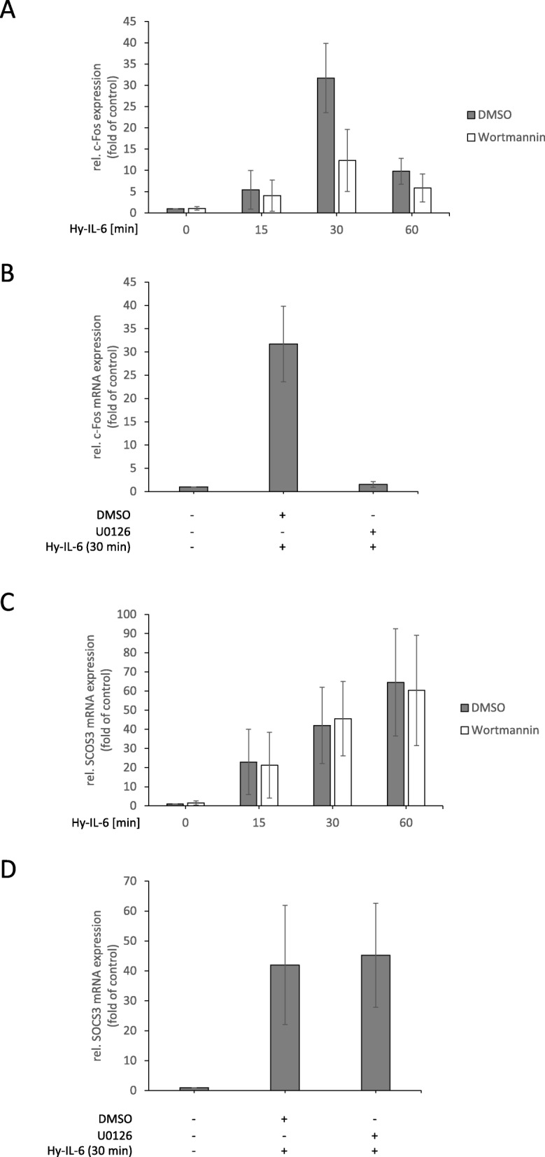
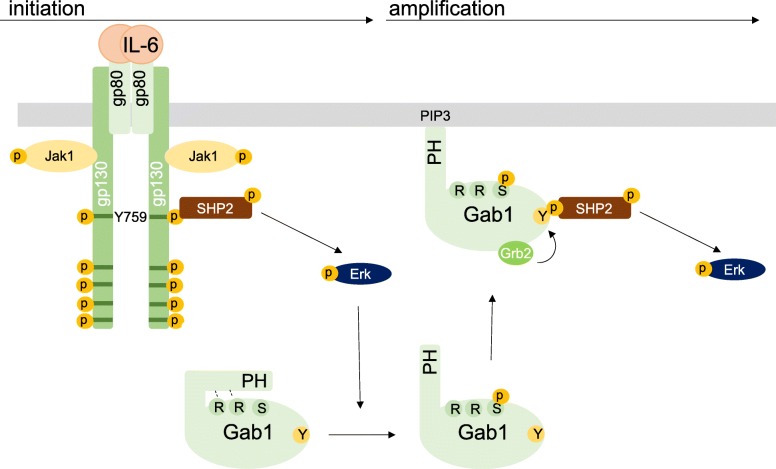
Results: Interleukin-6-induced MAPK pathway activation can be sub-divided into an early Gab1-independent and a subsequent Gab1-dependent phase. Early Gab1-independent MAPK activation is critical for the subsequent initiation of Gab1-dependent amplification of MAPK pathway activation and requires binding of SH2 domain-containing phosphatase 2 (SHP2) to the interleukin-6 receptor complex. Subsequent and coordinated recruitment of Grb2 and SHP2 to Gab1 is essential for Gab1-dependent amplification of IL-6-induced late MAPK pathway activation and subsequent gene expression.

Conclusions: Overall, we elaborated the molecular requirements for Gab1-dependent, spatiotemporal orchestration of interleukin-6-dependent MAPK signalling. We discriminated IL-6-induced Gab1-independent, early activation of MAPK signalling and Gab1-dependent, sustained activation of MAPK signalling.

Keywords: Cytokines; Erk; Gab1; IL-6; Interleukin-6; Jak; Janus kinase; MAPK; PI3K; SHP2; STAT; Signal orchestration; Signal transduction; c-Fos.


Conflict of interest statement

The authors declare that they have no competing interests.

Figures

Fig. 1
Fig. 1
IL-6-induced Erk1/2 pathway activation is amplified by Gab1 in murine embryonic fibroblasts. a Murine embryonic fibroblast (MEF-WT) and Gab1-deficient murine embryonic fibroblast (MEF-Gab1−/−) cells were seeded and cultivated for 24 h. On the following day, cells were serum-starved for 4 h and subsequently stimulated with Hy-IL-6 (100 ng/ml) for the indicated times. Subsequently, cell lysates were prepared and proteins separated by SDS-PAGE. After Western blotting, membranes were stained for (p)STAT3, STAT3, (p)Y627-Gab1, Gab1, (p)Erk1/2 and Erk1/2. Representative results of n = 3 independent experiments are shown. b For quantification of Gab1 Y627 phosphorylation signals of (p)Y627-Gab1 and Gab1 were analysed via densitometry. The diagram shows the ratio of (p)Y627-Gab1 to Gab1 for each time point. Maximal phosphorylation of Gab1 Y627 in each experiment was set to 100%. Data are given as mean of three independent experiments ± SD. c For quantification of Erk2 phosphorylation, signals of (p)Erk2 and Erk2 were analysed via densitometry. The diagram shows the ratio of (p)Erk2 to Erk2 for each time point. Maximal phosphorylation of Erk2 in each experiment was set to 100%. Data are given as mean of three independent experiments ± SD
Fig. 2
Fig. 2
Reconstitution of Gab1 expression in human Gab1-deficient cells rescues IL-6-induced Erk1/2 pathway activation. a HEK293, HEK293 Gab1-KO, and HEK293 Gab1-rec cells were seeded and cultivated for 24 h. On the following day, cells were serum-starved for 4 h and subsequently stimulated with Hy-IL-6 (50 ng/ml) for the times, indicated. Subsequently, cell lysates were prepared and proteins separated by SDS-PAGE. After Western blotting, membranes were stained for (p)STAT3, STAT3, (p)Y627-Gab1, Gab1, (p)Erk1/2, and Erk1/2. Representative results of n = 3 independent experiments are shown. b For quantification of Gab1 Y627 phosphorylation signals of (p)Y627-Gab1 and Gab1 were analysed via densitometry. The diagram shows the ratio of (p)Y627-Gab1 and Gab1 for each time point. Maximal phosphorylation of Gab1 in each experiment was set to 100%. Data are given as mean of three independent experiments ± SD. c For quantification of Erk2 phosphorylation signals of (p)Erk2 and Erk2 were analysed via densitometry. The diagram shows the ratio of (p)Erk2 and Erk2 for each time point. Maximal phosphorylation of Erk2 in each experiment was set to 100%. Data are given as mean of three independent experiments ± SD
Fig. 3
Fig. 3
IL-6-induced Erk pathway activation depends on phosphorylation of Y759 in gp130 .a HEK293, HEK293 Gab1-KO and HEK293 Gab1-rec cells were seeded and transfected with expression vectors (0.4 μg) for the EpoR/gp130 chimeric proteins EG (YYYYYY), EG (YFYYYY) or EG (FYFFFF). After 24 h, cells were serum-starved for 4 h and were stimulated with Epo (7 U/ml) for 15 min. Subsequently, cell lysates were prepared and proteins separated by SDS-PAGE. After Western blotting, membranes were stained for (p)STAT3, STAT3, (p)SHP2, SHP2, Gab1, (p)Erk1/2, and Erk1/2. Representative results of n = 5 independent experiments are shown. b For quantification of Erk2 phosphorylation, signals of (p)Erk2 and Erk2 were analysed via densitometry. The diagram shows the ratio of (p)Erk2 and Erk2. Maximal phosphorylation of Erk2 in each experiment was set to 100%. Data are given as mean of five independent experiments ± SD. Student’s t-test: n.s. = non-significant, *p < 0.05. c HEK293 Gab1-KO cells were seeded and transfected with expression vectors (0.4 μg) for the EpoR/gp130 chimeric proteins EG (YYYYYY) or EG (YFYYYY). After 24 h, cells were serum-starved for 4 h. Cells were stimulated with Epo (7 units/ml) for the indicated times. Cell lysates were prepared and proteins separated by SDS-PAGE. After Western blotting, membranes were stained for (p)STAT3, STAT3, (p)SHP2, SHP2, (p)Erk1/2, and Erk1/2. Representative results of n = 3 independent experiments are shown. d For quantification of Erk2 phosphorylation, signals of (p)Erk2 and Erk2 were analysed via densitometry. The diagram shows the ratio of (p)Erk2 and Erk2 for each time point. Maximal phosphorylation of Erk2 in each experiment was set to 100%. Data are given as mean of three independent experiments ± SD
Fig. 4
Fig. 4
SHP2 binding to gp130 is required for IL-6-induced phosphorylation of Gab1. a HEK293 and HEK293 Gab1-KO cells were seeded and transfected with expression vectors (0.4 μg) for IL-5Rα/gp130(YYFFFF) and IL-5Rβ/gp130(YYFFFF), for IL-5Rα/gp130(YFFFFF) and IL-5Rβ/gp130(YFFFFF), or for IL-5Rα/gp130(YFFFFF) and IL-5Rβ/gp130(YF)-SHP2 chimeric receptors. After 24 h, cells were serum-starved for 4 h. Cells were treated with IL-5 (100 ng/ml) for 10 min. Subsequently, cell lysates were prepared and proteins separated by SDS-PAGE. After Western blotting, membranes were stained for (p)Y627-Gab1, Gab1, (p)Erk1/2, and Erk1/2. Representative results of n = 5 independent experiments are shown. b For quantification of Erk2 phosphorylation, signals of (p)Erk2 and Erk2 were analysed via densitometry. The diagram shows the ratio of (p)Erk2 and Erk2. Maximal phosphorylation of Erk2 in each experiment was set to 100%. Data are given as mean of five independent experiments ± SD. Student’s t-test: n.s. = non-significant, *p < 0.05
Fig. 5
Fig. 5
Tyrosine 759 in gp130 is crucial for IL-6-induced Gab1 translocation and phosphorylation (a) HEK293 Gab1-KO cells were seeded on poly-L-lysine-coated glass cover slips and co-transfected with expression vectors for Gab1-WT-GFP and either EG (YYYYYY) or EG (YFYYYY). On the following day, cells were serum-starved for 4 h, subsequently placed into the incubation chamber of a laser scanning microscope and left for 30 min. Cells were treated with Epo (7 U/ml) to induce IL-6 signalling. Imaging was performed before and after treatment with Epo for up to 30 min as indicated. b HEK293 Gab1-KO cells were seeded and co-transfected with expression vectors for Gab1-WT-GFP and either EG (YYYYYY) or EG (YFYYYY). After 24 h, cells were serum-starved for 4 h. Cells were left untreated or treated with Epo (7 U/ml) for 15 min. Subsequently, cell lysates were prepared and proteins separated by SDS-PAGE. After Western blotting, membranes were stained for (p)STAT3, STAT3, (p)SHP2, SHP2, (p)Y627-Gab1, (p)S552-Gab1, Gab1, (p)Erk1/2, and Erk1/2. After stripping, membranes were stained for GFP. Representative results of n = 3 independent experiments are shown
Fig. 6
Fig. 6
IL-6-induced phosphorylation of Y627 in Gab1 is crucial for Gab1-dependent Erk1/2 pathway amplification. a HEK293 and HEK293 Gab1-rec cells were seeded and cultivated for 24 h. On the following day, cells were serum starved. HEK293 cells were left untreated whereas HEK293 Gab1-WT cells were treated with increasing amount of doxycycline (Dox) as indicated in the figure for 4 h to induce dose-dependently Gab1-WT expression. Subsequently, cells were stimulated with Hy-IL-6 (50 ng/ml) for 15 min. Subsequently, cell lysates were prepared and proteins separated by SDS-PAGE. After Western blotting, membranes were stained for (p)Y627-Gab1, Gab1, (p)STAT3, STAT3, (p)Erk1/2 and Erk1/2. Representative results of n = 3 independent experiments are shown. b HEK293 Gab1-rec and HEK293 Gab1-Y627/659F-rec cells were seeded and cultivated for 24 h. On the following day, all cells were serum starved and were additionally treated with doxycycline (0.5 ng/ml) for 4 h. Subsequently, cells were stimulated with Hy-IL-6 (50 ng/ml) for the times, indicated. Cell lysates were prepared and proteins separated by SDS-PAGE. After Western blotting, membranes were stained for (p)Y627-Gab1, Gab1, (p)STAT3, STAT3, (p)Erk1/2, and Erk1/2. Representative results of n = 3 independent experiments are shown. c For quantification of Erk2 phosphorylation signals of (p)Erk2 and Erk2 were analysed via densitometry. The diagram shows the ratio of (p)Erk2 and Erk2 for each time point. Maximal phosphorylation of Erk2 was set to 100%. Data are given as mean of three independent experiments ± SD
Fig. 7
Fig. 7
Loss of constitutive interaction of Gab1 and Grb2 impairs IL-6-induced phosphorylation of Y627 in Gab1 and Erk1/2 pathway activation. a HEK293 Gab1-rec and HEK293 Gab1-ΔGrb2-rec cells were seeded and cultivated for 24 h. On the following day, all cells were serum starved and additionally treated with doxycycline (0.5 ng/ml) for 4 h to induce expression of Gab1-WT or Gab1-ΔGrb2, respectively. Subsequently, cells were stimulated with Hy-IL-6 (50 ng/ml) for the times, indicated. Cell lysates were prepared and proteins separated by SDS-PAGE. After Western blotting, membranes were stained for (p)Y627-Gab1, Gab1, (p)STAT3, STAT3, (p)Erk1/2, and Erk1/2. Representative results of n = 3 independent experiments are shown. b For quantification of Erk2 phosphorylation signals of (p)Erk2 and Erk2 were analysed via densitometry. The diagram shows the ratio of (p)Erk2 and Erk2 for each time point. Maximal phosphorylation of Erk2 was set to 100%. Data are given as mean of three independent experiments ± SD. c HEK293 Gab1-KO cells were seeded and transfected with expression vectors (0.4 μg) for Gab1-WT-GFP or Gab1-ΔGrb2-GFP. 24 h after transfection, cells were serum-starved for 4 h. Cells were stimulated with Hy-IL-6 (50 ng/ml) for 15 min. Subsequently, cell lysates were prepared to be used for co-immunoprecipitation assays. As input controls (left panels) proteins were separated by SDS-PAGE. After Western blotting, membranes were stained for (p)SHP2, SHP2, (p)Y627-Gab1, (p)S552-Gab1, Gab1, and Grb2. After stripping, membranes were stained for GFP. For co-immunoprecipitation assays (right panels) the proteins were incubated with antibodies against the GFP-tag in Gab1-GFP. Immune complexes were isolated and proteins separated by SDS-PAGE. After Western blotting, membranes were stained for (p)SHP2, Grb2, and Gab1. After stripping, membranes were stained for SHP2 and GFP. Representative results of n = 3 independent experiments are shown
Fig. 8
Fig. 8
Gab1 signalling impacts IL-6-induced MAPK-dependent gene expression in a time-dependent manner. a HEK293, HEK293 Gab1-KO and HEK293 Gab1-rec cells were seeded and cultivated for 24 h. On the following day, cells were serum starved for 4 h and subsequently stimulated with Hy-IL-6 (50 ng/ml) for the times, indicated. Total mRNA was extracted and subjected to qRT-PCR analysis to monitor c-Fos mRNA expression. The expression of c-Fos mRNA was normalized to SDHA mRNA expression. Expression of mRNA is given in fold of mRNA expression in control (=untreated HEK293 cells, set as 1). Data are given as mean of three independent experiments ± SD. b Additionally, HEK293 cells were pre-treated with U0126 (10 μM) or its solvent control DMSO for 30 min prior to stimulation with Hy-IL-6 (50 ng/ml, 30 min). Total mRNA was extracted and subjected to qRT-PCR analysis to monitor c-Fos mRNA expression as described for (a). (c, d) SOCS3 mRNA expression was analysed as described for c-Fos mRNA in (a) and (b)
Fig. 9
Fig. 9
PI3K signalling impacts IL-6-induced Erk activation in normal human dermal fibroblasts in a time-dependent manner. a Normal human dermal fibroblasts (NHDF) were seeded and cultivated for 24 h. On the following day, cells were serum starved for 24 h. After serum starvation, NHDF were pre-treated with Wortmannin (100 nM) or its solvent control DMSO for 30 min. Subsequently, cells were stimulated with Hy-IL-6 (50 ng/ml) for the times, indicated. Cell lysates were prepared and proteins were separated by SDS-PAGE. After Western blotting, membranes were stained for (p)STAT3, STAT3, (p)Y627-Gab1, Gab1, (p)S473-Akt, Akt, (p)Erk1/2, and Erk1/2. Representative results of n = 3 independent experiments are shown. b For quantification of Gab1 Y627 phosphorylation signals of (p)Y627-Gab1 and Gab1 were analysed via densitometry. The diagram shows the ratio of (p)Y627-Gab1 to Gab1 for each time point. Maximal phosphorylation of Gab1 Y627 in each experiment was set to 100%. Data are given as mean of three independent experiments ± SD. c For quantification of Erk2 phosphorylation, signals of (p)Erk2 and Erk2 were analysed via densitometry. The diagram shows the ratio of (p)Erk2 to Erk2 for each time point. Maximal phosphorylation of Erk2 in each experiment was set to 100%. Data are given as mean of three independent experiments ± SD
Fig. 10
Fig. 10
Gab1 signalling impacts IL-6-induced MAPK-dependent gene expression in normal human dermal fibroblasts in a time-in dependent manner. a Normal human dermal fibroblasts (NHDF) were seeded and cultivated for 24 h. On the following day, cells were serum starved for 24 h. After serum starvation, NHDF were pre-treated with Wortmannin (100 nM) or its solvent control DMSO for 30 min. Subsequently, cells were stimulated with Hy-IL-6 (50 ng/ml) for the times, indicated. Total mRNA was extracted and subjected to qRT-PCR analysis to monitor c-Fos mRNA expression. The expression of c-Fos mRNA was normalized to SDHA mRNA expression. Expression of mRNA is given in fold of mRNA expression in control (=untreated NHDF, set as 1). Data are given as mean of three independent experiments ± SD. b Additionally, NHDF were pre-treated with U0126 (10 μM) or its solvent control DMSO for 30 min prior to stimulation with Hy-IL-6 (50 ng/ml, 30 min). Total mRNA was extracted and subjected to qRT-PCR analysis to monitor c-Fos mRNA expression as described for (a). (c, d) SOCS3 mRNA expression was analysed as described for c-Fos mRNA in (a) and (b)
Fig. 11
Fig. 11
Initiation- and amplification-phase model of IL-6-induced Erk1/2 pathway activation. Initial ligand-induced activation of gp130 leads to phosphorylation of tyrosine residues in gp130, enabling recruitment of SHP2 to phosphorylated tyrosine 759 to activate the Erk1/2 pathway in the initiation phase. Activated Erk phosphorylates serine 552 within Gab1. This phosphorylation releases the intramolecular block of the PH domain and enables recruitment of Gab1 to PIP3 at the plasma membrane. Gab1 recruited at the plasma membrane becomes tyrosine phosphorylated. Phosphorylated tyrosine 627 and 659 within Gab1 serve as binding sites for SHP2. Activation of Erk1/2 in the Gab1-dependent amplification phase requires binding of SHP2 and Grb2 to Gab1. Furthermore, binding of SHP2 to Gab1 depends on binding of Grb2 to Gab1

Similar articles

Cited by

References

    1. Heinrich PC, Behrmann I, Haan S, Hermanns HM, Müller-Newen G, Schaper F. Principles of interleukin (IL)-6-type cytokine signalling and its regulation. Biochem J. 2003;374:1–20. doi: 10.1042/bj20030407. - DOI - PMC - PubMed
    1. Heinrich PC, Behrmann I, Müller-Newen G, Schaper F, Graeve L. Interleukin-6-type cytokine signalling through the gp130/Jak/STAT pathway. Biochem J. 1998;334:297–314. doi: 10.1042/bj3340297. - DOI - PMC - PubMed
    1. Kamimura D, Ishihara K, Hirano T. IL-6 signal transduction and its physiological roles: the signal orchestration model. Rev Physiol Biochem Pharmacol. 2003;149:1–38. - PubMed
    1. Schiemann WP, Bartoe JL, Nathanson NM. Box 3-independent signaling mechanisms are involved in leukemia inhibitory factor receptor alpha- and gp130-mediated stimulation of mitogen-activated protein kinase. Evidence for participation of multiple signaling pathways which converge at Ras. J Biol Chem. 1997;272:16631–16636. doi: 10.1074/jbc.272.26.16631. - DOI - PubMed
    1. Wöhrle FU, Daly RJ, Brummer T. Function, regulation and pathological roles of the gab/DOS docking proteins. Cell Commun Signal. 2009;7:22. doi: 10.1186/1478-811X-7-22. - DOI - PMC - PubMed

MeSH terms

Substances