Review
. 2019 Oct 16;63(4):433-456.
doi: 10.1042/EBC20180038.

Understanding biochemistry: structure and function of nucleic acids

Affiliations
Review

Understanding biochemistry: structure and function of nucleic acids

Steve Minchin et al. Essays Biochem. .

Abstract

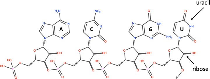
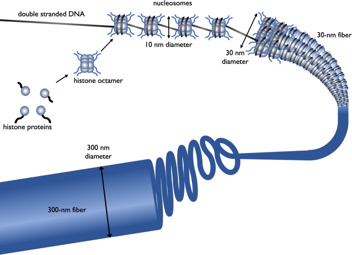
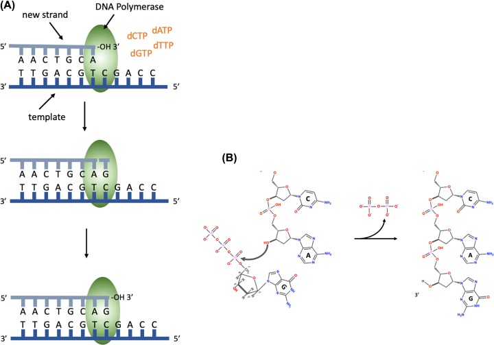
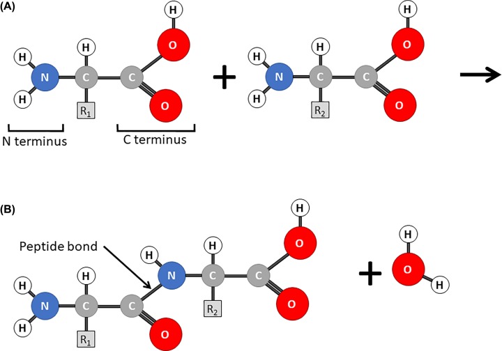
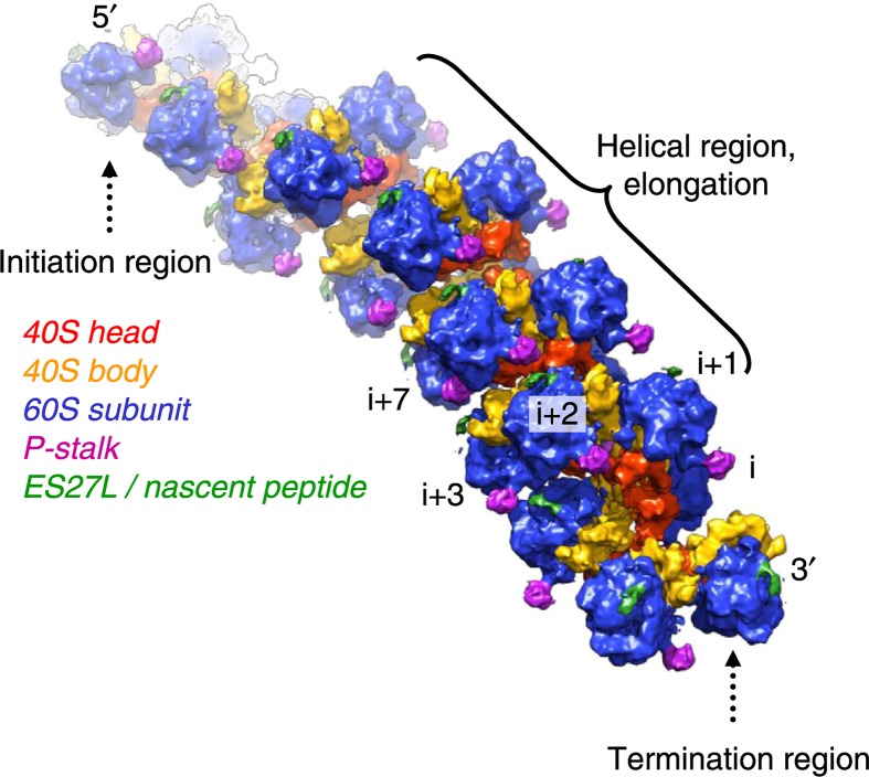
Nucleic acids, deoxyribonucleic acid (DNA) and ribonucleic acid (RNA), carry genetic information which is read in cells to make the RNA and proteins by which living things function. The well-known structure of the DNA double helix allows this information to be copied and passed on to the next generation. In this article we summarise the structure and function of nucleic acids. The article includes a historical perspective and summarises some of the early work which led to our understanding of this important molecule and how it functions; many of these pioneering scientists were awarded Nobel Prizes for their work. We explain the structure of the DNA molecule, how it is packaged into chromosomes and how it is replicated prior to cell division. We look at how the concept of the gene has developed since the term was first coined and how DNA is copied into RNA (transcription) and translated into protein (translation).

Keywords: Concept of the gene; DNA Replication; Nucleic acids; Transcription; Translation.


Conflict of interest statement

The authors declare that there are no competing interests associated with the manuscript.

Figures

Figure 1
Figure 1. The structure of DNA
(A) A nucleotide (guanosine triphosphate). The nitrogenous base (guanine in this example) is linked to the 1′ carbon of the deoxyribose and the phosphate groups are linked to the 5′ carbon. A nucleoside is a base linked to a sugar. A nucleotide is a nucleoside with one or more phosphate groups. (B) A DNA strand containing four nucleotides with the nitrogenous bases thymine (T), cytosine (C), adenine (A) and guanine (G) respectively. The 3′ carbon of one nucleotide is linked to the 5′ carbon of the next via a phosphodiester bond. The 5′ end is at the top and the 3′ end at the bottom.
Figure 2
Figure 2. DNA structure
(A) The DNA double helix, with the sugar phosphate backbone on the outside and the nitrogenous bases in the middle. (B) An A:T and a G:C base pair with the C1′ of the deoxyribose indicated by the arrow. Note that the C1′ of the deoxyribose is in the same position in all base pairs. In this figure, the atoms on the upper edge of the base pair face into the major groove and those facing lower edge face into the minor groove. The hydrogen bonds between the base pairs are indicated by the dotted line.
Figure 3
Figure 3. The structure of RNA
An RNA strand containing the four nucleotides with the nitrogenous bases: adenine (A), cytosine (C), guanine (G) and uracil (U) respectively. The 3′ carbon of the ribose of one nucleotide is linked to the 5′ carbon of the next via a phosphodiester bond. The 5′ end on the left and the 3′ end on the right.
Figure 4
Figure 4. The different levels of chromatin structure
Histone proteins (H2A, H2B, H3 and H4) associate to form a histone octamer. Approximately 147 bp of DNA wraps around histone octamer to form a nucleosome, generating a ‘beads on a string’ structure, the nucleosome together with histone H1 condense into the 30-nm fibre, there is further condensation to form the 300-nm fibre. During mitosis there is further compaction (not shown).
Figure 5
Figure 5. DNA synthesis
(A) DNA polymerase binds the template DNA and the new strand. The next nucleotide to be added to the 3′ end of the growing chain will contain guanine (G), this is complementary to the C on the template strand. DNA polymerase catalyses the formation of a phosphodiester bond. (B) The chemical reaction during the formation of a phosphodiester bond, showing the addition of a nucleotide containing guanine and the release of pyrophosphate.
Figure 6
Figure 6. DNA synthesis at a replication fork
A single replication fork showing the leading and lagging strands. The leading strand is synthesised continuously, reading the template 3′ to 5′, synthesising DNA in the 5′ to 3′ direction. The lagging strand is synthesised discontinuously, in short Okazaki fragments (1000 bases in prokaryotes and 100 bases in eukaryotes).
Figure 7
Figure 7. Telomeres and telomerase
(A) Following DNA replication and removal of the primer for the last Okazaki fragment of the lagging strand, there will be a region at the 3′ end that is not base paired, called a 3′ overhang. (B) Telomerase binds and uses the RNA it contains to act as a template to extend the 3′ overhang. This extends the 3′ end sufficiently for a new RNA primer to bind and the final Okazaki fragment to be made.
Figure 8
Figure 8. The flow of genetic information
The arrows represent steps where DNA or RNA is being used as a template to direct the synthesis of another polymer, either RNA or protein.
Figure 9
Figure 9. DNA sequence showing the amino acid translation underneath
(A) Wild-type sequence, (B) a single base insertion (shown in red) causes a frameshift so all subsequent amino acids are different from the wild-type, (C) insertion of three base pairs (shown in red) causes two incorrect amino acids to be incorporated into the protein but there is no frameshift so the rest of the protein has the wild-type sequence.
Figure 10
Figure 10. The structure of a protein-coding eukaryotic gene
The DNA includes an untranslated region at both the 5′ and 3′ ends as well as introns and exons. The codon where translation starts (green) and the stop codon (red) are shown. The DNA is transcribed into mRNA and is processed by addition of the 5′ cap, splicing out the introns and addition of the poly A tail. This mature mRNA is exported from the nucleus into the cytoplasm.
Figure 11
Figure 11. Schematic diagram of transcription
Figure 12
Figure 12. Control of transcription in prokaryotes
The different binding sites for transcription factors are shown on the DNA; ABS, activator binding site, RBS, repressor binding site. The left-hand panel indicates the presence of lactose and/or glucose in the environment, the right-hand panel indicates transcription levels.
Figure 13
Figure 13. Regulation of transcription in eukaryotes
(A) When a gene is in a silent state the surrounding DNA will be in condensed chromatin and the histones will epigenetic modifications which facilitate gene repression (red spheres). (B) A gene that is being transcribed will have activators bound to enhancer sequences, the activators recruit co-activators that acetylate the histone and add other epigenetic modifications that facilitate gene transcription (green spheres). The activator and co-activators will recruit RNA Polymerase and the GTFs to the core promoter.
Figure 14
Figure 14. Transfer RNA
(A) Tertiary structure of the phenylalanine tRNA from yeast showing the anticodon (grey), the acceptor stem (violet) with the nucleotides CAA at the 3′ OH end (yellow). Image modified from ‘TRNA-Phe yeast’ Yikrazuul (licensed under CC BY-SA 3.0). (B) Clover leaf representation of the secondary structure of tRNA.
Figure 15
Figure 15. The structure of the ribosome of Thermus thermophilus showing the small subunit (green), large subunit (blue), mRNA (red) and three tRNAs in the acceptor, peptidyl and exit sites (yellow)
In (A) the new tRNA is delivered to the ribosome by elongation factor EF-Tu (purple). In (B) the amino acid on the incoming tRNA is brought close to the amino acid on the tRNA in the peptidly site to facilitate peptide bond formation (bright green) (Adapted from Goodsell 2010, licensed under CC-BY-4.0 licence).
Figure 16
Figure 16. Protein synthesis
(A) During initiation, the mRNA recruits a tRNA charged with a methionine and the small ribosomal subunit, (B) the large subunit then docks to give the translation complex, (C) a tRNA with an amino acid attached enters the A site, (D) the peptide bond is formed between the amino acid in the P site and the one in the A site. The effect is that the growing peptide chain is transferred to the incoming aminoacyl tRNA in the A site leaving an empty tRNA in the P site. (E) Finally, everything moves along the mRNA by one codon in a process called translocation so the peptidyl tRNA with the growing peptide chain attached moves to the P site and the spent tRNA to the E site from where it leaves the ribosome. (F) When a stop codon is in the A site, a termination or release factor enters the A site, (G) the peptide is released from the ribosome and (H) the two subunits of the ribosome disassociate and are recycled.
Figure 17
Figure 17. Amino acids and peptide bonds
(A) Amino acids consist of a carbon atom with an amine group (the N terminus), a carboxylic acid group (the C terminus) and a variable R group. The simplest R group is a methyl group giving the amino acid alanine. (B) When two amino acids are joined together a peptide bond is formed between the N terminus of one amino acid and the C terminus of another. This is a condensation reaction releasing one molecule of water.
Figure 18
Figure 18. The polyribosome
Cyro-electron micrograph reconstruction of eukaryotic polyribosome. Reprinted from (Myasnikov 2014) by permission.

References

Recommended reading and key publications: Nobel lectures

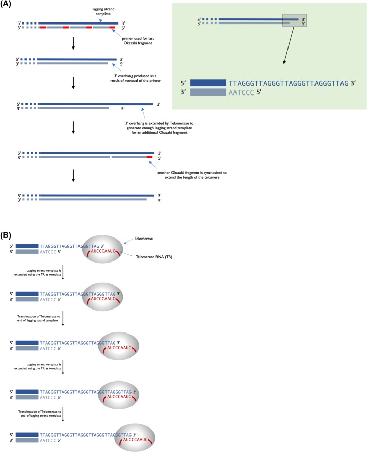
    1. Blackburn E.H. (2010) Telomeres and Telomerase: The Means to the End (Nobel Lecture) 49, Int. Ed., pp. 7405–7421, Angewandte Chemie - PubMed
    1. Ehrenberg M. (2009) Scientific Background on the Nobel Prize in Chemistry 2009 Structure and Function of the Ribosome, The Royal Swedish Academy of Sciences, https://www.nobelprize.org/uploads/2018/06/advanced-chemistryprize2009.pdf
    1. Kornberg R.D. (2007) The Molecular Basis of Eukaryotic Transcription (Nobel Lecture) 32, Int. Ed., pp. 12955–12961, Angewandte Chemie - PubMed

Review articles

    1. Afonina Z.A. and Shirokov V.A. (2018) Three dimensional organization of polyribosomes–a modern approach. Biochemistry (Moscow) 83, S48–S55 10.1134/S0006297918140055 - DOI - PubMed
    1. Gerstein M.B, Bruce C., Rozowsky J.S., Zheng D, Du J., Korbel J.O.. et al. (2007) What is a gene, post-ENCODE? History and updated definition. Genome Res. 17, 669–681 10.1101/gr.6339607 - DOI - PubMed
    1. Kruglyak L. and Stern D.L. (2007) An embarrassment of switches. Science 317, 758–759 10.1126/science.1146921 - DOI - PubMed
    1. Minchin S.D. and Busby S.J.W. (2013) Transcription factors. In Brenner’s Encyclopedia of Genetics(Maloy S. and Hughes K., eds), Elsevier, U.S.A.
    1. Roberts M. (2019) Recombinant DNA technology and DNA sequencing. Essays Biochem. 63, 10.1042/EBC20180039 - DOI - PubMed

Historical perspectives

    1. Dahm R. (2008) Discovering DNA: Friedrich Miescher and the early years of nucleic acid research. Hum. Genet. 122, 565–581 10.1007/s00439-007-0433-0 - DOI - PubMed
    1. McCarty M. (2003) Discovering genes are made of DNA. Nature 421, 406 10.1038/nature01398 - DOI - PubMed
    1. Maddox B. (2003) The double helix and the “wronged heroine”. Nature 421, 407–408 - PubMed
    1. Kemp M. (2003) The Mona Lisa of modern science. Nature 421, 416–420 10.1038/nature01403 - DOI - PubMed

Original research papers

    1. Crick F.H.C., Barnett L., Brenner S. and Watts-Tobin R.J. (1961) General nature of the genetic code for proteins. Nature 192, 1227–1232 10.1038/1921227a0 - DOI - PubMed
    1. Franklin R.E. and Gosling R.G. (1953) Molecular configuration in sodium thymonucleate. Nature 171, 740–741 10.1038/171740a0 - DOI - PubMed
    1. Meselson M. and Stahl F.W. (1958) The replication of DNA in Escherichia coli. Proc. Natl. Acad. Sci. U.S.A. 44, 671–682 10.1073/pnas.44.7.671 - DOI - PMC - PubMed
    1. Watson J. and Crick F. (1953) Molecular structure of nucleic acid. A structure for deoxyribose nucleic acid. Nature 171, 737–738 10.1038/171737a0 - DOI - PubMed

Citations for figures

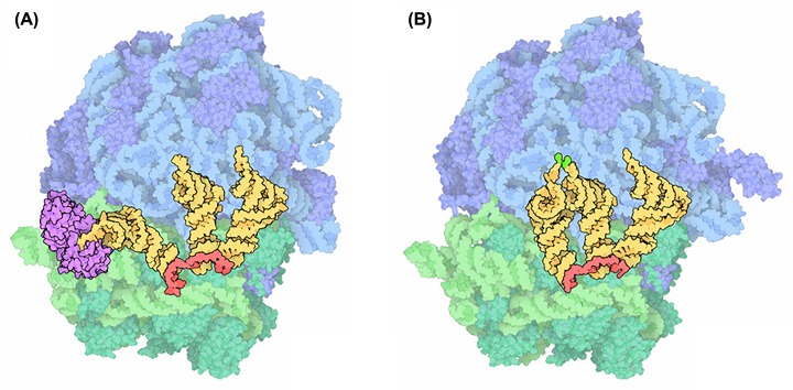
    1. Goodsell D. (2010) Molecule of the month: ribosome. https://pdb101.rcsb.org/motm/121
    1. Myasnikov A.G. (2014) The molecular structure of the left-handed supra-molecular helix of eukaryotic polyribosomes. Nat. Commun. 5, 5294 10.1038/ncomms6294 - DOI - PubMed
    1. Yikrazuul X.X. (2010) tRNA-Phe yeast. https://commons.wikimedia.org/wiki/File:TRNA-Phe_yeast_1ehz.png