. 2019 Oct 22;11(10):1611.
doi: 10.3390/cancers11101611.

Screening a Broad Range of Solid and Haematological Tumour Types for CD70 Expression Using a Uniform IHC Methodology as Potential Patient Stratification Method

Affiliations

Screening a Broad Range of Solid and Haematological Tumour Types for CD70 Expression Using a Uniform IHC Methodology as Potential Patient Stratification Method

Tal Flieswasser et al. Cancers (Basel). .

Abstract

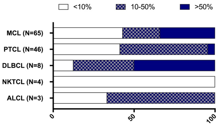
The constitutive expression of CD70 has been described in various haematological and solid tumour types. In addition, the co-expression of its receptor in tumours has been demonstrated, mediating tumour cell proliferation. Although CD70 expression is a prerequisite to enrol patients in solid tumour clinical trials using anti-CD70 immunotherapy, there is currently no standardised test to evaluate CD70 expression. These differences in immunohistochemistry (IHC) protocols make it challenging to compare the expression levels that were obtained in different studies, pointing out the need for one uniform methodology. In this retrospective study, over 600 tumour samples from different solid and haematological malignancies were analysed while using one validated IHC method. CD70 and CD27 expression was demonstrated in a broad range of tumour types. In solid tumours, 43% demonstrated CD70 positivity with the highest degree in renal cell carcinoma (79.5%). Kaposi sarcoma showed no CD70 expression on the tumour cells. In lymphoma samples, 58% demonstrated CD70 positivity. Moreover, the co-expression of CD70 and CD27 was observed in 39% of lymphoma samples. These findings highlight the need to further explore anti-CD70 therapies in a broad range of CD70 expressing tumour types and in doing so, implementing one standardised protocol to define CD70 overexpression to use it as a diagnostic tool.

Keywords: CD27; CD70; biomarker; immune checkpoint; immunohistochemistry.


Conflict of interest statement

The authors declare no conflict of interest. L.V.R., H.d.H., hold ownership interest (including patents) in argenx. V.R. is a consultant/advisory board member for Infinity Pharmaceuticals, Bristol-Myers Squibb, Eisai, PharmaMar, Gilead Sciences, NanoString Technologies, Incyte, BMS, MSD, Roche/Genentech, Epizyme, Astra Zeneca and Servier.

Figures

Figure 1
Figure 1
Proportion of CD70 staining among solid tumour types. CD70 staining was categorised in 3 different groups based on the percentage of tumour cells staining positive: expression levels ranging from 0 to 10% (empty bars); between 10 to 50% (patterned bars); and, expression levels above 50% (solid bars). * p < 0.05, **** p < 0.0001. For statistics, tumour types with N < 5 were not considered in the statistical analysis.
Figure 2
Figure 2
Micrographs of CD70 immunohistochemical stainings across various solid tumour types showing range of staining percentages. (A) >50% CD70 staining on tumour cells in renal cell carcinoma; (B) 10–50% staining in melanoma; (C) <10% staining in breast cancer; and, (D) CD70 staining in control tissue (tonsil). Magnitude 200×.
Figure 3
Figure 3
Representative micrographs of CD70 immunohistochemical staining on samples of patients with haematological malignancies. (A) CD70 staining in a mantle cell lymphoma; (B) cutaneous T-cell lymphoma; and, (C) diffuse large B cell lymphoma is shown. Magnitude 100×.
Figure 4
Figure 4
Proportion of CD70 staining among various lymphoma subsets. CD70 staining was categorised in three different groups: CD70 positive tumour cells ranging from 0 to 10% (white bars); between 10% to 50% (patterned bars); and expression levels above 50% (blue bars). ALCL, anaplastic large cell lymphoma; DLBCL, diffuse large B cell lymphoma; MCL, mantle cell lymphoma; NKTCL, NK/T cell lymphoma; PTCL, peripheral T-cell lymphoma.
Figure 5
Figure 5
Proportion of CD27 staining among various lymphoma subsets. CD27 staining was categorised in three different groups: CD27 positive tumour cells ranging from 0 to 10% (white bars); between 10 to 50% (patterned bars); and expression levels above 50% (red bars). ALCL, anaplastic large cell lymphoma; DLBCL, diffuse large B cell lymphoma; MCL, mantle cell lymphoma; NKTCL, NK/T cell lymphoma; PTCL, peripheral T-cell lymphoma.
Figure 6
Figure 6
Overview of CD70 and CD27 expression in 117 lymphoma samples. Each symbol represents a different patient’s sample and bars represent the mean. ALCL, anaplastic large cell lymphoma; DLBCL, diffuse large B cell lymphoma; MCL, mantle cell lymphoma; NKTCL, NK/T cell lymphoma PTCL, peripheral T-cell lymphoma. TS, tumour sample
Figure 7
Figure 7
Micrographs of immunohistochemical stainings in one MCL patient sample. Sequential cuts of one MCL patient sample showing staining of (A) HE; (B) CD70; (C) CD27; (D) PD-L1; (E) PD-1; and, (F) nuclear staining of MCL cells for cyclin D1. Magnitude 40×.
Figure 8
Figure 8
Expression levels of CD70, CD27, PD-1, and PD-L1 in MCL cohort. The expression of CD70 CD27, PD-1, and PD-L1 in a total of 65 MCL samples. Y axis shows percentage of positive cells for a particular protein. Each symbol represents a different patient. ** p < 0.01, **** p < 0.0001.

References

    1. Zou W., Wolchok J.D., Chen L. PD-L1 (B7-H1) and PD-1 pathway blockade for cancer therapy: Mechanisms, response, biomarkers and combinations. Sci. Transl. Med. 2016;8:328rv4. doi: 10.1126/scitranslmed.aad7118. - DOI - PMC - PubMed
    1. Cottrell T.R., Taube J.M. PD-L1 and Emerging Biomarkers in Immune Checkpoint Blockade Therapy. Cancer J. 2018;24:41–46. doi: 10.1097/PPO.0000000000000301. - DOI - PMC - PubMed
    1. Nolte M.A., van Olffen R.W., van Gisbergen K.P.J.M., van Lier R.A.W. Timing and tuning of CD27-CD70 interactions: The impact of signal strength in setting the balance between adaptive responses and immunopathology. Immunol. Rev. 2009;229:216–231. doi: 10.1111/j.1600-065X.2009.00774.x. - DOI - PubMed
    1. Laouar A., Haridas V., Vargas D., Zhinan X., Chaplin D., van Lier R.A.W., Manjunath N. CD70+ antigen-presenting cells control the proliferation and differentiation of T cells in the intestinal mucosa. Nat. Immunol. 2005;6:698–706. doi: 10.1038/ni1212. - DOI - PMC - PubMed
    1. Wajant H. Therapeutic targeting of CD70 and CD27. Expert Opin. Ther. Targets. 2016;20:959–973. doi: 10.1517/14728222.2016.1158812. - DOI - PubMed