. 2019 Oct 22;24(20):3809.
doi: 10.3390/molecules24203809.

Ivalin Induces Mitochondria-Mediated Apoptosis Associated with the NF-κB Activation in Human Hepatocellular Carcinoma SMMC-7721 Cells

Affiliations

Ivalin Induces Mitochondria-Mediated Apoptosis Associated with the NF-κB Activation in Human Hepatocellular Carcinoma SMMC-7721 Cells

Zhuo Han et al. Molecules. .

Abstract

Ivalin, a natural compound isolated from Carpesium divaricatum, showed excellent microtubule depolymerization activities among human hepatocellular carcinoma in our previous work. Here, we investigated its functions on mitochondria-mediated apoptosis in hepatocellular carcinoma SMMC-7721 cells. DAPI (4',6-diamidino-2-phenylindole) staining, annexin V-fluorexcein isothiocyanate (FITC) apoptosis detection, and western blotting were applied to explore the apoptotic effect of Ivalin. Next, the induction effect of Ivalin on the mitochondrial pathway was also confirmed via a series of phenomena including the damage of mitochondria membrane potential, mitochondria cytochrome c escape, cleaved caspase-3 induction, and the reactive oxygen species generation. In this connection, we understood that Ivalin induced apoptosis through the mitochondrial pathway and the overload of reactive oxygen species. Furthermore, we found that the activation of nuclear factor-κB (NF-κB) and subsequent p53 induction were associated with the apoptotic effect of Ivalin. These data confirmed that Ivalin might be a promising pro-apoptotic compound that can be utilized as a potential drug for clinical treatment.

Keywords: Carpesium divaricatum; Ivalin; NF-κB; hepatocellular carcinoma; mitochondria-mediated apoptosis.


Conflict of interest statement

The authors declare no conflicts of interest.

Figures

Figure 1
Figure 1
Structure of Ivalin.
Figure 2
Figure 2
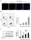
SMMC-7721 cells treated with Ivalin causing apoptosis. (A) Fluorescence micrographs of untreated and Ivalin treated SMMC-7721 cells with 4′,6-diamidino-2-phenylindole (DAPI). Magnification: 100×. (B) Results from the flow cytometry analysis, the quantification of the apoptotic cells after indicate treatment. (C) Western blot showed that Ivalin induced apoptosis by enhancing the Bax and declining the Bcl-2 expression. * p < 0.05; ** p < 0.01, *** p < 0.001 vs. the control group.
Figure 3
Figure 3
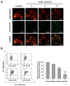
Effects of MMP generation in Ivalin-treated cells. (A,B) After Ivalin treatment for 24 h, flow cytometry and fluorescence microscope were used to detect cellular mitochondrial membrane potential. (A) Ivalin treatment decreased the red fluorescence intensity (aggregates) and increased green fluorescence intensity (monomers) in SMMC-7721 cells, indicating that Ivalin reduced the mitochondrial membrane potential, thereby leading to mitochondrial dysfunction. (B) Ivalin induced the loss of mitochondrial membrane potential as shown by flow cytometry. *** p < 0.01 vs. the control group.
Figure 4
Figure 4
Ivalin trigged apoptosis by means of the mitochondria activation. (A) The cytochrome c in mitochondria with the stimulation of Ivalin inflowed into the cytosol. (B) Cleaved caspase-3 was increased with the treatment of Ivalin. *** p < 0.001 vs. the control group.
Figure 5
Figure 5
Ivalin induced the generation of intracellular ROS. Graph shows the fluorescence intensities of DCF in the SMMC-7721 cells exposed to Ivalin in contrast to control. The fluorescence intensity of control group was set as 1. * p < 0.05; ** p < 0.01 vs. the control group.
Figure 6
Figure 6
Trends of Ivalin on the p53, NF-κB and IκB expression. (A) The expressions of relative proteins were measured by western blotting. * p < 0.05; ** p < 0.01, *** p < 0.001 vs. the control group. (B) Relative mRNA expression values were calculated by real-time PCR. The quantity of each mRNA was relative to the glyceraldehyde-3-phosphate dehydrogenase (GAPDH) mRNA levels.

References

    1. Siegel R.L., Miller K.D., Jemal A. Cancer statistics, 2019. Ca-A. Cancer J. Clin. 2019;69:7–34. doi: 10.3322/caac.21551. - DOI - PubMed
    1. Abdalla E.K., Vauthey J.N. Focus on treatment of large hepatocellular carcinoma. Ann. Surg. Oncol. 2004;11:1035–1036. doi: 10.1245/ASO.2004.10.906. - DOI - PubMed
    1. Yan M.-D., Yao C.-J., Chow J.-M., Chang C.-L., Hwang P.-A., Chuang S.-E., Whang-Peng J., Lai G.-M. Fucoidan Elevates MicroRNA-29b to Regulate DNMT3B-MTSS1 Axis and Inhibit EMT in Human Hepatocellular Carcinoma Cells. Mar. Drugs. 2015;13:6099–6116. doi: 10.3390/md13106099. - DOI - PMC - PubMed
    1. Mor E., Kaspa R.T., Sheiner P., Schwartz M. Treatment of hepatocellular carcinoma associated with cirrhosis in the era of liver transplantation. Ann. Intern. Med. 1998;129:643–653. doi: 10.7326/0003-4819-129-8-199810150-00013. - DOI - PubMed
    1. Li X., Zhao Y., Wu W.K.K., Liu S., Cui M., Lou H. Solamargine induces apoptosis associated with p53 transcription-dependent and transcription-independent pathways in human osteosarcoma U2OS cells. Life Sci. 2011;88:314–321. doi: 10.1016/j.lfs.2010.12.006. - DOI - PubMed

MeSH terms