Review
. 2020 Jul 1;10(7):a034835.
doi: 10.1101/cshperspect.a034835.

The Biology of B-Progenitor Acute Lymphoblastic Leukemia

Affiliations
Review

The Biology of B-Progenitor Acute Lymphoblastic Leukemia

Kathryn G Roberts et al. Cold Spring Harb Perspect Med. .

Abstract

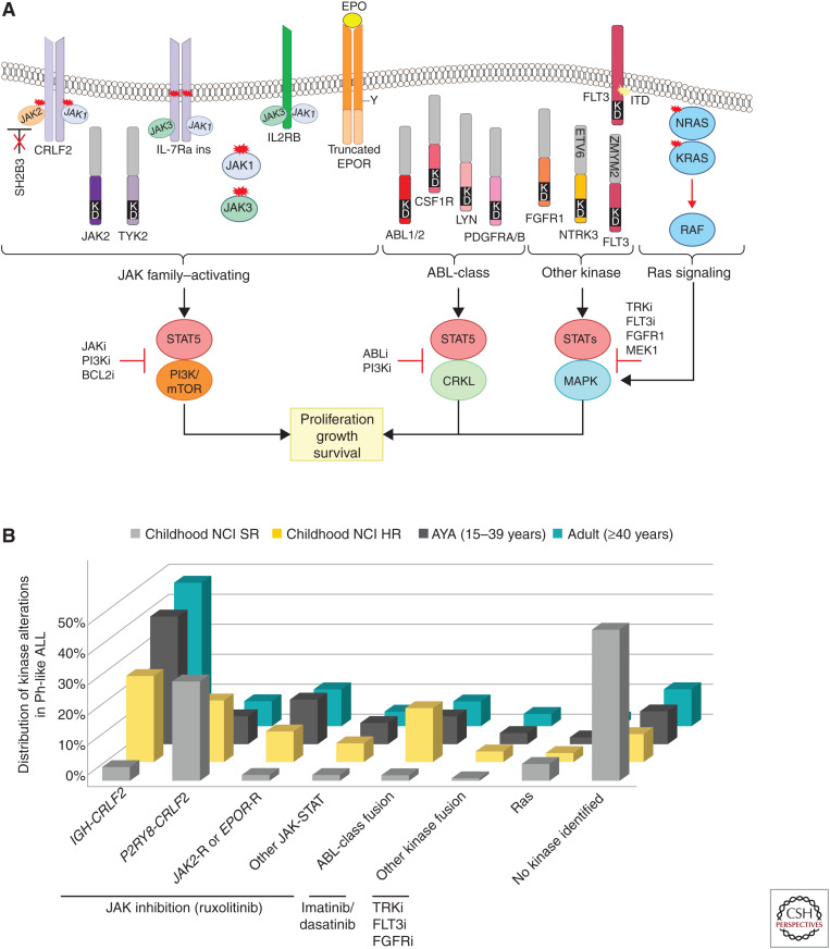
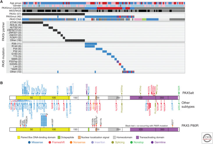
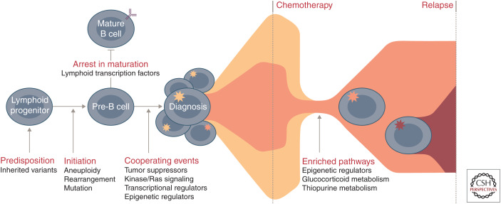
Genomic analyses have revolutionized our understanding of the biology of B-progenitor acute lymphoblastic leukemia (ALL). Studies of thousands of cases across the age spectrum have revised the taxonomy of B-ALL by identifying multiple new subgroups with diverse sequence and structural initiating events that vary substantially by age at diagnosis and prognostic significance. There is a growing appreciation of the role of inherited genetic variation in predisposition to ALL and drug responsiveness and of the nature of genetic variegation and clonal evolution that may be targeted for improved diagnostic, risk stratification, disease monitoring, and therapeutic intervention. This review provides an overview of the current state of knowledge of the genetic basis of B-ALL, with an emphasis on recent discoveries that have changed our approach to diagnosis and monitoring.


Figures

Figure 1.
Figure 1.
(A) tSNE plot showing B-progenitor acute lymphoblastic leukemia (B-ALL) subtypes based on RNA-seq gene expression profiling of 1988 cases. (B) Distribution of B-ALL subtypes within each age group. Subtypes are grouped as gross chromosomal abnormalities (aneuploidy or copy number gain), transcription factor (TF) rearrangement, other TF-driven, kinase-driven, and all others (Gu et al. 2019).
Figure 2.
Figure 2.
(A) Kinase alterations and signaling pathways dysregulated in Philadelphia chromosome-like (Ph-like) ALL. The majority of kinase and cytokine receptor alterations converge on two pathways that activate JAK-family member signaling or ABL signaling. Alterations that activate JAK-STAT signaling can be targeted with JAK and PI3K inhibitors. ABL-class alterations can be targeted with ABL-inhibitors such as dasatinib. Other kinase alterations and those that activate Ras signaling can be targeted with specific inhibitors including those that inactivate TRK, FLT3, FGFR1, and MEK for the MAPK pathway. (B) Distribution of kinase subtypes in Ph-like ALL within each age group (Roberts et al. 2014a, 2017a, 2018; Reshmi et al. 2017). Combined prevalence of Ph-like ALL subtypes in childhood National Cancer Institute (NCI) standard-risk (SR; age 1–9.99 yr and WBC < 50,000/µL), NCI high-risk (HR; age 10–15 yr or WBC ≥ 50,000/µL), adolescent and young adults (1639 yr), and adults (≥40 yr). Genomic subtypes include IGH-CRLF2, P2RY8-CRLF2, and ABL-class fusions (ABL1, ABL2, CSF1R, LYN, PDGFRA, and PDGFRB); JAK2 and EPOR rearrangements and other mutations in JAK–STAT signaling (JAK1/3, IL7R, SH2B3, TYK2, and IL2RB); and other kinase alterations (FLT3, FGFR1, NTRK3), Ras mutations (KRAS, NRAS, NF1, PTPN11, BRAF, and CBL), and unknown alterations.
Figure 3.
Figure 3.
(A) Genetic alterations of PAX5, including gene rearrangements (PAX5r), sequence mutations (PAX5mut). and focal intragenic amplifications (PAX5amp, pink in PAX5 CNA) observed in the PAX5alt cohort. (B) Protein domain plot of PAX5 showing the mutations detected in PAX5alt and other B-ALL subtypes (top panel) and in the PAX5 P80R subtype (bottom panel). (CNA) Copy number alteration.
Figure 4.
Figure 4.
Commonly altered pathways and stepwise progression of B-progenitor acute lymphoblastic leukemia (B-ALL). Common genetic polymorphisms (IKZF1, CDKN2A/B, PIP4K2A, GATA3, ARID5B, CEBPE, and ERG) and deleterious nonsilent inherited variants (PAX5, ETV6, IKZF1, TP53, and ERG) increase the risk of ALL susceptibility. Driving or founding lesions of ALL define genomic subtypes: aneuploidy and other chromosomal abnormalities (hyperdiploid, low-hypodiploid, near-haploid, iAMP21), rearrangements deregulating transcription factors (ETV6-RUNX1, ETV6-RUNX1-like, KMT2A, TCF3-PBX1, DUX4, ZNF384, MEF2D, NUTM1, TCF3-HLF, PAX5, BCL2/MYC) or kinase genes (Ph-like, BCR-ABL1), and specific mutations in lymphoid transcription factors (PAX5 P80R, IKZF1, N195Y). Deletion and loss of lymphoid transcription factors (e.g., IKZF1, PAX5, EBF1) coupled with the alteration of tumor suppressors and cell cycle regulators (CDKN2A/B, TP53), kinase signaling pathway genes (e.g., NRAS, KRAS, FLT3), other transcriptional regulators (e.g., ETV6, ERG), or epigenetic regulators (e.g., CREBBP, WHSC1, CTCF) result in the accumulation of immature lymphoid blasts and presentation at diagnosis. During treatment, the predominant diagnosis clone is commonly eradicated and relapse arises from a minor clone that already harbors and/or acquires additional genetic alterations that drive resistance. Pathways that are enriched at relapse include those involving epigenetic regulators (e.g., CREBBP, SETD2, KDM6A), the glucocorticoid response (e.g., CREBBP, NR3C1), and thiopurine metabolism (e.g., NT5C2, MSH6).

References

    1. Agraz Doblas A, Bueno C, Bashford-Rogers R, Roy A, Schneider P, Bardini M, Ballerini P, Cazzaniga G, Moreno T, Revilla C, et al. 2019. Unraveling the cellular origin and clinical prognostic markers of infant B-cell acute lymphoblastic leukemia using genome-wide analysis. Haematologica 104: 1176–1188. 10.3324/haematol.2018.206375 - DOI - PMC - PubMed
    1. Alekseyenko AA, Walsh EM, Wang X, Grayson AR, Hsi PT, Kharchenko PV, Kuroda MI, French CA. 2015. The oncogenic BRD4-NUT chromatin regulator drives aberrant transcription within large topological domains. Genes Dev 29: 1507–1523. 10.1101/gad.267583.115 - DOI - PMC - PubMed
    1. Alexander TB, Gu Z, Iacobucci I, Dickerson K, Choi JK, Xu B, Payne-Turner D, Yoshihara H, Loh ML, Horan J, et al. 2018. The genetic basis and cell of origin of mixed phenotype acute leukaemia. Nature 562: 373–379. 10.1038/s41586-018-0436-0 - DOI - PMC - PubMed
    1. Andersson AK, Ma J, Wang J, Chen X, Gedman AL, Dang J, Nakitandwe J, Holmfeldt L, Parker M, Easton J, et al. 2015. The landscape of somatic mutations in infant MLL-rearranged acute lymphoblastic leukemias. Nat Genet 47: 330–337. 10.1038/ng.3230 - DOI - PMC - PubMed
    1. Arber DA, Orazi A, Hasserjian R, Thiele J, Borowitz MJ, Le Beau MM, Bloomfield CD, Cazzola M, Vardiman JW. 2016. The 2016 revision to the World Health Organization classification of myeloid neoplasms and acute leukemia. Blood 127: 2391–2405. 10.1182/blood-2016-03-643544 - DOI - PubMed

Publication types

MeSH terms

LinkOut - more resources