. 2019 Nov 29;294(48):18488-18503.
doi: 10.1074/jbc.RA119.010178. Epub 2019 Oct 24.

Impaired tau-microtubule interactions are prevalent among pathogenic tau variants arising from missense mutations

Affiliations

Impaired tau-microtubule interactions are prevalent among pathogenic tau variants arising from missense mutations

Yuxing Xia et al. J Biol Chem. .

Abstract

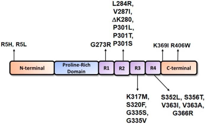
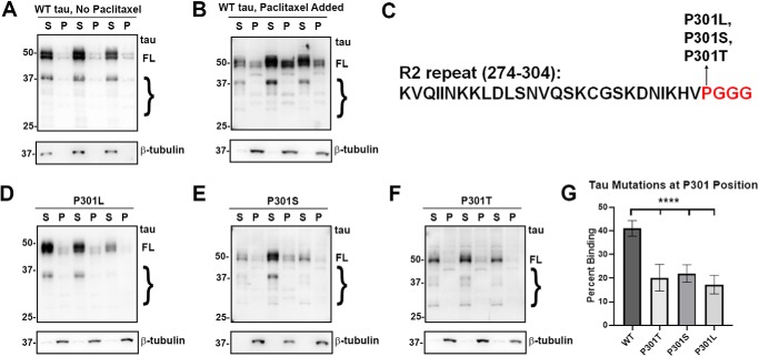
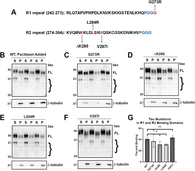
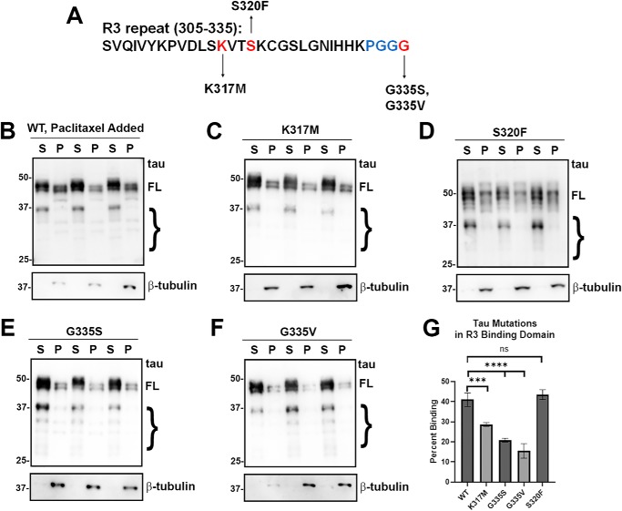
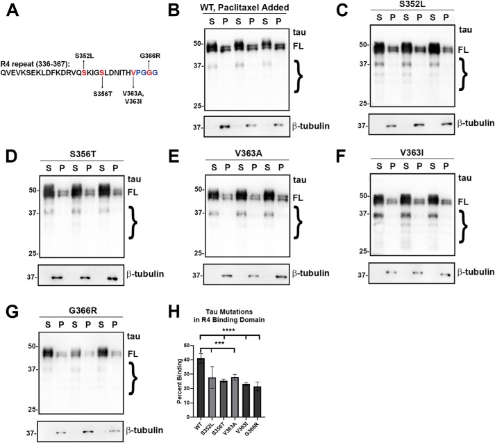
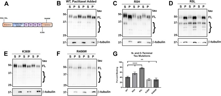
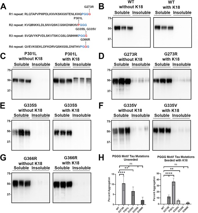
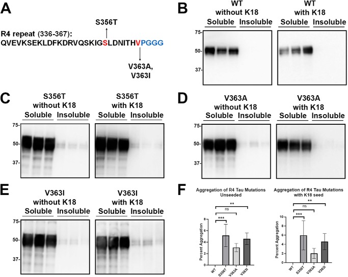
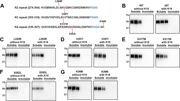
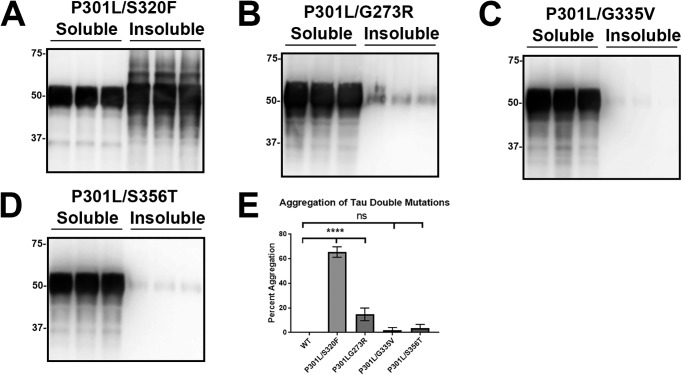
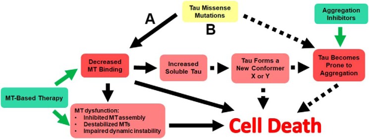
tau is a microtubule (MT)-associated protein that promotes tubulin assembly and stabilizes MTs by binding longitudinally along the MT surface. tau can aberrantly aggregate into pathological inclusions that define Alzheimer's disease, frontotemporal dementias, and other tauopathies. A spectrum of missense mutations in the tau-encoding gene microtubule-associated protein tau (MAPT) can cause frontotemporal dementias. tau aggregation is postulated to spread by a prion-like mechanism. Using a cell-based inclusion seeding assay, we recently reported that only a few tau variants are intrinsically prone to this type of aggregation. Here, we extended these studies to additional tau mutants and investigated their MT binding properties in mammalian cell-based assays. A limited number of tau variants exhibited modest aggregation propensity in vivo, but most tau mutants did not aggregate. Reduced MT binding appeared to be the most common dysfunction for the majority of tau variants due to missense mutations, implying that MT-targeting therapies could potentially be effective in the management of tauopathies.

Keywords: Alzheimer disease; aggregation; microtubule; prion; tau protein (tau); tauopathy.


Conflict of interest statement

The authors declare that they have no conflicts of interest with the contents of this article

Figures

Figure 1.
Figure 1.
Schematic of 4R tau protein depicting major structural domains. tau is an MT-associated protein that consists of an N-terminal region, a proline-rich domain, an MTBD composed of four MT-binding repeats in the 4R isoforms, and a C-terminal region. Indicated are the locations of the pathogenic tau missense mutants that were investigated herein; they were numbered relative to the longest 2N4R tau isoform expressed in the human brain. Most tau missense mutants are clustered within the MTBD.
Figure 2.
Figure 2.
tau mutants at the Pro-301 residue significantly impaired MT binding. A and B, cell-based MT-binding assay performed with HEK293T cells transfected to express WT tau with or without the presence of paclitaxel as described under “Experimental procedures.” Antibody specific for β-tubulin (clone TUB 2.1) was used to assay the polymerization of tubulins. 3026 is a polyclonal antibody against total tau. S = supernatants; P = pellet fractions. Without paclitaxel, the majority of tubulin is not polymerized and soluble, whereas with paclitaxel, the majority of tubulin is polymerized as MTs in the pellet fraction. C, P301L, P301S, and P301T tau mutants are in the PGGG motif of the R2 repeat. In the presence of paclitaxel, P301L (D), P301S (E), and P301T (F) all demonstrate significantly decreased MT binding when compared with WT tau. The relative molecular masses of protein markers are indicated on the left. On the right, FL is for full-length tau, and the brace indicates degradative tau bands. G, one-way ANOVA with Dunnett's test was performed with n = 18 for WT tau and n = 3 for each of these tau mutants. ****, p < 0.0001.
Figure 3.
Figure 3.
tau mutants in the R1 and R2 repeats had varied effects on MT binding. A, tau mutants G273R, ΔK280, L284R, and V287I are present within the R1 or R2 repeat as indicated. B–F, MT-binding assay was performed in the presence of paclitaxel as described under “Experimental procedures.” Relative to WT tau, ΔK280 and L284R tau demonstrated decreased MT binding, whereas G273R tau and V287I tau did not have a significant effect. Immunoblots were probed with anti-β-tubulin antibody TUB 2.1 or total tau antibody 3026 as indicated. S = supernatants; P = pellet fractions. The relative molecular masses of protein markers are indicated on the left. On the right, FL is for full-length tau, and the brace indicates degraded tau bands. G, one-way ANOVA with Dunnett's test was performed with n = 18 for WT tau and n = 3 for each of these tau mutants. *, p < 0.05; ns, no statistical significance.
Figure 4.
Figure 4.
Most tau mutants in the R3 repeat decreased MT binding. A, tau mutants K317M, S320F, G335S, and G335V are located within the R3 repeat. B–F, MT-binding assay was performed in the presence of paclitaxel as described under “Experimental procedures.” HEK293T cells were transfected with WT or the indicated tau mutations. K317M, G335S, and G335V tau mutants significantly decreased MT binding, whereas the S320F tau had no major change. Immunoblots were probed with anti-β-tubulin antibody TUB 2.1 or total tau antibody 3026 as indicated. S = supernatants; P = pellet fractions. The relative molecular masses of protein markers are indicated on the left. On the right, FL is for full-length tau, and the brace indicates degradative tau bands. G, one-way ANOVA with Dunnett's test was performed with n = 18 for WT tau and n = 3 for each tau mutant. ***, p < 0.001; ****, p < 0.0001; and ns, no statistical significance.
Figure 5.
Figure 5.
Most of the tau variants mutants in the R4 repeat significantly reduced MT binding. A, tau mutants S352L, S356T, V363I, V363A, and G366R are located within the R4 repeat. B–G, MT-binding assay was performed in the presence of paclitaxel as described under “Experimental procedures.” HEK293T cells were transfected with WT or the indicated tau mutations. S352L, S356T, V363I, V363A, and G366R tau mutants significantly decreased MT binding. Immunoblots were probed with anti-β-tubulin antibody TUB 2.1 or total tau antibody 3026 as indicated. S = supernatants; P = pellet fractions. The relative molecular masses of protein markers are indicated on the left. On the right, FL is for full-length tau, and the brace indicates degradative tau bands. H, one-way ANOVA with Dunnett's test was performed with n = 18 for WT tau and n = 3 for each tau mutant. ***, p < 0.001; ****, p < 0.0001.
Figure 6.
Figure 6.
tau mutants in the N and C termini had differential effects on MT binding. A, diagram shows N- and C-terminal tau mutants. B–F, MT-binding assay was performed in the presence of paclitaxel as described under “Experimental procedures.” HEK293T cells were transfected with WT or the indicated tau mutations. Both the R5H and R5L tau mutants in the N terminus significantly increased MT binding, whereas the mutants near the C terminus, K369I and R406W, decreased MT binding. Immunoblots were probed with anti-β-tubulin antibody TUB 2.1 or total tau antibody 3026 as indicated. S = supernatants; P = pellet fractions. The relative molecular masses of protein markers are indicated on the left. On the right, FL is for full-length tau, and the brace indicates degradative tau bands. G, one-way ANOVA with Dunnett's test was performed with n = 18 for WT and n = 3 for each tau mutant. **, p < 0.01; ****, p < 0.0001.
Figure 7.
Figure 7.
tau mutants in the PGGG motif showed varied susceptibility to prion-like seeding. A, diagram depicts tau mutations in the PGGG motif within R1, R2, R3, and R4 repeats. HEK293T cells were transfected with WT or tau mutants and assessed for aggregation with or without exogenous K18 seeds. B, WT tau did not aggregate by itself or in the presence of K18 seeds. C, P301L did not aggregate without seeding but had robust induced aggregation in the presence of K18 seeds. D and F, G273R and G335V demonstrated modest levels of self-aggregation with or without exogenous K18 seeds. E, interestingly, G335S did not aggregate, despite being at the same location as G335V. G, G366R showed low levels of aggregation, but this was not statistically significant. Immunoblots were probed with total tau antibody 3026. The relative molecular masses of protein markers are indicated on the left. H, one-way ANOVA with Dunnett's test was performed with n = 9 for WT tau and n = 3 for each tau mutant. ****, p < 0.0001; **, p < 0.01; and ns, no statistical significance.
Figure 8.
Figure 8.
Some tau mutants in the R4 repeat modestly aggregated without exogenous seeding. A, tau mutants S356T, V363A, and V363I are located within the R4 repeat. HEK293T cells were transfected with WT or tau mutants indicated and assessed for aggregation with or without exogenous K18 fibrillar seeds. Compared with WT tau (B), S356T (C) and V363I (E) mutants modestly self-aggregated without and with seeding. D, V363A did not significantly aggregate with or without K18 seeds. Immunoblots were probed with total tau antibody 3026. The relative molecular masses of protein markers are indicated on the left. One-way ANOVA with Dunnett's test was performed with n = 9 for WT tau and n = 3 for each tau mutant (F). *, p < 0.05; **, p < 0.01; and ns, no statistical significance.
Figure 9.
Figure 9.
Additional tau mutants did not show any propensity to aggregate. A, diagram depicts tau mutations L284R, V287I, K317M, S352L, and K369I within R2, R3, and R4 repeats. HEK293T cells were transfected with WT or tau mutants and were assessed for aggregation with or without exogenous K18 fibrillar seeds. Compared with WT tau (B), L284R (C), V287I (D), K317M (E), S352L (F), and K369I (G) did not aggregate with or without exogenous K18 seeds. Immunoblots were probed with total tau antibody 3026. The relative molecular masses of protein markers are indicated on the left.
Figure 10.
Figure 10.
P301L/S320F tau was uniquely capable of robust self-aggregation compared with other double tau mutants. HEK293T cells were transfected with different tau double mutations and were fractionated into Triton-soluble and Triton-insoluble fractions as described under “Experimental procedures.” A, P301L/S320F tau double mutant presented extensive intrinsic aggregation. B, P301L/G273R tau modestly aggregated but did not show an enhanced effect from the addition of P301L. P301L/G335V tau (C) and P301L/S356T tau (D) did not significantly aggregate. Immunoblots were probed with total tau antibody 3026. The relative molecular masses of protein markers are indicated on the left. E, one-way ANOVA with Dunnett's test was performed with n = 9 for WT tau and n = 3 for each tau mutant. ****, p < 0.0001; and ns, statistical significance.
Figure 11.
Figure 11.
Proposed mechanisms of tau pathogenesis. A, model for the majority of tau variants due to missense mutations that predominantly impair MT binding, resulting in increased unbound tau as depicted by arrows with solid lines. These altered tau properties may result in aberrant tau cellular distribution, reduced MT assembly/stability, and MT dynamics. An increased pool of unbound tau can convert into new conformers/oligomers that can be toxic and/or permissive to aggregated pathological inclusions. For these mutants, therapies that directly target MT dysfunctions and restore function are more likely to be beneficial. B, model associated with tau variants due to missense mutations that robustly potentiate tau aggregation, but also impair MT interactions. These mutants can lead to neuronal demise through two independent pathways depicted by arrows with solid lines and dashed lines. Therapies that target MT dysfunctions and tau aggregation are likely to have an impact on disease course for these types of mutants, but it is still unclear which would be the most beneficial.

References

    1. Kovacs G. G. (2018) Tauopathies. Handb. Clin. Neurol. 145, 355–368 10.1016/B978-0-12-802395-2.00025-0 - DOI - PubMed
    1. Jack C. R. Jr., Bennett D. A., Blennow K., Carrillo M. C., Dunn B., Haeberlein S. B., Holtzman D. M., Jagust W., Jessen F., Karlawish J., Liu E., Molinuevo J. L., Montine T., Phelps C., Rankin K. P., et al. (2018) NIA-AA research framework: toward a biological definition of Alzheimer's disease. Alzheimers Dement. 14, 535–562 10.1016/j.jalz.2018.02.018 - DOI - PMC - PubMed
    1. Nelson P. T., Alafuzoff I., Bigio E. H., Bouras C., Braak H., Cairns N. J., Castellani R. J., Crain B. J., Davies P., Del Tredici K., Duyckaerts C., Frosch M. P., Haroutunian V., Hof P. R., Hulette C. M., et al. (2012) Correlation of Alzheimer disease neuropathologic changes with cognitive status: a review of the literature. J. Neuropathol. Exp. Neurol. 71, 362–381 10.1097/NEN.0b013e31825018f7 - DOI - PMC - PubMed
    1. Nelson P. T., Jicha G. A., Schmitt F. A., Liu H., Davis D. G., Mendiondo M. S., Abner E. L., and Markesbery W. R. (2007) Clinicopathologic correlations in a large Alzheimer disease center autopsy cohort: neuritic plaques and neurofibrillary tangles “do count” when staging disease severity. J. Neuropathol. Exp. Neurol. 66, 1136–1146 10.1097/nen.0b013e31815c5efb - DOI - PMC - PubMed
    1. Hutton M., Lendon C. L., Rizzu P., Baker M., Froelich S., Houlden H., Pickering-Brown S., Chakraverty S., Isaacs A., Grover A., Hackett J., Adamson J., Lincoln S., Dickson D., Davies P., et al. (1998) Association of missense and 5′-splice-site mutations in tau with the inherited dementia FTDP-17. Nature 393, 702–705 10.1038/31508 - DOI - PubMed

Publication types

LinkOut - more resources