

18F-FAC PET Visualizes Brain-Infiltrating Leukocytes in a Mouse Model of Multiple Sclerosis
- PMID: 31653711
- PMCID: PMC7198381
- DOI: 10.2967/jnumed.119.229351
18F-FAC PET Visualizes Brain-Infiltrating Leukocytes in a Mouse Model of Multiple Sclerosis
Abstract
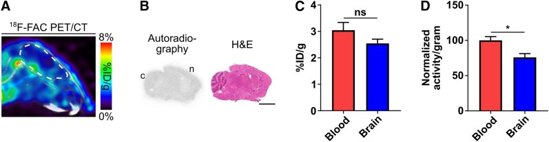
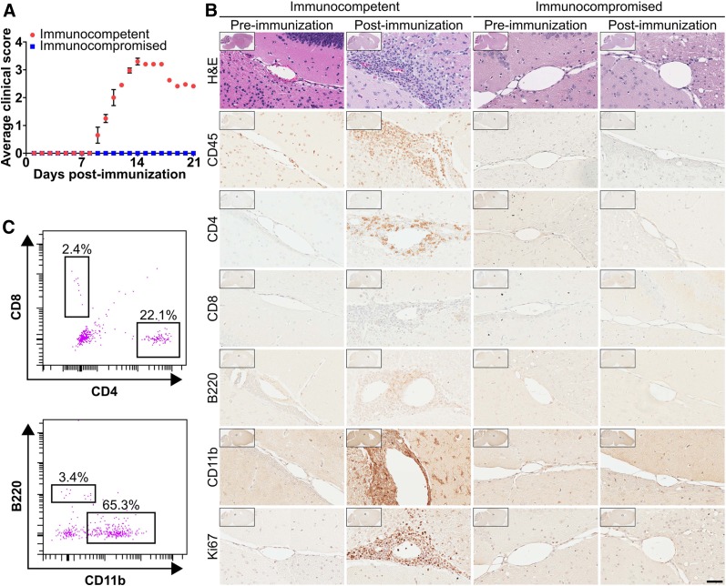
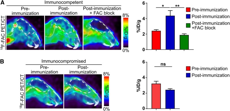
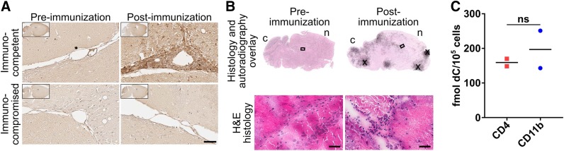
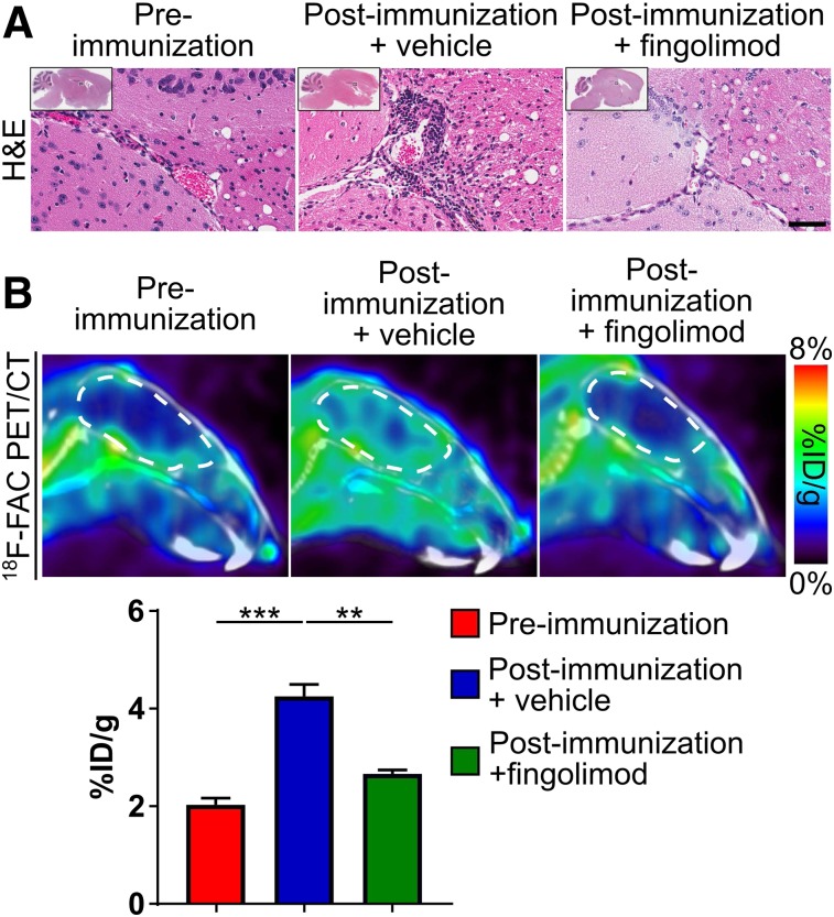
Brain-infiltrating leukocytes contribute to multiple sclerosis (MS) and autoimmune encephalomyelitis and likely play a role in traumatic brain injury, seizure, and stroke. Brain-infiltrating leukocytes are also primary targets for MS disease-modifying therapies. However, no method exists for noninvasively visualizing these cells in a living organism. 1-(2'-deoxy-2'-18F-fluoroarabinofuranosyl) cytosine (18F-FAC) is a PET radiotracer that measures deoxyribonucleoside salvage and accumulates preferentially in immune cells. We hypothesized that 18F-FAC PET could noninvasively image brain-infiltrating leukocytes. Methods: Healthy mice were imaged with 18F-FAC PET to quantify if this radiotracer crosses the blood-brain barrier (BBB). Experimental autoimmune encephalomyelitis (EAE) is a mouse disease model with brain-infiltrating leukocytes. To determine whether 18F-FAC accumulates in brain-infiltrating leukocytes, EAE mice were analyzed with 18F-FAC PET, digital autoradiography, and immunohistochemistry, and deoxyribonucleoside salvage activity in brain-infiltrating leukocytes was analyzed ex vivo. Fingolimod-treated EAE mice were imaged with 18F-FAC PET to assess if this approach can monitor the effect of an immunomodulatory drug on brain-infiltrating leukocytes. PET scans of individuals injected with 2-chloro-2'-deoxy-2'-18F-fluoro-9-β-d-arabinofuranosyl-adenine (18F-CFA), a PET radiotracer that measures deoxyribonucleoside salvage in humans, were analyzed to evaluate whether 18F-CFA crosses the human BBB. Results:18F-FAC accumulates in the healthy mouse brain at levels similar to 18F-FAC in the blood (2.54 ± 0.2 and 3.04 ± 0.3 percentage injected dose per gram, respectively) indicating that 18F-FAC crosses the BBB. EAE mice accumulate 18F-FAC in the brain at 180% of the levels of control mice. Brain 18F-FAC accumulation localizes to periventricular regions with significant leukocyte infiltration, and deoxyribonucleoside salvage activity is present at similar levels in brain-infiltrating T and innate immune cells. These data suggest that 18F-FAC accumulates in brain-infiltrating leukocytes in this model. Fingolimod-treated EAE mice accumulate 18F-FAC in the brain at 37% lower levels than control-treated EAE mice, demonstrating that 18F-FAC PET can monitor therapeutic interventions in this mouse model. 18F-CFA accumulates in the human brain at 15% of blood levels (0.08 ± 0.01 and 0.54 ± 0.07 SUV, respectively), indicating that 18F-CFA does not cross the BBB in humans. Conclusion:18F-FAC PET can visualize brain-infiltrating leukocytes in a mouse MS model and can monitor the response of these cells to an immunomodulatory drug. Translating this strategy into humans will require exploring additional radiotracers.
Keywords: PET imaging; autoimmune disease; brain; leukocytes.
© 2020 by the Society of Nuclear Medicine and Molecular Imaging.
Figures
References
-
- Compston A, Coles A. Multiple sclerosis. Lancet. 2008;372:1502–1517. - PubMed
-
- Dendrou CA, Fugger L, Friese MA. Immunopathology of multiple sclerosis. Nat Rev Immunol. 2015;15:545–558. - PubMed
-
- Traugott U, Reinherz EL, Raine CS. Multiple sclerosis: distribution of T cell subsets within active chronic lesions. Science. 1983;219:308–310. - PubMed
-
- Wingerchuk DM, Carter JL. Multiple sclerosis: current and emerging disease-modifying therapies and treatment strategies. Mayo Clin Proc. 2014;89:225–240. - PubMed
Publication types
MeSH terms
Substances
Grants and funding
LinkOut - more resources
Full Text Sources
Other Literature Sources
Medical