. 2019 Dec 13;86(1):e01993-19.
doi: 10.1128/AEM.01993-19. Print 2019 Dec 13.

Comparative Genomics of Rumen Butyrivibrio spp. Uncovers a Continuum of Polysaccharide-Degrading Capabilities

Affiliations

Comparative Genomics of Rumen Butyrivibrio spp. Uncovers a Continuum of Polysaccharide-Degrading Capabilities

Nikola Palevich et al. Appl Environ Microbiol. .

Abstract

Plant polysaccharide breakdown by microbes in the rumen is fundamental to digestion in ruminant livestock. Bacterial species belonging to the rumen genera Butyrivibrio and Pseudobutyrivibrio are important degraders and utilizers of lignocellulosic plant material. These bacteria degrade polysaccharides and ferment the released monosaccharides to yield short-chain fatty acids that are used by the ruminant for growth and the production of meat, milk, and fiber products. Although rumen Butyrivibrio and Pseudobutyrivibrio species are regarded as common rumen inhabitants, their polysaccharide-degrading and carbohydrate-utilizing enzymes are not well understood. In this study, we analyzed the genomes of 40 Butyrivibrio and 6 Pseudobutyrivibrio strains isolated from the plant-adherent fraction of New Zealand dairy cows to explore the polysaccharide-degrading potential of these important rumen bacteria. Comparative genome analyses combined with phylogenetic analysis of their 16S rRNA genes and short-chain fatty acid production patterns provide insight into the genomic diversity and physiology of these bacteria and divide Butyrivibrio into 3 species clusters. Rumen Butyrivibrio bacteria were found to encode a large and diverse spectrum of degradative carbohydrate-active enzymes (CAZymes) and binding proteins. In total, 4,421 glycoside hydrolases (GHs), 1,283 carbohydrate esterases (CEs), 110 polysaccharide lyases (PLs), 3,605 glycosyltransferases (GTs), and 1,706 carbohydrate-binding protein modules (CBM) with predicted activities involved in the depolymerization and transport of the insoluble plant polysaccharides were identified. Butyrivibrio genomes had similar patterns of CAZyme families but varied greatly in the number of genes within each category in the Carbohydrate-Active Enzymes database (CAZy), suggesting some level of functional redundancy. These results suggest that rumen Butyrivibrio species occupy similar niches but apply different degradation strategies to be able to coexist in the rumen.IMPORTANCE Feeding a global population of 8 billion people and climate change are the primary challenges facing agriculture today. Ruminant livestock are important food-producing animals, and maximizing their productivity requires an understanding of their digestive systems and the roles played by rumen microbes in plant polysaccharide degradation. Members of the genera Butyrivibrio and Pseudobutyrivibrio are a phylogenetically diverse group of bacteria and are commonly found in the rumen, where they are a substantial source of polysaccharide-degrading enzymes for the depolymerization of lignocellulosic material. Our findings have highlighted the immense enzymatic machinery of Butyrivibrio and Pseudobutyrivibrio species for the degradation of plant fiber, suggesting that these bacteria occupy similar niches but apply different degradation strategies in order to coexist in the competitive rumen environment.

Keywords: Butyrivibrio; CAZy; Pseudobutyrivibrio; bacteria; enolase; genome; polysaccharide; rumen.


Figures

FIG 1
FIG 1
FGD of Butyrivibrio (B.) and Pseudobutyrivibrio (P.) genomes. The predicted ORFeomes of all 46 genomes were subjected to an FGD analysis, and the resulting distance matrix was imported into MEGA6 (82). The functional distribution was visualized using the UPGMA method (113, 114). The tree is drawn to scale, with the branch lengths being in the same units as those of the functional distances used to infer the distribution tree. The bar represents the number of nucleotide substitutions per site.
FIG 2
FIG 2
Flower plot diagram of unique, group-specific, and core gene families in the Butyrivibrio and Pseudobutyrivibrio genomes. The core genome is shown in the center circle. Each colored segment represents the number of gene families shared among the four species groups, and the outer petals represent unique gene families for individual genomes.
FIG 3
FIG 3
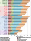
Distribution of each CAZyme class and family in Butyrivibrio and Pseudobutyrivibrio genomes. Colored bars represent the total numbers of genomes that contain members of the specific CAZyme family present in their genomes.
FIG 4
FIG 4
Comparative analysis of annotated Butyrivibrio and Pseudobutyrivibrio CAZymes. The numbers and types of CAZyme modules or domains are represented as colored horizontal bars.
FIG 5
FIG 5
Heat map of normalized relative abundances for CAZyme families determined for the Butyrivibrio and Pseudobutyrivibrio genomes. The relative normalized inferred CAZy gene family abundances per genome (Z-score) are shown using a heat color scheme (red to green), indicating low to high relative abundance. The Bray-Curtis distances (119) of compositional dissimilarity and hierarchical cluster analysis using Ward’s method (120) were used to calculate normalized abundance. Genome names are colored to represent Butyrivibrio cluster 3 in green, Butyrivibrio cluster 2 in red, Butyrivibrio cluster 1 in blue, and Pseudobutyrivibrio in purple.
FIG 6
FIG 6
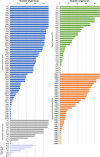
Comparisons of gene presence/absence for enzymes involved in the carbohydrate metabolic pathways in Butyrivibrio leading to the formation of butyrate, formate, acetate, and lactate. All metabolic pathways were compiled using information from the MetaCyc (121) and KEGG (122) databases. The presence or absence of genes encoding particular enzymes within genomes is indicated by full or empty cells, respectively in the panels. The order of genomes in the panels, from left to right, are as follows: row 1, Butyrivibrio cluster 3 (green) strains AB2020, FE2007, MD2001, ND3005, WTE3004, YRB2005, MC2013, and NC3005; row 2, Butyrivibrio cluster 2 (red) strains AE3006, MB2005, WCD3002, AD3002, WCD2001, VCD2006, AE3004, LC3010, WCE2006, AC2005, FC2001, XPD2002, and NC2002; row 3, Butyrivibrio cluster 1 (blue) strains NK4A153, AE2005, AE3003, LB2008, MB2003, AE2015, P6B7, B316T, FD2007, VCB2006, XBB1001, AE3009, MC2021, XPD2006, VCB2001, FCS006, NC2007, FCS014, and AE2032; and row 4, Pseudobutyrivibrio (purple) strains MA3014, HUN009, CF1b, AD2017, LB2011, and MD2005. The enolase-catalyzed reaction is shown in red, as the gene was absent from a number of Butyrivibrio strains. Color schemes for the metabolism pathways are as follows: the formation of formate in blue, acetate in green, butyrate in purple, l-lactate in red, and d-lactate by the proposed methylglyoxal shunt in orange (69). Abbreviations: DHAP, dihydroxyacetone phosphate; DKI, 5-keto-4-deoxyuronate; DKII, 2,5-diketo3-deoxygluconate; KDG, 2-keto-3-deoxygluconate; KDGP, 2-keto-3-deoxy-gluconate phosphate. Abbreviations for sugar transport systems are as follows: ABC, ATP binding cassette; MFS, major facilitator superfamily.

References

    1. Godfray HCJ, Beddington JR, Crute IR, Haddad L, Lawrence D, Muir JF, Pretty J, Robinson S, Thomas SM, Toulmin C. 2010. Food security: the challenge of feeding 9 billion people. Science 327:812–818. doi:10.1126/science.1185383. - DOI - PubMed
    1. Eisler MC, Lee MRF, Tarlton JF, Martin GB, Beddington J, Dungait JAJ, Greathead H, Liu J, Mathew S, Miller H, Misselbrook T, Murray P, Vinod VK, Van Saun R, Winter M. 2014. Agriculture: steps to sustainable livestock. Nature 507:32–34. doi:10.1038/507032a. - DOI - PubMed
    1. Hess M, Sczyrba A, Egan R, Kim T-W, Chokhawala H, Schroth G, Luo S, Clark DS, Chen F, Zhang T, Mackie RI, Pennacchio LA, Tringe SG, Visel A, Woyke T, Wang Z, Rubin EM. 2011. Metagenomic discovery of biomass-degrading genes and genomes from cow rumen. Science 331:463–467. doi:10.1126/science.1200387. - DOI - PubMed
    1. Koike S, Yoshitani S, Kobayashi Y, Tanaka K. 2003. Phylogenetic analysis of fiber-associated rumen bacterial community and PCR detection of uncultured bacteria. FEMS Microbiol Lett 229:23–30. doi:10.1016/S0378-1097(03)00760-2. - DOI - PubMed
    1. McAllister TA, Bae HD, Jones GA, Cheng KJ. 1994. Microbial attachment and feed digestion in the rumen. J Anim Sci 72:3004–3018. doi:10.2527/1994.72113004x. - DOI - PubMed

Publication types

LinkOut - more resources