. 2019 Oct 25;9(1):15302.
doi: 10.1038/s41598-019-51636-5.

IL-21 regulates SOCS1 expression in autoreactive CD8+ T cells but is not required for acquisition of CTL activity in the islets of non-obese diabetic mice

Affiliations

IL-21 regulates SOCS1 expression in autoreactive CD8+ T cells but is not required for acquisition of CTL activity in the islets of non-obese diabetic mice

Andrew P R Sutherland et al. Sci Rep. .

Abstract

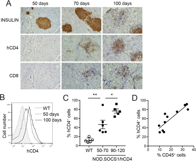
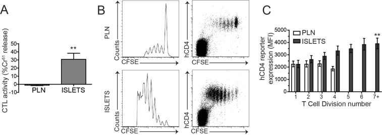
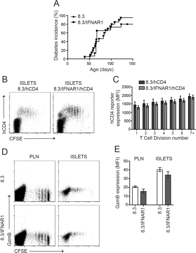
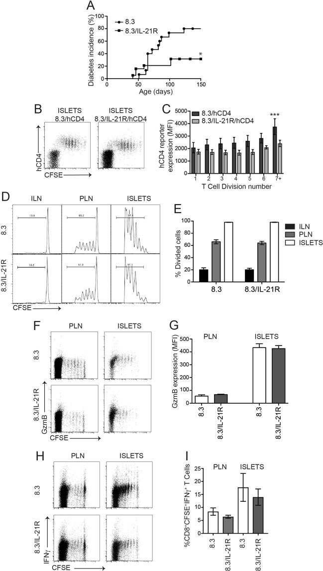
In type 1 diabetes, maturation of activated autoreactive CD8+ T cells to fully armed effector cytotoxic T lymphocytes (CTL) occurs within the islet. At present the signals required for the maturation process are poorly defined. Cytokines could potentially provide the necessary "third signal" required to generate fully mature CTL capable of killing insulin-producing β-cells. To determine whether autoreactive CTL within islets respond to cytokines we generated non-obese diabetic (NOD) mice with a reporter for cytokine signalling. These mice express a reporter gene, hCD4, under the control of the endogenous regulatory elements for suppressor of cytokine signalling (SOCS)1, which is itself regulated by pro-inflammatory cytokines. In NOD mice, the hCD4 reporter was expressed in infiltrated islets and the expression level was positively correlated with the frequency of infiltrating CD45+ cells. SOCS1 reporter expression was induced in transferred β-cell-specific CD8+ 8.3T cells upon migration from pancreatic draining lymph nodes into islets. To determine which cytokines induced SOCS1 promoter activity in islets, we examined hCD4 reporter expression and CTL maturation in the absence of the cytokine receptors IFNAR1 or IL-21R. We show that IFNAR1 deficiency does not confer protection from diabetes in 8.3 TCR transgenic mice, nor is IFNAR1 signalling required for SOCS1 reporter upregulation or CTL maturation in islets. In contrast, IL-21R-deficient 8.3 mice have reduced diabetes incidence and reduced SOCS1 reporter activity in islet CTLs. However IL-21R deficiency did not affect islet CD8+ T cell proliferation or expression of granzyme B or IFNγ. Together these data indicate that autoreactive CD8+ T cells respond to IL-21 and not type I IFNs in the islets of NOD mice, but neither IFNAR1 nor IL-21R are required for islet intrinsic CTL maturation.


Conflict of interest statement

The authors declare no competing interests.

Figures

Figure 1
Figure 1
SOCS1 reporter expression in islets of NOD mice. (A) Serial pancreas sections from 50-, 70 and 100-day old female NOD.SOCS1/hCD4 mice were stained with antibodies to insulin, human CD4 (hCD4) and murine CD8. Representative sections from n = 3 mice/age are shown. Magnification 200x. (BD) Islets were isolated from female NOD.SOCS1/hCD4 mice or wild-type littermates at 50–70 days of age and 90–120 days of age. The proportion of CD45+ cells and hCD4+ cells in the islets was determined by flow cytometry. (B) Representative histograms of hCD4 staining in CD45 islet cells of wild-type (WT) and NOD.SOCS1/hCD4 mice at 50- and 100-days of age. (C) Pooled data showing mean ± SEM of the %CD45hCD4+ cells in the islets of individual mice (n = 5–6/group). **p = 0.0056, *p = 0.016 one-way ANOVA with Sidak’s post-test for multiple comparisons. (D) Correlation of %CD45hCD4+ cells with %CD45+ cells in the islets of NOD.SOCS1/hCD4 mice. R2 = 0.763, p = 0.0004, linear regression analysis.
Figure 2
Figure 2
SOCS1 reporter expression is increased during CD8+ CTL differentiation in NOD islets. (A) Pancreatic lymph nodes and islets were isolated 5 days after 8.3 splenocyte transfer, CFSE positive cells were isolated by cell sorting and CTL activity quantified in vitro. Data show mean + SEM from 3 independent experiments. **p < 0.01. (B) Representative plots of pancreatic lymph nodes (PLN) and islets isolated 5 days after 8.3/hCD4 splenocyte transfer showing expression of hCD4 and CFSE dilution. (C) Expression of hCD4 (mean fluorescence intensity, MFI) on CD8+CFSE+ T cells for each cell division was calculated using CFSE dilution profiles. Data show mean ± SEM for n = 6 mice from 3 independent experiments. **p < 0.01, one-way ANOVA comparing division 1 and division 7.
Figure 3
Figure 3
IFNAR1 is not required for SOCS1 upregulation or CTL differentiation in 8.3 CD8+ T cells in NOD islets. (A) Diabetes incidence in 8.3/IFNAR1 and 8.3 controls (n = 15–20 mice per group). (B) Representative plots of islets isolated 5 days after 8.3/hCD4 or 8.3/IFNAR1/hCD4 splenocyte transfer showing expression of hCD4 and CFSE dilution. (C) Expression of hCD4 (MFI) on CD8+CFSE+ T cells for each cell division calculated using CFSE dilution profiles. Data show mean ± SEM for n = 7–8 mice from 3 independent experiments. Difference between MFI in division 1 and MFI in division 7 for wild-type (p = 0.03) and IFNAR1 (p = 0.0003) cells, one-way ANOVA with Sidak’s post-test for multiple comparisons. (D) Representative plots of pancreatic lymph nodes (PLN) and islets isolated 5 days after 8.3 or 8.3/IFNAR1 splenocyte transfer showing expression of granzyme B and CFSE dilution. (E) Mean granzyme B expression (MFI) on CD8+ T cells in pancreatic lymph nodes and islets. Data show mean ± SEM for n = 4–5 mice from 3 independent experiments. Difference between 8.3 and 8.3/IFNAR1 not statistically significant.
Figure 4
Figure 4
IL-21R is required for maximal SOCS1 upregulation but not CTL differentiation in 8.3 CD8+ T cells in NOD islets. (A) Diabetes incidence in 8.3/IL-21R and 8.3 controls (n = 15–19 mice per group, p < 0.05 Log-rank (Mantel-Cox) test). (B) Representative plots from islets isolated 5 days after 8.3/IL-21R/hCD4 splenocyte transfer showing expression of hCD4 and CFSE dilution. (C) Expression of hCD4 (MFI) on CD8+CFSE+ T cells for each cell division calculated using CFSE dilution profiles. Data show mean ± SEM for n = 3–8 mice from 3 independent experiments. Difference between genotypes ***p < 0.001 two-way ANOVA. Difference between MFI in division 1 and MFI in division 7 for wild-type p = 0.02, one-way ANOVA with Sidak’s post-test for multiple comparisons. (D) Representative histograms of inguinal lymph node (ILN), pancreatic lymph nodes (PLN) and islets isolated 5 days after 8.3/IL-21R splenocyte transfer showing CFSE dilution. (E) Mean ± SEM frequency of CD8+ T cell division in inguinal lymph nodes, pancreatic lymph nodes and islets of n = 12 mice from 3 independent experiments. Genotypes not statistically different. (F) Representative plots of pancreatic lymph nodes and islets isolated 5 days after 8.3/IL-21R splenocyte transfer showing expression of granzyme B and CFSE dilution. (G) Expression of granzyme B (MFI) on CD8+ T cells in pancreatic lymph nodes and islets. Data show mean ± SEM of n = 8 mice from 4 independent experiments. Genotypes not statistically different. (H) Representative plots of pancreatic lymph nodes and islets isolated 5 days after 8.3/IL-21R splenocyte transfer, restimulated with IGRP for 6 hours in vitro showing expression of IFNγ and CFSE dilution. (I) Frequency of CD8+CFSE+IFNγ+ cells (%) in pancreatic lymph nodes and islets. Data show mean ± SEM of n = 5–10 mice from 3 independent experiments. Genotypes not statistically different.

References

    1. Anderson MS, Bluestone JA. The NOD mouse: a model of immune dysregulation. Annu Rev Immunol. 2005;23:447–485. doi: 10.1146/annurev.immunol.23.021704.115643. - DOI - PubMed
    1. Graham KL, et al. Pathogenic mechanisms in type 1 diabetes: the islet is both target and driver of disease. Rev Diabet Stud. 2012;9:148–168. doi: 10.1900/RDS.2012.9.148. - DOI - PMC - PubMed
    1. Graham KL, et al. Autoreactive cytotoxic T lymphocytes acquire higher expression of cytotoxic effector markers in the islets of NOD mice after priming in pancreatic lymph nodes. Am J Pathol. 2011;178:2716–2725. doi: 10.1016/j.ajpath.2011.02.015. - DOI - PMC - PubMed
    1. Graham KL, et al. Intra-islet proliferation of cytotoxic T lymphocytes contributes to insulitis progression. Eur J Immunol. 2012;42:1717–1722. doi: 10.1002/eji.201242435. - DOI - PubMed
    1. Sutherland RM, et al. Cognate antigen engagement on parenchymal cells stimulates CD8(+) T cell proliferation in situ. Nature communications. 2017;8:14809. doi: 10.1038/ncomms14809. - DOI - PMC - PubMed

Publication types

MeSH terms

Substances