. 2019 Oct 25;9(1):15303.
doi: 10.1038/s41598-019-51698-5.

Retinal artery occlusion is associated with compositional and functional shifts in the gut microbiome and altered trimethylamine-N-oxide levels

Affiliations

Retinal artery occlusion is associated with compositional and functional shifts in the gut microbiome and altered trimethylamine-N-oxide levels

Denise C Zysset-Burri et al. Sci Rep. .

Abstract

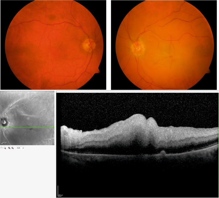
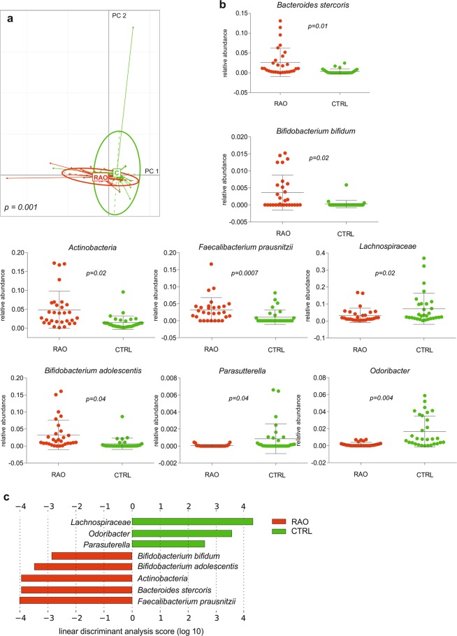
Retinal artery occlusion (RAO) is a sight threatening complication of cardiovascular disease and commonly occurs due to underlying atherosclerosis. As cardiovascular disease and atherosclerosis in particular has been associated with compositional alterations in the gut microbiome, we investigated this association in patients with clinically confirmed non-arteritic RAO compared to age- and sex-matched controls. On the phylum level, the relative abundance of Bacteroidetes was decreased in patients with RAO compared to controls, whereas the opposite applied for the phylum of Proteobacteria. Several genera and species such as Actinobacter, Bifidobacterium spp., Bacteroides stercoris, Faecalibacterium prausnitzii were relatively enriched in patients with RAO, whereas others such as Odoribacter, Parasutterella or Lachnospiraceae were significantly lower. Patient's gut microbiomes were enriched in genes of the cholesterol metabolism pathway. The gut derived, pro-atherogenic metabolite trimethylamine-N-oxide (TMAO) was significantly higher in patients with RAO compared to controls (p = 0.023) and a negative correlation between relative abundances of genera Parasutterella and Lachnospiraceae and TMAO levels and a positive correlation between relative abundance of genus Akkermansia and TMAO levels was found in study subjects. Our findings proposes that RAO is associated with alterations in the gut microbiome and with elevated TMAO levels, suggesting that RAO could be targeted by microbiome-altering interventions.


Conflict of interest statement

The authors declare no competing interests.

Figures

Figure 1
Figure 1
Representative images of a patient with central retinal artery occlusion. Top: Representative color photographs of the unaffected right eye and the left eye with typical features of a central artery occlusion with ischemia and edema of the retina and a cherry red spot. Bottom: Optical coherence tomography (OCT) images of the same patient showing a scan through the right retina. Swelling of the inner retinal layers is evident due to occlusion of the central retinal artery.
Figure 2
Figure 2
Taxonomic characterization of the gut microbiome. Relative abundances of microbiota at phyla level in all study subjects (a) and averaged for study groups (b). There is a decrease in relative abundance of Bacteroidetes with respect to Proteobacteria in the RAO cohort. Relative abundances of microbiota at genus level in all study subjects (c) and averaged for study goups (d). There is a decrease of Bacteroides in the RAO group compared to controls. CTRL, c ontrol (n = 30); RAO, retinal artery occlusion (n = 29).
Figure 3
Figure 3
Gut enterotypes in the cohort. Three enterotypes exist in the cohort based on the abundance of microbial genera. Patients (n = 29) and controls (n = 30) are denoted by squares (□) and triangles (∆), respectively (a). Boxplots represent the abundance of Bacteroides, Eubacterium and Prevotella, the proposed drivers of the three enterotypes (b). Blue is enterotype 1, purple is enterotype 2 and orange is enterotype 3.
Figure 4
Figure 4
Distinct microbial composition between retinal artery occlusion (RAO) patients and control. (a) Principal component analysis of microbial species abundance grouped patients and controls separately, with PERMANOVA confirming a significant difference between the groups (p = 0.001). (b) Box plots representing the mean abundance +/− s.d. of bacterial genera and species associated with RAO (Kruskal-Wallis test, p < 0.05). (c) LDA (linear discriminant analysis) score plot of differentially abundant taxonomic features among groups (LDA score for discriminative features >2.0). Red is patients (RAO, n = 29), green is controls (CTRL, n = 30).
Figure 5
Figure 5
Species-specific microbial pathways associated with retinal artery occlusion (RAO). (a) Principal component analysis of microbial pathway abundance grouped patients and controls separately, with PERMANOVA confirming a significant difference between the groups (p = 0.003). (b) Box plots representing the abundance of pathways associated with RAO and illustration of the respective pathways which differed in patients with RAO and control subjects (Kruskal-Wallis test, p < 0.05). Red is patients (RAO, n = 29), green is controls (CTRL, n = 30).
Figure 6
Figure 6
Trimethylamine-N-oxide (TMAO) concentration associated with retinal artery occlusion (RAO). (a) The TMAO concentration was significantly higher in a subgroup of 12 RAO patients compared to 11 controls (Fisher’s exact test, p = 0.023). (b) No significant difference in the TMAO concentration among the three enterotypes has been found (Kruskal Wallis test, p > 0.05). Abundance of carnitine oxygenase (cntA, c) and choline trimethylamine-lyase (cutC, d) in patients (RAO, n = 29) and controls (CTRL, n = 30) as well as in the TMAO subgroup of 12 patients (RAO TMAO) and 11 controls (CTRL TMAO) as percentage of total reads. Obtained p values (Fisher’s exact test) are indicated. Red is patient (RAO), green is controls (CTRL), blue is enterotype 1, purple is enterotype 2 and orange is enterotype 3.

References

    1. Hayreh SS. Acute retinal arterial occlusive disorders. Progress in retinal and eye research. 2011;30:359–394. doi: 10.1016/j.preteyeres.2011.05.001. - DOI - PMC - PubMed
    1. Dunlap AB, Kosmorsky GS, Kashyap VS. The fate of patients with retinal artery occlusion and Hollenhorst plaque. Journal of vascular surgery. 2007;46:1125–1129. doi: 10.1016/j.jvs.2007.07.054. - DOI - PubMed
    1. Hayreh SS, Podhajsky PA, Zimmerman MB. Retinal artery occlusion: associated systemic and ophthalmic abnormalities. Ophthalmology. 2009;116:1928–1936. doi: 10.1016/j.ophtha.2009.03.006. - DOI - PMC - PubMed
    1. The Lipid Research Clinics Coronary Primary Prevention Trial results I. Reduction in incidence of coronary heart disease. Jama. 1984;251:351–364. doi: 10.1001/jama.1984.03340270029025. - DOI - PubMed
    1. Frost PH, et al. Serum lipids and incidence of coronary heart disease. Findings from the Systolic Hypertension in the Elderly Program (SHEP) Circulation. 1996;94:2381–2388. doi: 10.1161/01.CIR.94.10.2381. - DOI - PubMed

MeSH terms

Supplementary concepts