Review
. 2019 Dec;48(4):765-778.
doi: 10.1016/j.ecl.2019.08.007. Epub 2019 Sep 19.

Renovascular Hypertension

Affiliations
Review

Renovascular Hypertension

Sandra M Herrmann et al. Endocrinol Metab Clin North Am. 2019 Dec.

Abstract

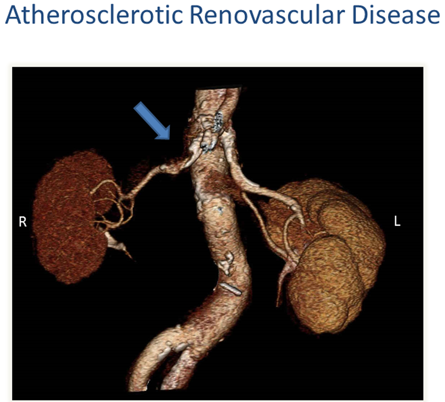
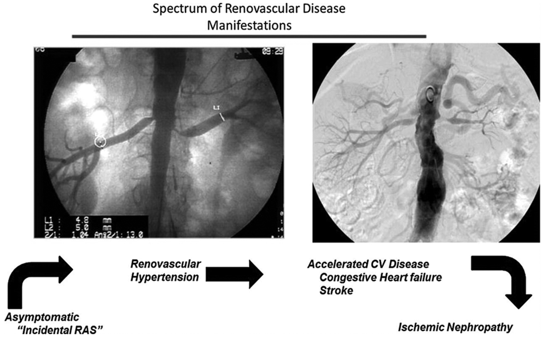
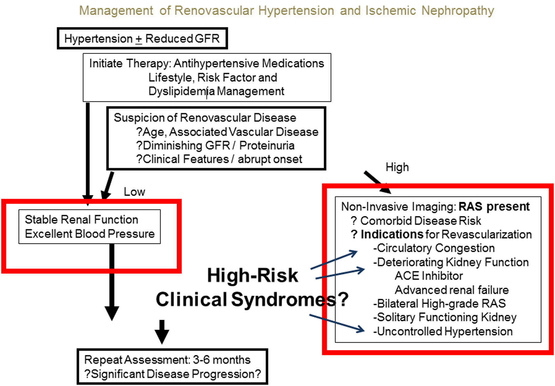
Renovascular disease (RVD) is a major cause of secondary hypertension. Atherosclerotic renal artery stenosis is the most common type of RVD followed by fibromuscular dysplasia. It has long been recognized as the prototype of angiotensin-dependent hypertension. However, the mechanisms underlying the physiopathology of hypertensive occlusive vascular renal disease are complex and distinction between the different causes of RVD should be made. Recognition of these distinct types of RVD with different degrees of renal occlusive disease is important for management. The greatest challenge is to individualize and implement the best approach for each patient in the setting of widely different comorbidities.

Keywords: Hypertension; Ischemic nephropathy; Renal artery stenosis; Renin and angiotensin; Renovascular disease.


Figures

Figure 1:
Figure 1:
CT angiography depicting moderate atherosclerotic renovascular disease affecting the right kidney (arrow). Right (R); Left (L)
Figure 2:
Figure 2:
Studies from human subjects with translesional pressure gradients indicate that an aortic-renal pressure gradient of 10% to 20% is necessary to detect renin release. Mean aortic pressure (Pd); mean pressure distal to the renal artery stenosis (Pa). (From De Bruyne B, Manoharan G, Pijls NHJ, et al. Assessment of renal artery stenosis severity by pressure gradient measurements. J Am Coll Cardiol. 2006; 48:1851–1855)
Figure 3:
Figure 3:
Oxygenation is threatened lower in the post-stenotic kidney as compared to the nonstenotic contralateral kidney: Representative coronal blood oxygen level-dependent images. The R2* map (reflecting the level of deoxyhemoglobin) of the stenotic kidney shows a hypoxic cortical zone and widespread areas of elevated deoxyhemoglobin in the medullary segments (red). The unaffected contralateral kidney depicts the R2* map with a lower (blue) cortical zone and more gradual development of deeper medullary areas of hypoxia, and it is close to the appearance of a normal nonatherosclerotic renal artery stenosis kidney.(From Herrmann SM, Saad A, Eirin A, et al. Differences in GFR and Tissue Oxygenation, and Interactions between Stenotic and Contralateral Kidneys in Unilateral Atherosclerotic Renovascular Disease. Clin J Am Soc Nephrol. 2016 Mar 7; 11(3):458–69)
Figure 4:
Figure 4:
Spectrum of atherosclerotic renovascular disease: Left panel, Aortogram obtained during coronary angiography demonstrating moderate incidental stenosis of both renal arteries in a 67-year-old man with symptomatic coronary disease. Right panel, More severe occlusive disease observed in a 68-year-old woman presenting with severe hypertension and episodes of flash pulmonary edema. Different clinical manifestations depending on severity of vascular occlusion, from asymptomatic hypertension to ischemic nephropathy. As occlusion progresses this leads to accelerated hypertension, circulatory congestion, and ultimately threatens viability of the kidney. (From Herrmann SM, Saad A, Textor SC. Management of Atherosclerotic Renovascular Disease after Cardiovascular Outcomes in Renal Atherosclerotic Lesions (CORAL). Nephrol Dial Transplant. 2015 Mar;30(3):366–75.)
Figure 5:
Figure 5:
Management of RVH and ischemic nephropathy. The goal is to reduce morbidity associated with hypertension by controlling blood pressure and preserving kidney function. If medical therapy fails or renovascular disease progresses, revascularization of renal artery should be considered. (From Herrmann SM, Saad A, Textor SC. Management of atherosclerotic renovascular disease after Cardiovascular Outcomes in Renal Atherosclerotic Lesions (CORAL). Nephrol Dial Transplant. 2015 Mar;30(3):366–75)

References

    1. Herrmann SM, Textor SC. Current Concepts in the Treatment of Renovascular Hypertension. American Journal of Hypertension 2018;31:139–49. - PMC - PubMed
    1. Goldblatt H, Lynch J, Hanzal RF, Summerville WW. Studies on Experimental Hypertension: I. The Production of Persistent Elevation of Systolic Blood Pressure by Means of Renal Ischemia. The Journal of Experimental Medicine 1934;59:347–79. - PMC - PubMed
    1. Basso N, Terragno NA. History about the discovery of the renin-angiotensin system. Hypertension (Dallas, Texas : 1979) 2001;38:1246–9. - PubMed
    1. Stanley JC. David M. Hume memorial lecture. Surgical treatment of renovascular hypertension. American Journal of Surgery 1997;174:102–10. - PubMed
    1. Drieghe B, Madaric J, Sarno G, et al. Assessment of renal artery stenosis: side-by-side comparison of angiography and duplex ultrasound with pressure gradient measurements. European Heart Journal 2008;29:517–24. - PubMed

Publication types

MeSH terms

Supplementary concepts