. 2019;207(2):97-113.
doi: 10.1159/000503280. Epub 2019 Oct 25.

Investigating the Osteoinductive Potential of a Decellularized Xenograft Bone Substitute

Affiliations

Investigating the Osteoinductive Potential of a Decellularized Xenograft Bone Substitute

Daniel N Bracey et al. Cells Tissues Organs. 2019.

Abstract

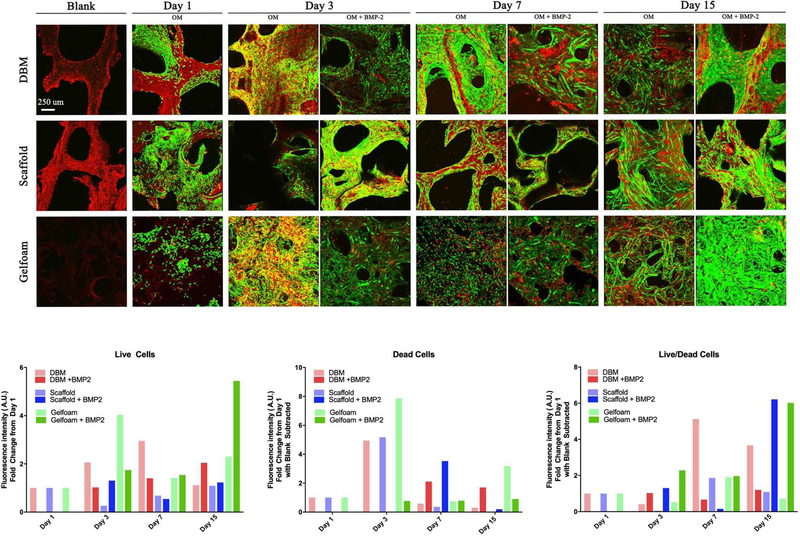
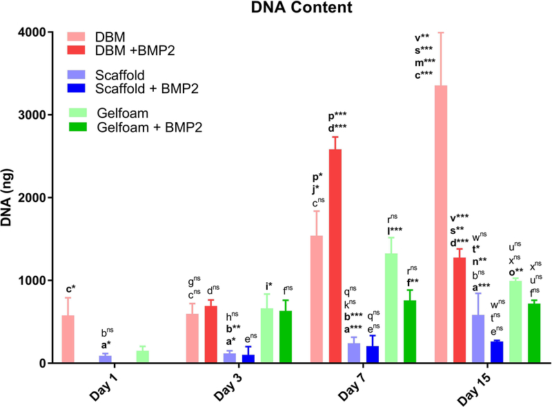
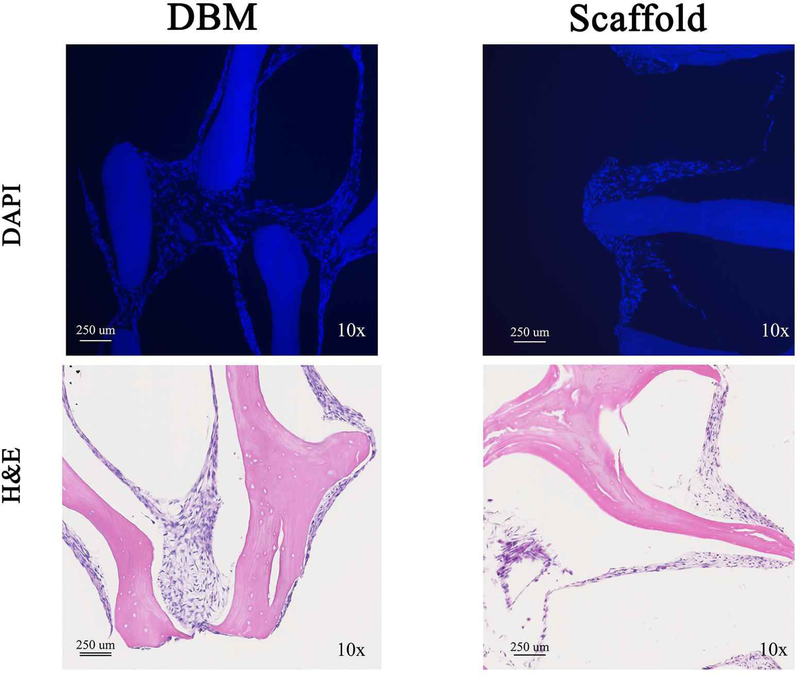
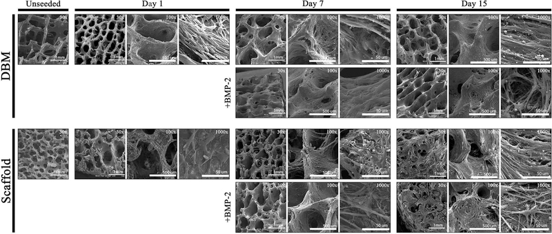
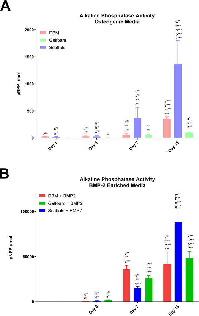
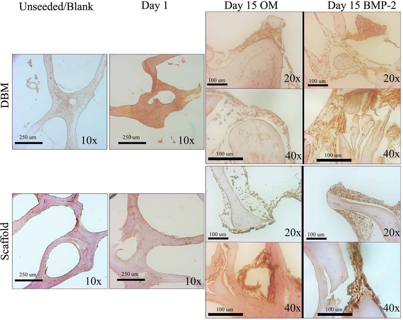
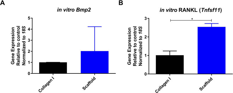
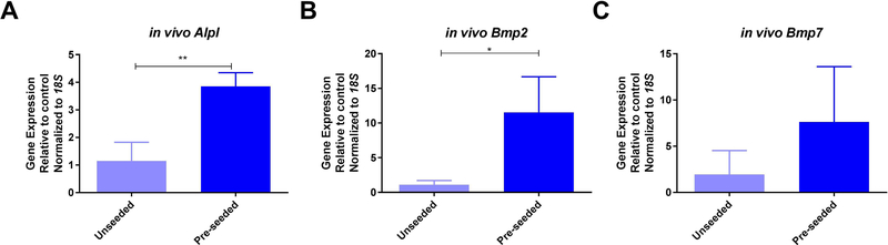
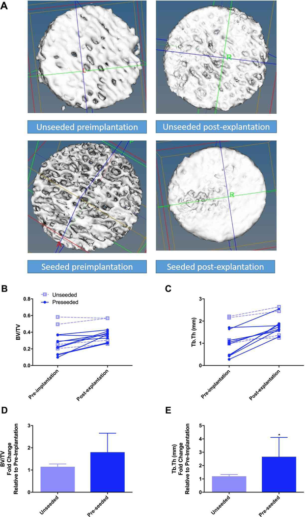
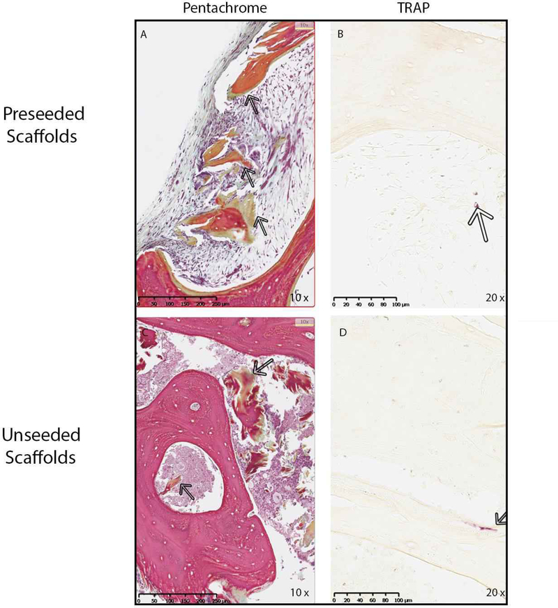
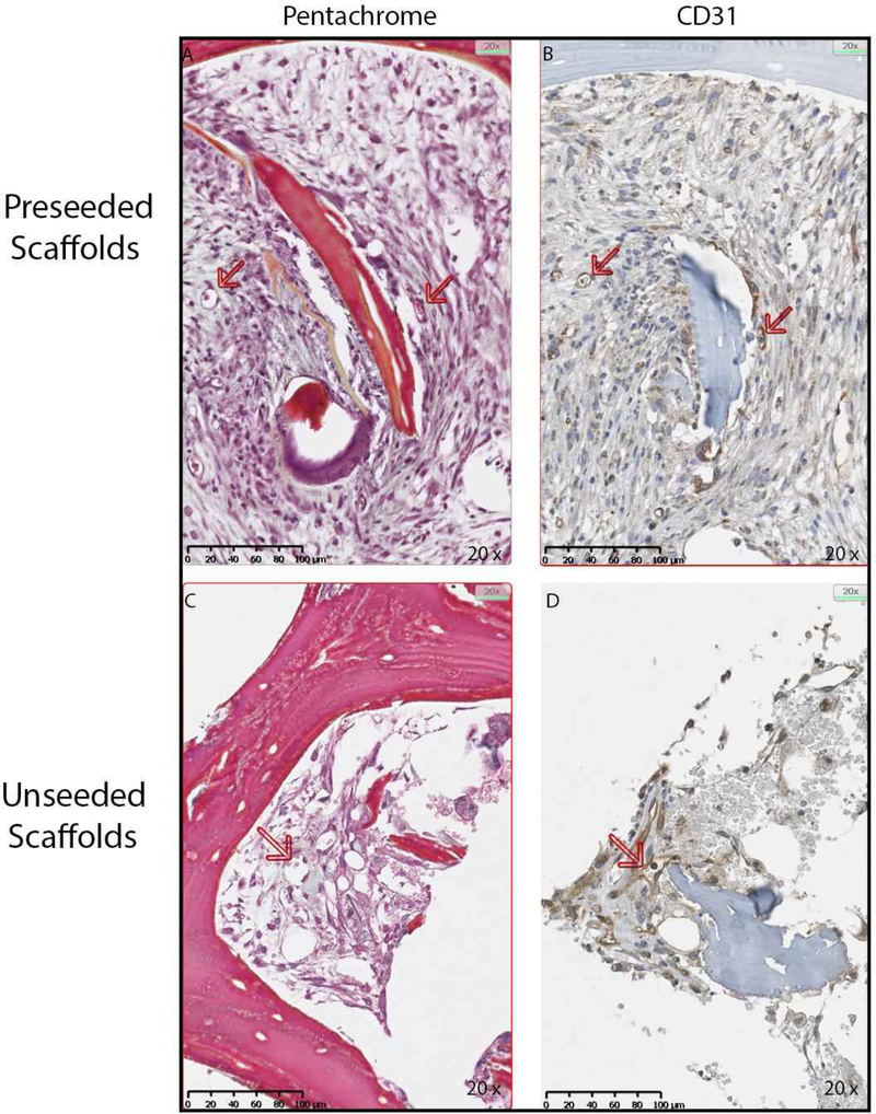
Bone grafting is the second most common tissue transplantation procedure worldwide. One of the alternative methods for bone repair under investigation is a tissue-engineered bone substitute. An ideal property of tissue-engineered bone substitutes is osteoinductivity, defined as the ability to stimulate primitive cells to differentiate into a bone-forming lineage. In the current study, we use a decellularization and oxidation protocol to produce a porcine bone scaffold and examine whether it possesses osteoinductive potential and can be used to create a tissue-engineered bone microenvironment. The decellularization protocol was patented by our lab and consists of chemical decellularization and oxidation steps using combinations of deionized water, trypsin, antimicrobials, peracetic acid, and triton-X100. To test if the bone scaffold was a viable host, preosteoblasts were seeded and analyzed for markers of osteogenic differentiation. The osteoinductive potential was observed in vitro with similar osteogenic markers being expressed in preosteoblasts seeded on the scaffolds and demineralized bone matrix. To assess these properties in vivo, scaffolds with and without preosteoblasts preseeded were subcutaneously implanted in mice for 4 weeks. MicroCT scanning revealed 1.6-fold increased bone volume to total volume ratio and 1.4-fold increase in trabecular thickness in scaffolds after implantation. The histological analysis demonstrates new bone formation and blood vessel formation with pentachrome staining demonstrating osteogenesis and angiogenesis, respectively, within the scaffold. Furthermore, CD31+ staining confirmed the endothelial lining of the blood vessels. These results demonstrate that porcine bone maintains its osteoinductive properties after the application of a patented decellularization and oxidation protocol developed in our laboratory. Future work must be performed to definitively prove osteogenesis of human mesenchymal stem cells, biocompatibility in large animal models, and osteoinduction/osseointegration in a relevant clinical model in vivo. The ability to create a functional bone microenvironment using decellularized xenografts will impact regenerative medicine, orthopedic reconstruction, and could be used in the research of multiple diseases.

Keywords: Angiogenesis; Bone microenvironment; Bone scaffold; Osteoinductivity; Tissue engineering.


Figures

Figure 1.
Figure 1.. C2C12 cells are viable on the decellularized bone scaffold.
Demineralized Bone Matrix (DBM), scaffold, and gelfoam matrices were seeded with 1 million C2C12 pre-osteoblast cells, incubated for 1, 3, 7, or 15 days. On day 1, matrices were switched into osteogenic media (OM) enriched with 100 ng/mL BMP-2. Representative micrographs show green fluorophore (calcein AM) staining of live cells and red fluorophore (ethidium) staining of dead cells. Constructs had notable autofluorescence with ethidium staining as shown in the “blank” images. Cross-sectional images were captured at 10X magnification and overlaid to create the shown 3D projections. Cell density increased with time on all three matrices, consistent with DNA quantification results. Fluorescence in each channel was quantified and represented as fold change in fluorescence intensity from Day 1 ± SD.
Figure 2.
Figure 2.. C2C12 cells proliferate slowly on the decellularized bone constructs.
DBM (red bars), bone scaffold (blue bars), or gelfoam (green bars) constructs were treated with control media, osteogenic media (OM), or OM with 100 ng/mL BMP-2 starting on day 2. Constructs were harvested at days 1, 3, 7, and 15, DNA content was measured with the PicoGreen® assay, and represented as mean DNA mass ± SD. * represents p<0.05, ** represents <0.01, and *** represents p<0.001 by two-way ANOVA for construct and treatment and one-way ANOVA for time within each construct and treatment. Comparisons are annotated as below: (a) scaffold vs DBM; (b) scaffold vs gelfoam; (c) DBM vs gelfoam (d) DBM OM vs OM + BMP-2; (e) scaffold OM vs OM + BMP-2 (f) gelfoam OM vs OM + BMP-2; (g) DBM day 1 vs 3; (h) scaffold day 1 vs 3; (i) gelfoam day 1 vs 3; (j) DBM day 1 vs 7; (k) scaffold day 1 vs 7; (l) gelfoam day 1 vs 7; (m) DBM day 1 vs 15; (n) scaffold day 1 vs 15; (o) gelfoam day 1 vs 15; (p) DBM day 3 vs 7; (q) scaffold day 3 vs 7; (r) gelfoam day 3 vs 7 (s) DBM day 3 vs 15; (t) scaffold day 3 vs 15; (u) gelfoam day 3 vs 15 (v) DBM day 7 vs 15; (w) scaffold day 7 vs 15; (x) gelfoam day 7 vs 15
Figure 3.
Figure 3.. C2C12 cell density is increased on DBM compared with scaffolds.
C2C12 cells were seeded on DBM (left panels) or decellularized bone scaffold (right panels). Seeded scaffolds were sectioned and stained for DAPI (top panels) or H&E (bottom panels). Representative images are shown taken at 10X.
Figure 4.
Figure 4.. C2C12 attachment and spreading is equivalent on bone scaffolds and DBM
C2C12-seeded DBM and bone scaffolds were analyzed by scanning electron microscopy at days 1, 7, or 15 and compared to unseeded scaffolds. Comparison with 100 ng/mL BMP-2 treated scaffolds are shown. Representative images at 30X (scale bar represents 1 mm), 100X (scale bar represents 500 μm), and 1000X (scale bar represents 50 μm) are shown.
Figure 5.
Figure 5.. Scaffolds induce ALP activity in C2C12 cells.
C2C12-seeded constructs: DBM (red bars), bone scaffolds (blue bars), or Gelfoam (green bars), were incubated in osteogenic media (A) or in the presence of 100 ng/mL BMP-2 (B) with treatments starting on day 2 and analyzed for enzymatic ALP activity represented as mean total p-nitrophenyl phosphate (pNPP) content ± SD. * represents p<0.05, ** represents <0.01, and *** represents p<0.001 by two-way ANOVA for construct and treatment and one-way ANOVA for time within each construct and treatment. Comparisons are annotated as described below: (a) scaffold vs DBM; (b) scaffold vs gelfoam; (c) DBM vs gelfoam (d) DBM OM vs OM + BMP-2; (e) scaffold OM vs OM + BMP-2 (f) gelfoam OM vs OM + BMP-2; (g) DBM day 1 vs 3; (h) scaffold day 1 vs 3; (i) gelfoam day 1 vs 3; (j) DBM day 1 vs 7; (k) scaffold day 1 vs 7; (l) gelfoam day 1 vs 7; (m) DBM day 1 vs 15; (n) scaffold day 1 vs 15; (o) gelfoam day 1 vs 15; (p) DBM day 3 vs 7; (q) scaffold day 3 vs 7; (r) gelfoam day 3 vs 7 (s) DBM day 3 vs 15; (t) scaffold day 3 vs 15; (u) gelfoam day 3 vs 15 (v) DBM day 7 vs 15; (w) scaffold day 7 vs 15; (x) gelfoam day 7 vs 15
Figure 6.
Figure 6.. ALP expression increases over time in both DBM and scaffolds.
DBM (top panels) or decellularized bone (bottom panels) scaffolds were sectioned unseeded, after 1-day culture of C2C12 cells or after 15 days in OM or OM with 100 ng/mL BMP-2. Sections were stained for ALP expression by immunohistochemistry, and representative images are shown at 10X, 20X, and 40X. Images were taken at different locations and did not represent subsets of each other.
Figure 7.
Figure 7.. Scaffolds induce MC3T3-E1 osteogenic differentiation.
MC3T3-E1 cells were seeded on 10 μg/mL collagen I (black bars) or decellularized bone scaffolds (blue bars) for one week. Gene expression of Bmp2 (A) and RANKL (Tnsfs11) (B) were measured and represented as mean fold change normalized to 18S ± SD. * represents p<0.05 by t-test.
Figure 8.
Figure 8.. Implantation of scaffolds in vivo induces osteogenic gene expression.
Bone scaffolds were seeded with MC3T3-E1 pre-osteoblast cells (light blue bars) or left unseeded (dark blue bars) and implanted subcutaneously in syngeneic mice. After four weeks scaffolds were removed, processed, and analyzed for gene expression of ALP (Alpl) (A), Bmp2 (B), and Bmp7 (C) represented as mean fold change normalized to 18S ± SD. * represents p<0.05 by t-test.
Figure 9.
Figure 9.. New bone formation occurs in implanted bone constructs.
Bone scaffold structure was analyzed by microCT (pre-implantation) and then implanted subcutaneously in mice either unseeded (open squares) or pre-seeded with MC3T3-E1 pre-osteoblast cells (closed circles). Representative microCT 3D projections of the bone scaffolds prior to implantation (left) and after explantation (right) from unseeded (top row) and seeded (bottom row), grossly demonstrating an appearance of bone formation are shown in A. Implants were removed after four weeks and scanned again by microCT (post-explantation). Bone volume ratio (BV/TV, B) and trabecular thickness (Tb.Th, C) were calculated, and the change between individual scaffolds is shown. Fold change in BV/TV (D) and Tb.Th (E) are shown for unseeded (light blue bars) and preseeded (dark blue bars) as mean ± SD. * represents p<0.05 by paired t-test.
Figure 10.
Figure 10.. Implantation of bone scaffolds induces a functional bone microenvironment.
MC3T3-E1 seeded and unseeded scaffolds were removed from mice after four weeks and processed for histology. Sections were stained with Movat’s pentachrome (A and C) to visualize the extracellular matrix deposition in the scaffolds. Arrows show the area of new bone formation in scaffold which stains green in representative photos captured at 10X. (B and D) Sections were also stained for osteoclasts using TRAP enzyme activity. Arrows point to purple staining demonstrating small TRAP-positive cells in the pores and along the bone surface in representative photos taken at 20X.
Figure 11.
Figure 11.. Angiogenesis occurs in the implanted bone scaffolds
MC3T3-E1 seeded and unseeded scaffolds were removed from mice after four weeks and processed for immunohistochemistry (IHC) for the endothelial marker CD31. In the representative micrographs at 20X, Movat’s pentachrome (A and C) demonstrate small vessels indicated with arrows, and corresponding IHC for CD31 confirms that these are endothelial cells (B and D).

References

    1. Albrektsson T, Johansson C (2001) Osteoinduction, osteoconduction and osseointegration. Eur Spine J 10 Suppl 2: S96–101. - PMC - PubMed
    1. Ansari S, Moshaverinia A, Pi SH, Han A, Abdelhamid AI, Zadeh HH (2013) Functionalization of scaffolds with chimeric anti-BMP-2 monoclonal antibodies for osseous regeneration. Biomaterials 34(38): 10191–10198. - PMC - PubMed
    1. Araujo-Gomes N, Romero-Gavilan F, Garcia-Arnaez I, Martinez-Ramos C, Sanchez-Perez AM, Azkargorta M, Elortza F, de Llano JJM, Gurruchaga M, Goni I, Suay J (2018) Osseointegration mechanisms: a proteomic approach. Journal of biological inorganic chemistry : JBIC : a publication of the Society of Biological Inorganic Chemistry. - PubMed
    1. Arca T, Proffitt J, Genever P (2011) Generating 3D tissue constructs with mesenchymal stem cells and a cancellous bone graft for orthopaedic applications. Biomed Mater 6(2): 025006. - PubMed
    1. Berner A, Boerckel JD, Saifzadeh S, Steck R, Ren J, Vaquette C, Zhang JQ, Nerlich M, Guldberg RE, Hutmacher DW, Woodruff MA (2012) Biomimetic tubular nanofiber mesh and platelet rich plasma-mediated delivery of BMP-7 for large bone defect regeneration. Cell and tissue research 347(3): 603–612. - PubMed

Publication types

Substances