. 2020 Jan;24(1):214-226.
doi: 10.1111/jcmm.14703. Epub 2019 Oct 27.

Glycyrrhizin mitigates radiation-induced acute lung injury by inhibiting the HMGB1/TLR4 signalling pathway

Affiliations

Glycyrrhizin mitigates radiation-induced acute lung injury by inhibiting the HMGB1/TLR4 signalling pathway

Lei Zheng et al. J Cell Mol Med. 2020 Jan.

Abstract

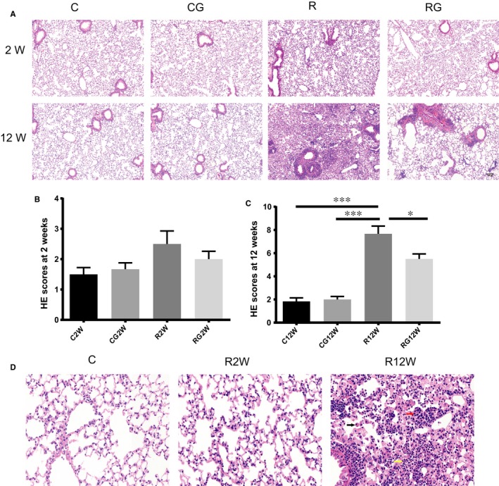
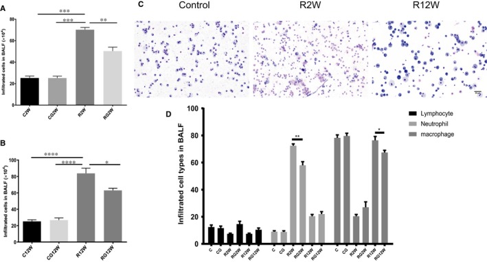
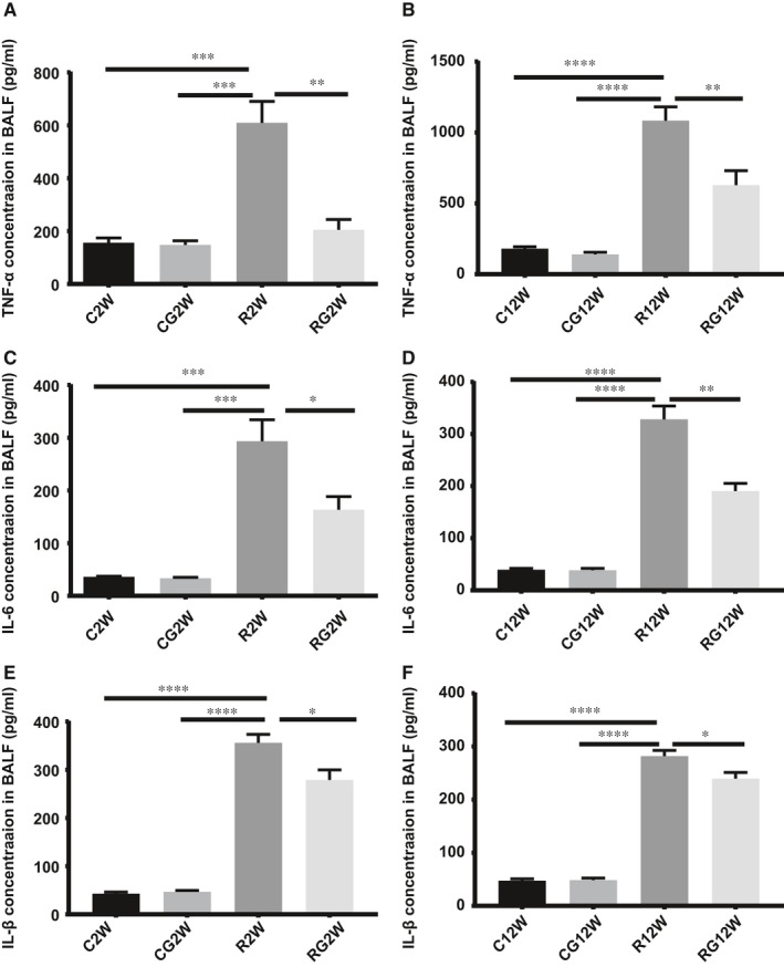
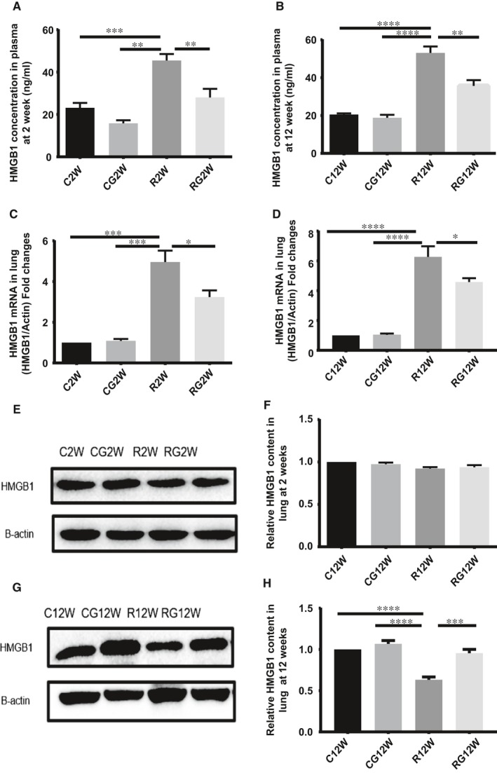
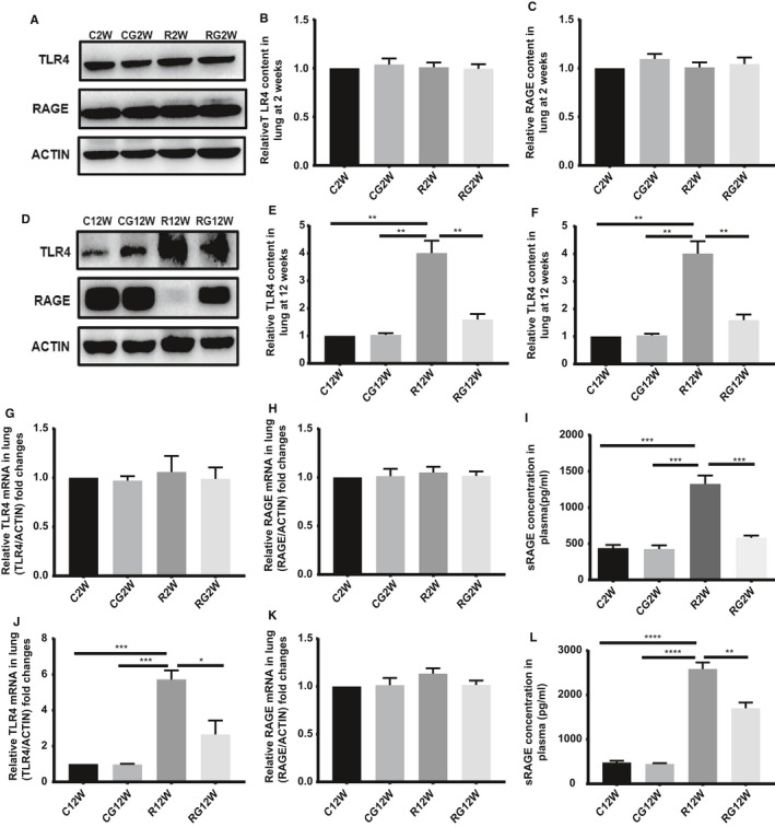
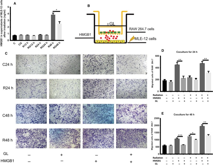
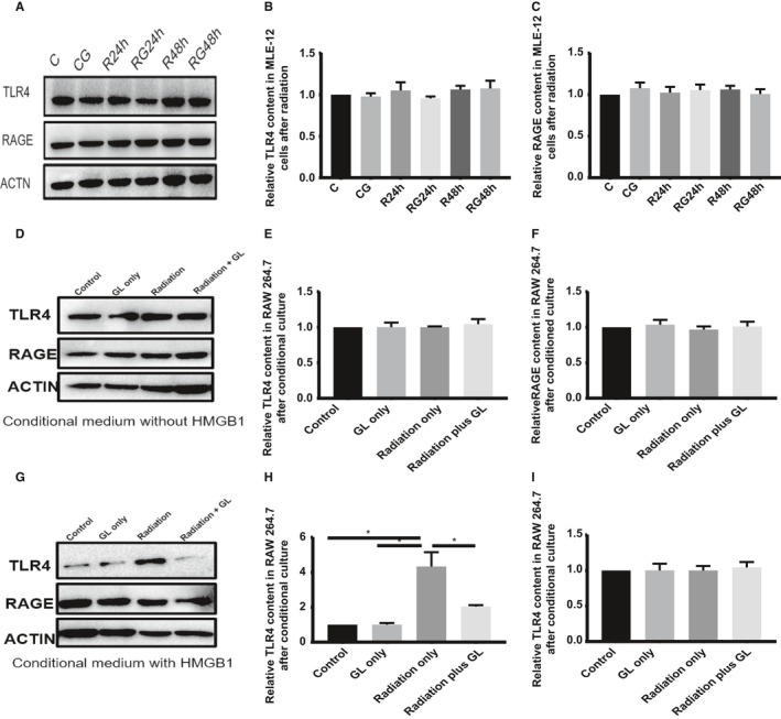
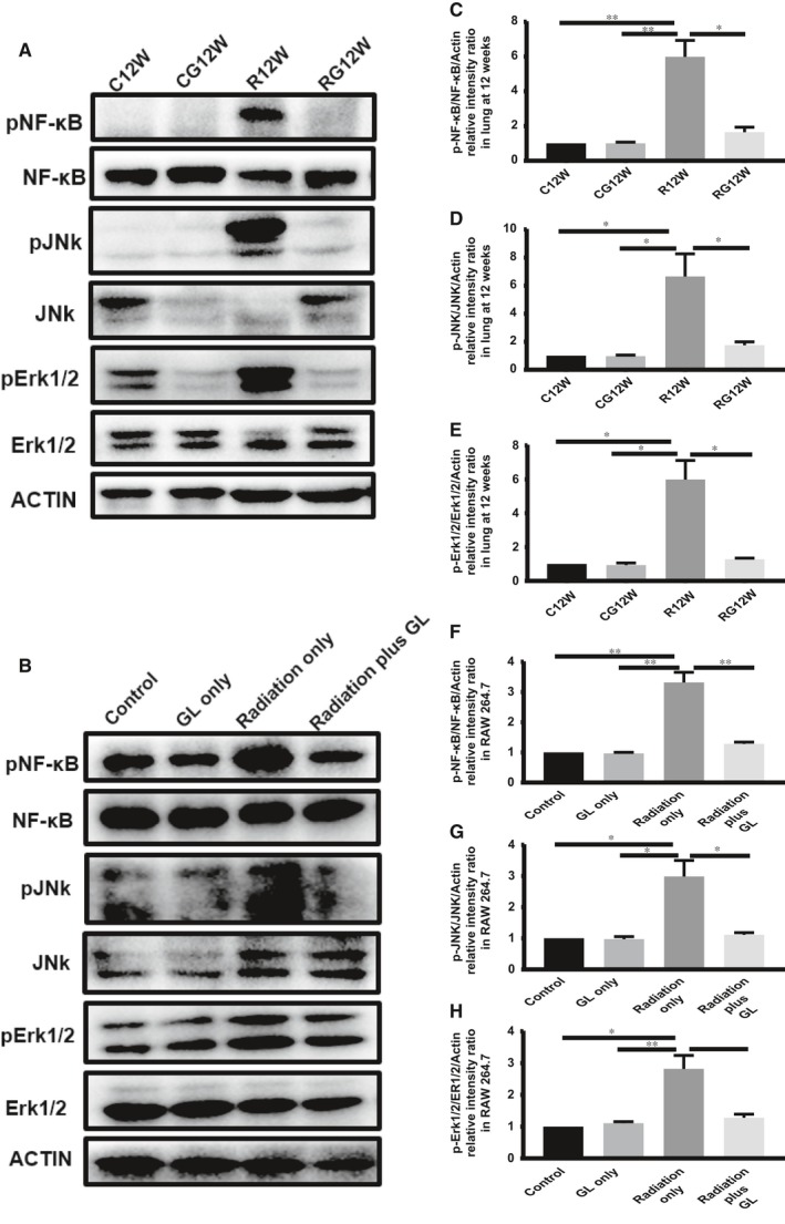
Radiation-induced lung injury (RILI) is the major complication of thoracic radiation therapy, and no effective treatment is available. This study explored the role of high-mobility group box 1 (HMGB1) in acute RILI and the therapeutic effect of glycyrrhizin, an inhibitor of HMGB1, on RILI. C57BL/6 mice received a 20 Gy dose of X-ray radiation to the whole thorax with or without administration of glycyrrhizin. Severe lung inflammation was present 12 weeks after irradiation, although only a mild change was noted at 2 weeks and could be alleviated by administration of glycyrrhizin. Glycyrrhizin decreased the plasma concentrations of HMGB1 and sRAGE as well as TNF-α, IL-1β and IL-6 levels in the bronchoalveolar lavage fluid (BALF). The expression of RAGE was decreased while that of TLR4 was significantly increased at 12 weeks, but not 2 weeks, after irradiation in mouse lung tissue. In vitro, the expression of TLR4 increased in RAW 264.7 cells after conditioning with the supernatant from the irradiated MLE-12 cells containing HMGB1 but showed no change when conditioned medium without HMGB1 was used. However, conditioned culture had no effect on RAGE expression in RAW 264.7 cells. Glycyrrhizin also inhibited the related downstream transcription factors of HMGB/TLR4, such as NF-κB, JNK and ERK1/2, in lung tissue and RAW 264.7 cells when TLR4 was activated. In conclusion, the HMGB1/TLR4 pathway mediates RILI and can be mitigated by glycyrrhizin.

Keywords: HMGB1; RAGE; TLR4; glycyrrhizin; radiation-induced lung injury (RILI).


Conflict of interest statement

The authors declare that they have no competing interests.

Figures

Figure 1
Figure 1
GL alleviates lung inflammation after radiation. A, Representative images of H&E staining at 2 and 12 wk, Scale bar = 100 µm. B,C, Lung tissue inflammation scores at 2 and 12 wk after irradiation (n = 6, *P < .5, **P < .01, ***P < .001). D, Magnified images of H&E staining of no radiation and 2/12 weeks after irradiation. Main infiltrating cell types in lung tissue after 12 weeks are marked by arrows: black arrow, macrophages; red arrow, lymphocytes; yellow arrow, neutrophils. Scale bar = 50 µm
Figure 2
Figure 2
Infiltrating cells in the BALF at 2 and 12 wk. A,B, Total numbers of infiltrating cells at 2 and 12 wk in the BALF. C, Representative Diff‐Quick staining images of cells in the BALF of no radiation and at 2 and 12 wk after irradiation. Scale bar = 50 µm. D, Statistical analysis results of three main cell types in the BALF after irradiation. (*P < .5, **P < .01, ***P < .001)
Figure 3
Figure 3
Cytokine concentrations in the BALF. TNF‐α, IL‐6 and IL‐1β concentrations in the BALF were measured by ELISA. A,C,E, 2 wk after irradiation; B, D, F, 12 wk after irradiation. (*P < .5, **P < .01, ***P < .001)
Figure 4
Figure 4
Glycyrrhizin inhibits HMGB1 synthesis and release. A,B, Plasma level of HMGB1 detected by ELISA at 2 and 12 wk after irradiation. C,D, Relative mRNA expression of HMGB1 in the lung tissue after irradiation. E,G, Western blotting analysis of HMGB1 in the lung tissues at 2 and 12 wk after irradiation. F,H, Relative qualification of HMGB1 content in the lung tissue after irradiation. (*P < .5, **P < .01, ***P < .001)
Figure 5
Figure 5
Expression of HMGB1 receptors in the lung tissue during different stages after radiation. A,D, Western blotting analysis of TLR4 and RAGE in the lung tissues at 2 and 12 wk after irradiation. B,C,E,F, Relative qualification of RAGE and TLR4 content in the lung tissue in 2 and 12 wk after irradiation. G,H,J,K, Relative mRNA expression of RAGE and TLR4 in the lung tissue at 2 and 12 wk after irradiation. I,L, ELISA results of sRAGE in plasma during different stages after irradiation. (*P < .5, **P < .01, ***P < .001)
Figure 6
Figure 6
GL blocks the chemotaxis of HMGB1 in vitro. A, HMGB1 in supernatants from MLE‐12 cells after irradiation at different times by ELISA. HMGB1 was increased only at 48 h after irradiation. B, Scheme of the transwell assay. MLE‐12 cells were seeded in the lower compartment and underwent irradiation, while RAW 264.7 cells were seeded in the upper chamber and co‐cultured for 24 and 48 h, respectively. C, Representative images of migrated cells stained with crystal violet after co‐culturing for different times. 100× D,E, Result of migrated cell counts after co‐culturing for 24 and 48 h. (*P < .5, **P < .01, ***P < .001)
Figure 7
Figure 7
GL inhibits TLR4 expression in immunocytes after conditioned culture. A, Western blotting analysis of TLR4 and RAGE in MLE‐12 cells at 24 and 48 h after irradiation in different groups. B,C, Relative qualification of RAGE and TLR4 content in MLE‐12 cells after irradiation. D,G, Western blotting analysis of TLR4 and RAGE in RAW 264.7 cells after conditioned culture with (48‐h supernatants of MLE‐12 cells after irradiation) or without (24‐h supernatants of MLE‐12 cells after irradiation) released HMGB1. E,H, Relative qualification of RAGE in RAW 264.7 cells after conditioned medium. F,I, Relative qualification of TLR4 in RAW 264.7 cells after conditioned medium
Figure 8
Figure 8
GL inhibits the downstream of HMGB1/TLR4 signalling pathway in vivo and in vitro. A, Western blotting analysis of NF‐κB, JNK and ERK1/2 in the lung tissue at 12 wk after irradiation. C,D,E, Relative qualification of pNF‐κB, pJNK and pErk1/2 in lung tissue at 12 wk. B, Western blotting analysis of NF‐κB, JNK and ERK1/2 in RAW 264.7 cells after conditioned culture. F,G,H, Relative qualification of pNF‐κB, pJNK and pErk1/2 in RAW 264.7 cells after conditioned culture

References

    1. Simone CB 2nd. Thoracic radiation normal tissue injury. Semin Radiat Oncol. 2017;27:370‐377. - PubMed
    1. Rube CE, Uthe D, Schmid KW, et al. Dose‐dependent induction of transforming growth factor beta (TGF‐beta) in the lung tissue of fibrosis‐prone mice after thoracic irradiation. Int J Radiat Oncol Biol Phys. 2000;47:1033‐1042. - PubMed
    1. Minami‐Shimmyo Y, Ohe Y, Yamamoto S, et al. Risk factors for treatment‐related death associated with chemotherapy and thoracic radiotherapy for lung cancer. J Thorac Oncol. 2012;7:177‐182. - PubMed
    1. Citrin DE, Mitchell JB. Mechanisms of normal tissue injury from irradiation. Semin Radiat Oncol. 2017;27:316‐324. - PMC - PubMed
    1. Hekim N, Cetin Z, Nikitaki Z, et al. Radiation triggering immune response and inflammation. Cancer Lett. 2015;368:156‐163. - PubMed

Publication types

MeSH terms