. 2020 Feb 1;77(2):225-233.
doi: 10.1001/jamaneurol.2019.3606.

Selective Vulnerability of the Nucleus Basalis of Meynert Among Neuropathologic Subtypes of Alzheimer Disease

Affiliations

Selective Vulnerability of the Nucleus Basalis of Meynert Among Neuropathologic Subtypes of Alzheimer Disease

Fadi S Hanna Al-Shaikh et al. JAMA Neurol. .

Erratum in

Abstract

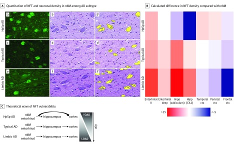
Importance: Corticolimbic patterns of neurofibrillary tangle (NFT) accumulation define neuropathologic subtypes of Alzheimer disease (AD), which underlie the clinical heterogeneity observed antemortem. The cholinergic system, which is the target of acetylcholinesterase inhibitor therapy, is selectively vulnerable in AD.

Objective: To investigate the major source of cholinergic innervation, the nucleus basalis of Meynert (nbM), in order to determine whether there is differential involvement of NFT accumulation or neuronal loss among AD subtypes.

Design, setting, and participants: In this cross-sectional study, retrospective abstraction of clinical records and quantitative assessment of NFTs and neuron counts in the nbM was completed in January 2019 at the Mayo Clinic using the Florida Autopsied Multi-Ethnic (FLAME) cohort, which had been accessioned from 1991 until 2015. The FLAME cohort is derived from the deeded autopsy program funded throughout the State of Florida's memory disorder clinic referral services. Of the 2809 consecutively accessioned FLAME cohort, 1464 were identified as neuropathologically diagnosed AD cases and nondemented normal controls available for clinicopathologic assessment. Quantification of NFTs and neuronal density in the anterior nbM was performed blinded to neuropathologic groupings.

Main outcomes and measures: Demographic and clinical characteristics, including cognitive decline measured using the Mini-Mental State Examination score (range, 0-30), were evaluated. The anterior nbM was investigated quantitatively for neuronal loss and NFT accumulation.

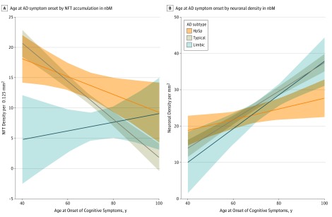
Results: In total, 1361 AD subtypes and 103 nondemented controls were assessed. The median (interquartile range) age at death was 72 (66-80) years in hippocampal sparing (HpSp) AD, 81 (76-86) years in typical AD, and 86 (82-90) years in limbic predominant AD. The median (interquartile range) count per 0.125 mm2 of thioflavin S-positive NFTs was highest in the nbM of HpSp AD (14 [9-20]; n = 163), lower in typical AD (10 [5-16]; n = 937), and lowest in limbic predominant AD (8 [5-11], n = 163) (P < .001). The median (interquartile range) neuronal density per millimeters squared was lowest in HpSp AD cases (22 [17-28]; n = 148), higher in typical AD (25 [19-30]; n = 727), and highest in limbic predominant AD (26 [19-32]; n = 127) (P = .002). Multivariable regression modeling of clinical and demographic variables was performed to assess overlap in NFT accumulation and neuronal density differences among AD subtypes. Higher NFT accumulation in the nbM was associated with younger age at onset for HpSp AD (β, -1.5; 95% CI, -2.9 to -0.15; P = .03) and typical AD (β, -3.2; 95% CI, -3.9 to -2.4; P < .001). In addition, higher NFT accumulation in the nbM of typical AD cases was associated with female sex (β, 2.5; 95% CI, 1.4-3.5; P < .001), apolipoprotein E ε4 allele (β, 1.3; 95% CI, 0.15-2.5; P = .03), and lower Mini-Mental State Examination scores (β, -1.8; 95% CI, -3.2 to -0.31; P = .02). Demographic and clinical progression variables were not associated with NFT accumulation in the nbM of limbic predominant AD cases.

Conclusions and relevance: These data provide supportive evidence that NFT accumulation in the nbM may underlie more widespread and severe cholinergic deficits in young-onset AD, in particular in patients with HpSp AD. Moreover, these findings underscore the importance of considering age at onset, sex, and apolipoprotein E genotype when assessing outcomes in AD.


Conflict of interest statement

Conflict of Interest Disclosures: Dr Duara reported receiving grants from the Florida Department of Health during the conduct of the study; grants from the National Institute on Aging; and personal fees from Med Learning Group, Eli Lilly and Company, and Piramal Imaging outside the submitted work. Dr Ertekin-Taner reported that her husband is the Chief Strategy Officer of Magnolia Health Solutions, LLC, although she has no role in the company and the present study has no connections with the company. Dr Graff-Radford reported receiving grants from Biogen, Novartis, AbbVie, and Eli Lilly and Company outside the submitted work. Dr Murray reported receiving grants from the National Institute on Aging, the Alzheimer’s Association, and the State of Florida during the conduct of the study. No other disclosures were reported.

Figures

Figure 1.
Figure 1.. Selective Vulnerability of the Nucleus Basalis of Meynert (nbM) and Corticolimbic Structures to Neurofibrillary Tangles (NFTs) Among Neuropathologic Subtypes of Alzheimer Disease (AD)
A. Thioflavin S microscopy (a, c, and e) shows greater NFT accumulation in the nbM of hippocampal sparing (HpSp) AD (a) compared with typical AD (c) and limbic predominant (limbic) AD (e). Hematoxylin-eosin–stained sections of the nbM (b, d, and f) were digitally quantified (b′, d′, and f′, respectively). Fewer neurons are observed in HpSp AD (b) compared with typical AD (d) and limbic predominant AD (f). Scale bar represents 50 μm. B. Heatmap of differences calculated between brain region of interest and the nbM, as exampled by the more severe involvement of the entorhinal cortex compared with the nbM in limbic predominant AD, shown in warmer colors, and the less severe involvement of the hippocampus (Hipp) in HpSp AD compared with the nbM, shown in cooler colors. C. We hypothesize that, although both the nbM and entorhinal cortex (ctx) are involved early among AD subtypes and across aging, the cortex may be more vulnerable in HpSp AD. By contrast, the pattern of greater vulnerability of limbic structures is manifested in both limbic predominant AD and perhaps as a function of older age. LOAD indicates late-onset AD; YOAD, young-onset AD.
Figure 2.
Figure 2.. Differences in Neurofibrillary Tangle (NFT) and Neuronal Density in the Nucleus Basalis of Meynert (nbM) Among Alzheimer Disease (AD) Subtypes
Data are displayed as jitter plots overlaying box plots of the 25th to 75th percentile, with the middle horizontal line representing the median. Nondemented normal controls are displayed for reference. Within each AD subtype, individuals younger than 65 years of age presenting with cognitive problems are displayed on the left and individuals 65 years or older are displayed on the right (lighter color). A, The NFT density in the nbM was measured using thioflavin S fluorescence microscopy. HpSp AD cases have the greatest accumulation of NFTs compared with typical AD, which is greater than limbic predominant (limbic) AD. B, Neuronal density was measured using a custom-designed digital pathology macro on hematoxylin-eosin–stained sections of the nbM. Hippocampal sparing (HpSp) AD has fewer remaining neurons compared with typical AD, which has fewer compared with limbic predominant AD. Pairwise comparisons were performed using Mann-Whitney rank sum test.
Figure 3.
Figure 3.. Association Between Age at Onset of Cognitive Symptoms and Neurofibrillary Tangle (NFT) Density and Neuronal Density in the Nucleus Basalis of Meynert (nmB) Among Alzheimer Disease (AD) Subtypes
Best-fit lines represent the association of age at onset per AD subtype while adjusting for other covariates in the regression models found in Table 2. A, Younger age at onset of cognitive symptoms is significantly associated with greater NFT accumulation measured in the nbM of hippocampal sparing (HpSp) AD and typical AD but not limbic predominant (limbic) AD. B, The association between age at onset of cognitive symptoms and neuronal density measured in hematoxylin-eosin–stained sections of the nbM among AD subtypes is significant among all of the AD subtypes.

References

    1. Montine TJ, Phelps CH, Beach TG, et al. ; National Institute on Aging; Alzheimer’s Association . National Institute on Aging-Alzheimer’s Association guidelines for the neuropathologic assessment of Alzheimer’s disease: a practical approach. Acta Neuropathol. 2012;123(1):1-11. doi:10.1007/s00401-011-0910-3 - DOI - PMC - PubMed
    1. Davies P, Maloney AJF. Selective loss of central cholinergic neurons in Alzheimer’s disease. Lancet. 1976;2(8000):1403. doi:10.1016/S0140-6736(76)91936-X - DOI - PubMed
    1. Mesulam M, Shaw P, Mash D, Weintraub S. Cholinergic nucleus basalis tauopathy emerges early in the aging-MCI-AD continuum. Ann Neurol. 2004;55(6):815-828. doi:10.1002/ana.20100 - DOI - PubMed
    1. Schmitz TW, Nathan Spreng R; Alzheimer’s Disease Neuroimaging Initiative . Basal forebrain degeneration precedes and predicts the cortical spread of Alzheimer’s pathology. Nat Commun. 2016;7:13249. doi:10.1038/ncomms13249 - DOI - PMC - PubMed
    1. Cavedo E, Grothe MJ, Colliot O, et al. ; Hippocampus Study Group . Reduced basal forebrain atrophy progression in a randomized Donepezil trial in prodromal Alzheimer’s disease. Sci Rep. 2017;7(1):11706. doi:10.1038/s41598-017-09780-3 - DOI - PMC - PubMed

Publication types