. 2020 Mar;29(3):670-685.
doi: 10.1002/pro.3758. Epub 2019 Nov 21.

Structural diversity in the Mycobacteria DUF3349 superfamily

Affiliations

Structural diversity in the Mycobacteria DUF3349 superfamily

Garry W Buchko et al. Protein Sci. 2020 Mar.

Abstract

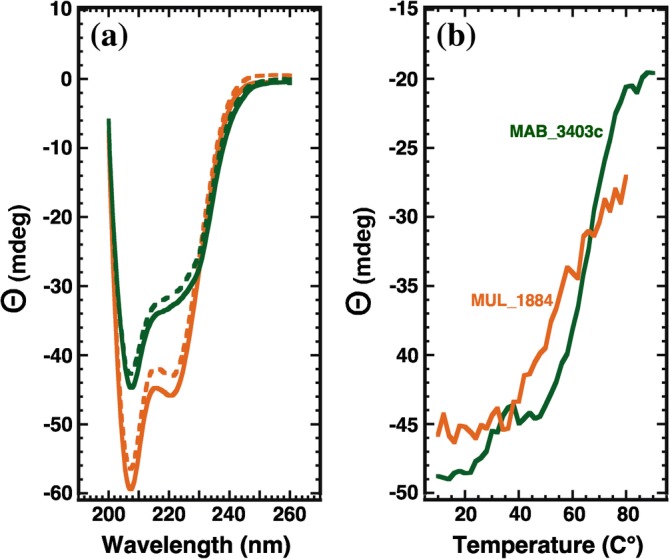
A protein superfamily with a "Domain of Unknown Function,", DUF3349 (PF11829), is present predominately in Mycobacterium and Rhodococcus bacterial species suggesting that these proteins may have a biological function unique to these bacteria. We previously reported the inaugural structure of a DUF3349 superfamily member, Mycobacterium tuberculosis Rv0543c. Here, we report the structures determined for three additional DUF3349 proteins: Mycobacterium smegmatis MSMEG_1063 and MSMEG_1066 and Mycobacterium abscessus MAB_3403c. Like Rv0543c, the NMR solution structure of MSMEG_1063 revealed a monomeric five α-helix bundle with a similar overall topology. Conversely, the crystal structure of MSMEG_1066 revealed a five α-helix protein with a strikingly different topology and a tetrameric quaternary structure that was confirmed by size exclusion chromatography. The NMR solution structure of a fourth member of the DUF3349 superfamily, MAB_3403c, with 18 residues missing at the N-terminus, revealed a monomeric α-helical protein with a folding topology similar to the three C-terminal helices in the protomer of the MSMEG_1066 tetramer. These structures, together with a GREMLIN-based bioinformatics analysis of the DUF3349 primary amino acid sequences, suggest two subfamilies within the DUF3349 family. The division of the DUF3349 into two distinct subfamilies would have been lost if structure solution had stopped with the first structure in the DUF3349 family, highlighting the insights generated by solving multiple structures within a protein superfamily. Future studies will determine if the structural diversity at the tertiary and quaternary levels in the DUF3349 protein superfamily have functional roles in Mycobacteria and Rhodococcus species with potential implications for structure-based drug discovery.

Keywords: NMR spectrometry; circular dichroism; structural genomics; structure-based drug discovery; tuberculosis.


Conflict of interest statement

The authors declare they have no conflict of interest with the contents of this publication.

Figures

Figure 1
Figure 1
Assigned 1H‐15N HSQC spectrum of 13C‐,15N‐labeled (a) MSMEG_1063 and (b) MAB_3403c (~1.0 mM) collected at a proton resonance frequency of 600 MHz, 20°C, in 100 mM NaCl, 20 mM Tris, 1 mM DTT, pH 7.1. Amide cross peaks for non‐native and native residues are colored magenta and blue, respectively, with assigned side chain resonance pairs connected with a dashed line and colored red. Amide cross peaks that are folded into the spectrum are underlined. Significant unidentified cross peaks are labeled with a question mark. For MSMEG_1063 (a) two tryptophan ring amide cross peaks, W88 (10.36/130.4 ppm) and W11 (9.38/131.8 ppm), along with the backbone amide cross peak for G15 (7.70/103.6 ppm) are outside the displayed region of the spectrum. For MAB_3403c (b), a tryptophan ring amide cross peak, W97 (10.25/128.3 ppm), is outside the displayed region of the spectrum
Figure 2
Figure 2
Solution structures for MSMEG_1063 and MAB_3403c. (a) Superposition of the final ensemble of 17 structures calculated for MSMEG_1063 (http://bioinformatics.org/firstglance/fgij//fg.htm?mol=2LKY). The α‐helices and the 310‐helix are colored blue and magenta, respectively. (b) Superposition of the structure closest to the average for Rv0543c (green) and MSMEG_1063 (blue) with the α‐helices shown as cylinders. (c) Superposition of the final ensemble of 20 structures calculated for MAB_3403c (http://bioinformatics.org/firstglance/fgij//fg.htm?mol=2M0N) with the α‐helices colored cyan. (d) Superposition of the three C‐terminal helices in the structure closest to the average for MSMEG_1063 (blue) and MAB_3403c (cyan) with the α‐helices shown as cylinders. In some of the figures parts of the unstructured N‐ and C‐terminals are not shown for clarity
Figure 3
Figure 3
Clustal Omega sequence alignment of MSMEG_1066, MSMEG_1063, Rv0543c, MAB_3403c, and MUL_1884. The identical and conserved residues, colored blue and red, respectively, are enlarged in bold. The α‐helical regions observed in the solved structures are shaded gray, the 310‐helices are shaded in magenta, and the residues truncated from the native sequence of MAB_3403c are shaded green. The numbering scheme for the helices observed in MSMEG_1066 and MSMEG_1063 are provided on the bottom. For comparative purposes, the first two α‐helices in MAB_3403c are labeled α3 and α3' and then sequentially α4 and α5. A structure has not been determined for MUL_1884
Figure 4
Figure 4
MSMEG_1066 adopts a tetrameric quaternary structure. (a) Crystal structure of MSMEG_1066 (http://bioinformatics.org/firstglance/fgij//fg.htm?mol=3OL3) with each unit in the tetramer colored differently, the α‐helices shown as cylinders, and the solvent‐accessible surface transparent. Due to symmetry properties, two different interfacial regions exist at opposing positions on the eclipse. (b) One interface is formed by α1 stacking over α2′ and α2 stacking over α1′ in a skewed “X” arrangement (c) The other interface is formed by α3 stacking against the C‐terminal of α5′ plus W96′ and the twofold symmetrical pair. In both (b) and (c), the side chains at the interfaces are illustrated in stick representation and labeled. (d) Solvent‐accessible surface of the ConSurf generated map for MSMEG_1066 (http://bioinformatics.org/firstglance/fgij//fg.htm?mol=3OL3) with highly conserved residues colored magenta and pink and poorly conserved residues colored cyan. (e) Electrostatic potentials at the solvent‐accessible surface of MSMEG_1066 (http://bioinformatics.org/firstglance/fgij//fg.htm?mol=3OL3) with positive regions colored red and negative regions colored blue. The face shown in (d) and (e) is the same face as illustrated in (a). Due to symmetry only one face of the tetramer is illustrated
Figure 5
Figure 5
Comparison of the fold adopted by the five α‐helices in MSMEG_1066 (http://bioinformatics.org/firstglance/fgij//fg.htm?mol=3OL3) with the five α‐helix fold in MSMEG_1063 (http://bioinformatics.org/firstglance/fgij//fg.htm?mol=2LKY) and the three α‐helix fold in MAB_3403c (3M0N). (a) Cartoon representation of a single protomer of MSMEG_1066 (http://bioinformatics.org/firstglance/fgij//fg.htm?mol=3OL3, gold) observed in the crystal structure. (b) Superposition of the structure closest to the average for MSMEG_1063 (blue) and a single structure of MSMEG_1066 (gold) with the α‐helices shown as cylinders. The five‐helical bundle observed in the crystal structure of the tetramer folds differently than the five‐helical bundle observed in the NMR structure of the monomer. Secondary structure diagram for (c) a single protomer of MSMEG_1066 (http://bioinformatics.org/firstglance/fgij//fg.htm?mol=3OL3, gold) and (d) the structure closest to the average for MSMEG_1063 (blue) with the α‐helices drawn as rectangles
Figure 6
Figure 6
Comparison of the fold adopted by the five α‐helices in MSMEG_1066 (3OL3) with the three α‐helix fold in MAB_3403c (3M0N). Superposition of the structure closest to the average for MAB_3403c (cyan) and a single structure of MSMEG_1066 (gold) with the α‐helices shown as cylinders. The residues remaining after the removal of the N‐terminal 18 residues of MAB_3403c fold similarly to the corresponding regions in the single protomer in the crystal structure of MSMEG_1066
Figure 7
Figure 7
Thermal stability of MSMEG_1063 and MSMEG_1066. (a) Steady‐state CD wavelength spectra for MSMEG_1066 (black) and MSMEG_1063 (blue) at 25°C before (solid line) and after (dashed line) heating the samples to 90°C. (b) CD thermal melts for MSMEG_1066 (black) and MSMEG_1063 (blue) obtained by measuring the ellipticity at 220 nm in 2.0°C intervals between 10 and 90°C
Figure 8
Figure 8
GREMLIN‐based phylogenic tree for the family of DUF3349 proteins. Proteins studied here are labeled. Based on the quaternary structures of the labeled proteins, the two major branches have been divided into proteins predicted to exist in solution as monomers and tetramers
Figure 9
Figure 9
Amino acid sequence logos of the two DUF3349 subfamilies of proteins suggested by the GREMLIN‐based phylogenic tree illustrated in Figure 8. The top alignment is for the monomer family with the α‐helices observed in the NMR structure for MSMEG_1063 illustrated with red rectangles above the amino acid conservation logo. The C‐terminal 310‐helix observed in MSMEG_1063 is indicated with the magenta rectangle. The bottom alignment is for the tetramer family with the α‐helices observed in the crystal structure of MSMEG_1066 illustrated with orange rectangles above the amino acid conservation logo. Residues of the side chains observed at the intermolecular interfaces in the crystal structure of MSMEG_1066 are indicated with a “hat” (^) symbol. The sequence numbering on the y‐axis is different from the numbering in Figure 3
Figure 10
Figure 10
Thermal stability of MAB3403c and MUL_1884. (a) Steady‐state CD wavelength spectra for MAB3403c (green) and MUL_1884 (orange) at 25°C before (solid line) and after (dashed line) heating the samples to 80°C (MUL_1884) or 90°C (MAB_3403c). (b) CD thermal melts for MAB_3403c (green) and MUL_1884 (orange) obtained by measuring the ellipticity at 220 nm in 2.0°C intervals

References

    1. Myler PJ, Stacy R, Stewart LJ, Staker BL, Van Voorhis WC, Buchko GW. Seattle Structural Genomics Center for Infectious Disease (SSGCID). Infect Dis Drug Targets. 2009;9:493–506. - PMC - PubMed
    1. Stacy R, Begley DW, Phan I, et al. Structural genomics of infectious disease drug targets: The SSGCID. Acta Crystallogr. 2011;F67:979–984. - PMC - PubMed
    1. Staker BL, Buchko GW, Myler PJ. Recent contributions of structure‐based drug design to the development of antibacterial compounds. Curr Opin Microbiol. 2015;27:133–138. - PMC - PubMed
    1. Baragana B, Forte B, Choi R, et al. Lysyl‐tRNA synthetase as a drug target in malaria and cryptosporidiosis. Proc Natl Acad Sci U S A. 2019;116:7015–7020. - PMC - PubMed
    1. Abendroth J, Frando A, Phan IQ, et al. Mycobacterium tuberculosis Rv3651 is a triple sensor‐domain protein. Protein Sci. 2018;27:568–572. - PMC - PubMed

Publication types

MeSH terms

Substances

LinkOut - more resources