Review
. 2019 Oct 10;11(10):1523.
doi: 10.3390/cancers11101523.

Targeting Lysophosphatidic Acid in Cancer: The Issues in Moving from Bench to Bedside

Affiliations
Review

Targeting Lysophosphatidic Acid in Cancer: The Issues in Moving from Bench to Bedside

Yan Xu. Cancers (Basel). .

Abstract

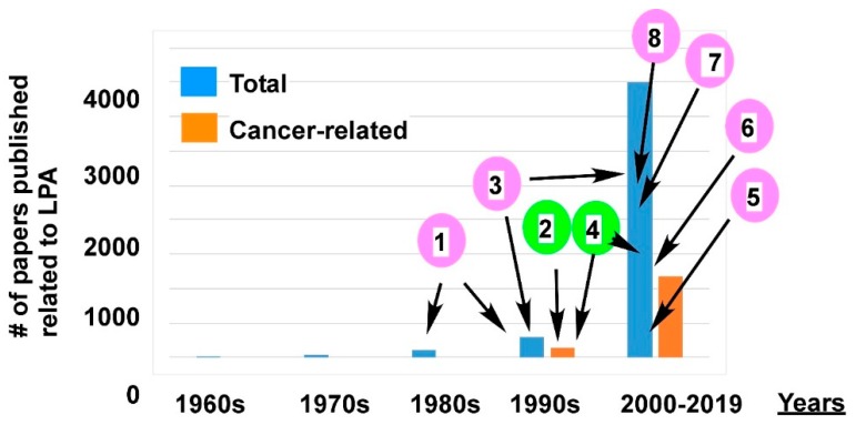
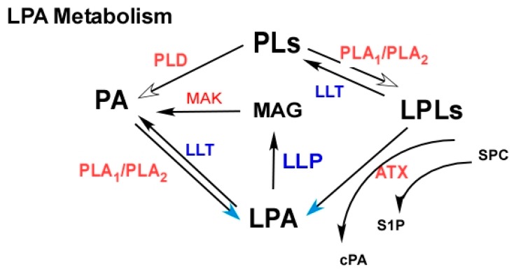
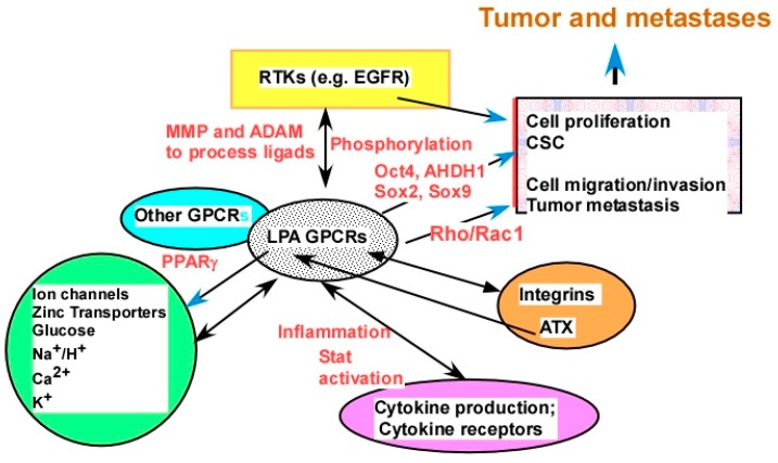
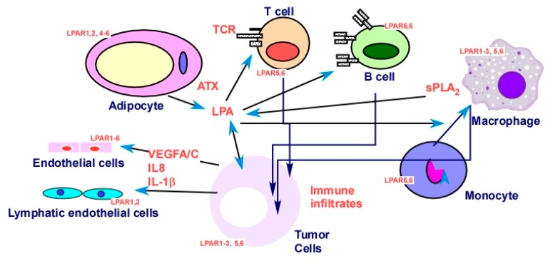
Since the clear demonstration of lysophosphatidic acid (LPA)'s pathological roles in cancer in the mid-1990s, more than 1000 papers relating LPA to various types of cancer were published. Through these studies, LPA was established as a target for cancer. Although LPA-related inhibitors entered clinical trials for fibrosis, the concept of targeting LPA is yet to be moved to clinical cancer treatment. The major challenges that we are facing in moving LPA application from bench to bedside include the intrinsic and complicated metabolic, functional, and signaling properties of LPA, as well as technical issues, which are discussed in this review. Potential strategies and perspectives to improve the translational progress are suggested. Despite these challenges, we are optimistic that LPA blockage, particularly in combination with other agents, is on the horizon to be incorporated into clinical applications.

Keywords: Autotaxin (ATX); G-protein coupled receptor (GPCR); cancer stem cell (CSC); electrospray ionization tandem mass spectrometry (ESI-MS/MS); lipid phosphate phosphatase enzymes (LPPs); lysophosphatidic acid (LPA); nuclear receptor peroxisome proliferator-activated receptor (PPAR); ovarian cancer (OC); phospholipase A2 enzymes (PLA2s); sphingosine-1 phosphate (S1P).


Conflict of interest statement

The author declares no conflict of interest.

Figures

Figure 1
Figure 1
Lysophosphatidic acid (LPA)-related papers published in decades and the milestones in LPA research. The blue bars are the total number of LPA-related papers published in each decade. The orange bars are the numbers of LPA studies related to cancer. The pink circled numbers are milestones related to LPA research in general, and the green circled numbers are cancer-related milestones. (1) LPA’s mitogen and growth factor like activity, as well as G-protein-mediated signaling mechanisms were discovered in the late 1980s and early 1990s [35,53,54,55,56,57,58,59]. (2) In 1995, the pathological significance of LPA in cancer was first reported [8,9,10]. (3) From 1996 to 2009, six LPA G-protein coupled receptors (GPCRs) were identified and cloned [60,61,62,63,64,65,66,67,68,69,70,71]. (4) From 1998 to the present, LPA as a putative cancer marker was reported [22,72,73,74,75,76,77,78,79,80,81,82]. (5) From 2000 to the present, new technologies, including the electrospray ionization tandem mass spectrometry (ESI-MS/MS) methods, were developed for LPA analyses [73,83,84,85,86,87]. In addition, LPA antibodies were developed and further improved. In 2008, Lpath Inc. successfully humanized an anti-LPA antibody. (6) In 2002, the major LPA-producing enzyme ATX was identified and cloned [88,89]. (7) From 2011 to 2017, ATX and LPA G-protein coupled receptors (GPCRs) were crystalized with their structures determined [90,91,92,93,94]. (8) From 2013 to the present, FDA-approved ATX and LPA receptor (LPAR) inhibitors entered clinical trials for fibrosis [95].
Figure 2
Figure 2
LPA metabolism as potential targets. Phospholipids (PLs), phosphatidic acid (PA), lysophospholipids (LPLs). The enzymes in red color, autotaxin (ATX), phospholipase A1 (PLA2), phospholipase D (PLD), and monoacylglycerol kinase (MAK), need to be inhibited to reduced LPA. ATX inhibitors are currently in clinical trials. The enzymes in blue, lipid phosphate phosphatase enzymes (LPPs), lysophospholipase transacylase (LLPT), or LPA acyltransferase (LPAT), need to be enhanced to increased LPA degradation. However, these enzymes are also involved in the metabolism of other lipid molecules, and the overall outcome may be complex. ATX may have multiple functions. It also produces sphingosine-1 phosphate (S1P) from sphingosylphosphorylcholine (SPC) and cyclic phosphatidic acids from lysophospholipids (LPLs). Cyclic PAs (cPAs) have anti-tumor activities [176].
Figure 3
Figure 3
LPA receptors as targets. In general, the EDG family LPA receptors (LPAR1–3) are coupled to Gi, Gq, and G12/13 proteins [61,189] and are more involved in tumor-promoting activities. The purinergic family LPA receptors (LPAR4–6) are all coupled to G12/13 and other trimeric proteins [61,185,186,187]. Their anti-tumor effects may be mediated by their ability to elevate cyclic adenosine monophosphate (cAMP) levels [149].
Figure 4
Figure 4
LPA cross-talk as potential targets. LPA interacts with major types of plasma membrane receptors, including ion channels, metal ion transporters, other transporters, receptor tyrosine kinases (RTKs), other GPCRs, integrins, and cytokine receptors. Examples from each category of receptors are discussed in the Section 2.2.4. Certain potential mechanisms of cross-talk are presented by words in red, including ligand production and/or processing, receptor phosphorylation, and production of downstream molecules mediating the cross-talk.
Figure 5
Figure 5
LPA in tumor cells and in the tumor microenvironment (TME). Tumor, stromal, and immune cells in the TME express LPA receptors, and they produce and/or respond to LPA [34,119,185,202,247,256,289,290,291,292,293,294]. The overall effects produce a tumor-promoting environment as detailed in Section 2.2.5 and in recent reviews [7,21,135].

References

    1. Hanahan D., Weinberg R.A. The hallmarks of cancer. Cell. 2000;100:57–70. doi: 10.1016/S0092-8674(00)81683-9. - DOI - PubMed
    1. Hanahan D., Weinberg R.A. Hallmarks of cancer: The next generation. Cell. 2011;144:646–674. doi: 10.1016/j.cell.2011.02.013. - DOI - PubMed
    1. Mills G.B., Moolenaar W.H. The emerging role of lysophosphatidic acid in cancer. Nat. Rev. Cancer. 2003;3:582–591. doi: 10.1038/nrc1143. - DOI - PubMed
    1. Sengupta S., Wang Z., Tipps R., Xu Y. Biology of LPA in health and disease. Semin. Cell Dev. Biol. 2004;15:503–512. doi: 10.1016/j.semcdb.2004.05.003. - DOI - PubMed
    1. Leblanc R., Peyruchaud O. New insights into the autotaxin/LPA axis in cancer development and metastasis. Exp. Cell Res. 2015;333:183–189. doi: 10.1016/j.yexcr.2014.11.010. - DOI - PubMed

LinkOut - more resources