. 2019 Oct 10;17(10):574.
doi: 10.3390/md17100574.

Naturally Drug-Loaded Chitin: Isolation and Applications

Affiliations

Naturally Drug-Loaded Chitin: Isolation and Applications

Valentine Kovalchuk et al. Mar Drugs. .

Abstract

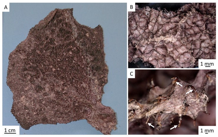
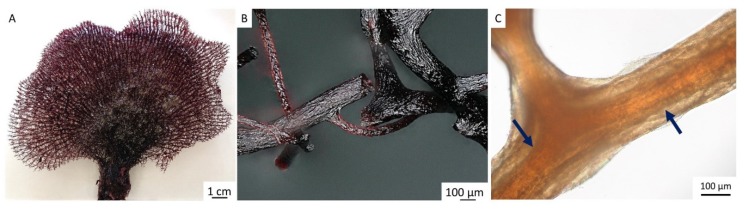
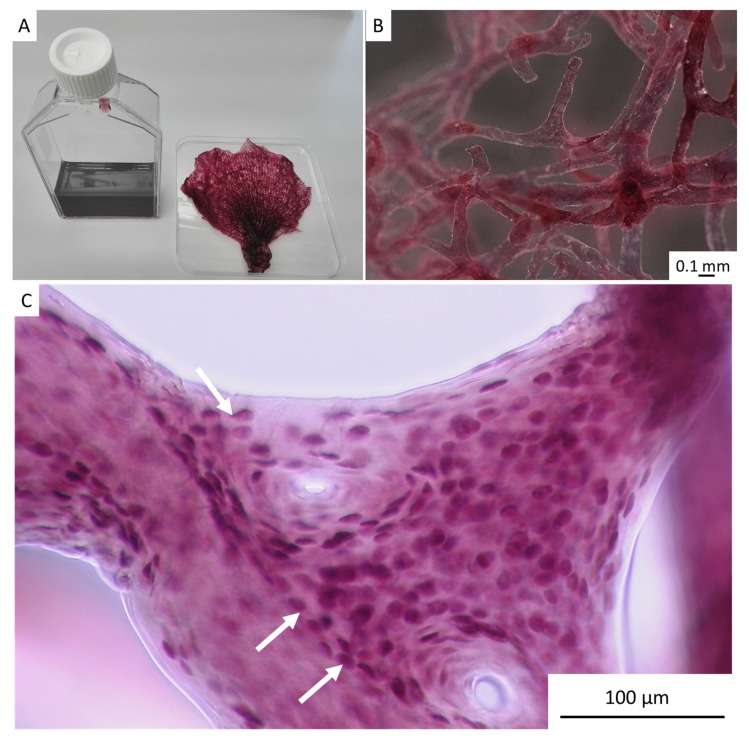
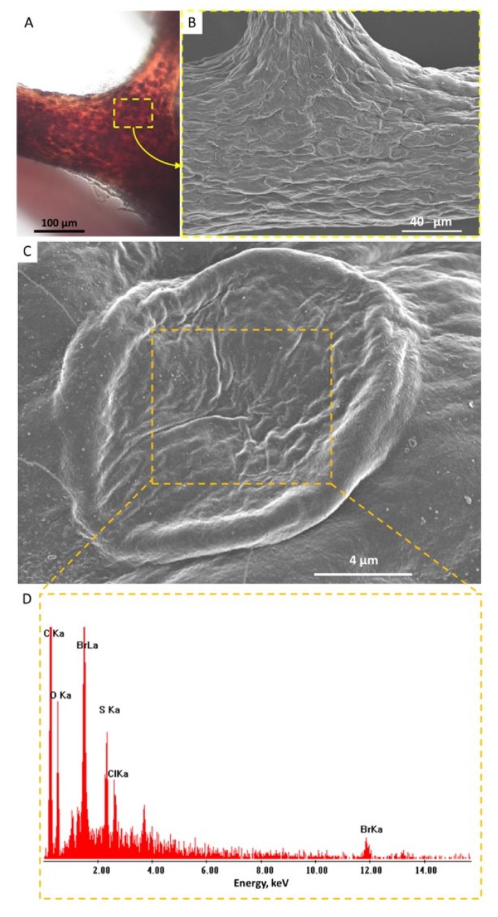
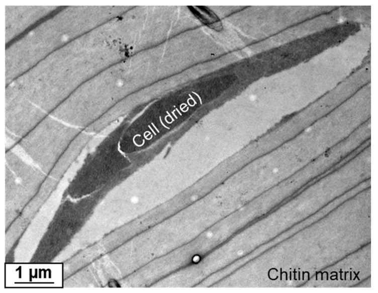
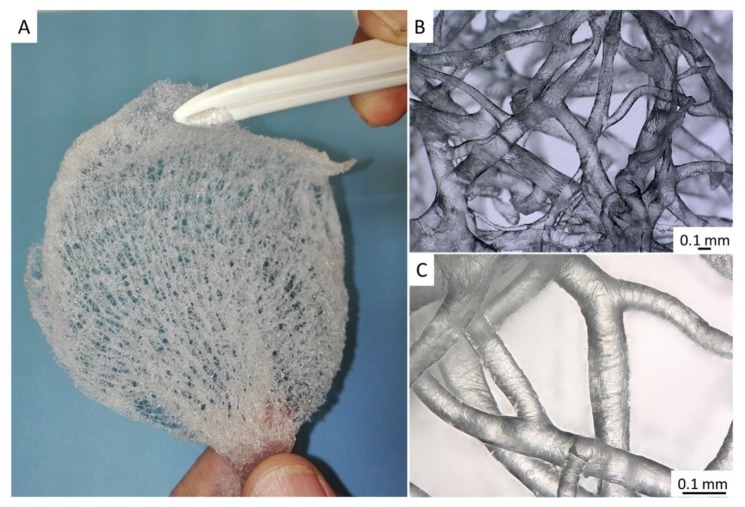
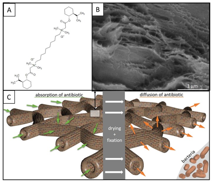
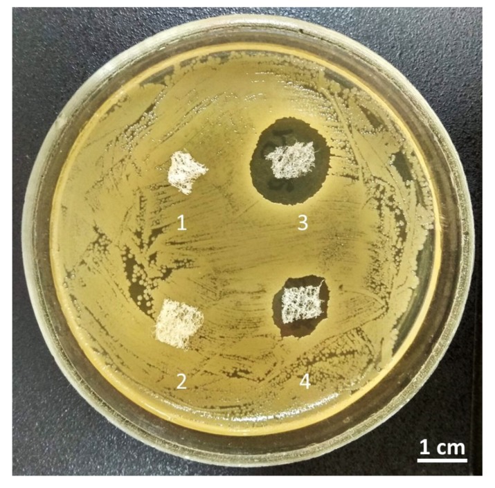
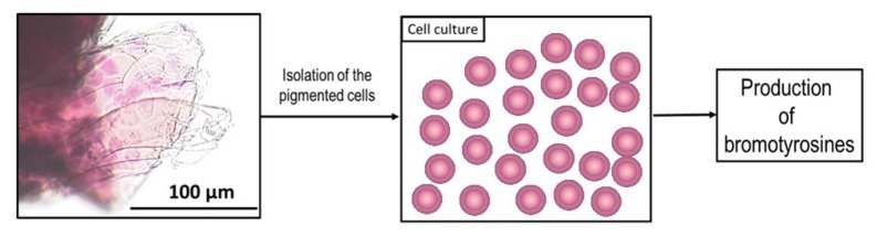
Naturally occurring three-dimensional (3D) biopolymer-based matrices that can be used in different biomedical applications are sustainable alternatives to various artificial 3D materials. For this purpose, chitin-based structures from marine sponges are very promising substitutes. Marine sponges from the order Verongiida (class Demospongiae) are typical examples of demosponges with well-developed chitinous skeletons. In particular, species belonging to the family Ianthellidae possess chitinous, flat, fan-like fibrous skeletons with a unique, microporous 3D architecture that makes them particularly interesting for applications. In this work, we focus our attention on the demosponge Ianthella flabelliformis (Linnaeus, 1759) for simultaneous extraction of both naturally occurring ("ready-to-use") chitin scaffolds, and biologically active bromotyrosines which are recognized as potential antibiotic, antitumor, and marine antifouling substances. We show that selected bromotyrosines are located within pigmental cells which, however, are localized within chitinous skeletal fibers of I. flabelliformis. A two-step reaction provides two products: treatment with methanol extracts the bromotyrosine compounds bastadin 25 and araplysillin-I N20 sulfamate, and a subsequent treatment with acetic acid and sodium hydroxide exposes the 3D chitinous scaffold. This scaffold is a mesh-like structure, which retains its capillary network, and its use as a potential drug delivery biomaterial was examined for the first time. The results demonstrate that sponge-derived chitin scaffolds, impregnated with decamethoxine, effectively inhibit growth of the human pathogen Staphylococcus aureus in an agar diffusion assay.

Keywords: Ianthella; bromotyrosines; chitin; decamethoxine; demosponges; drug delivery; pigmental cells; scaffolds.


Conflict of interest statement

The authors declare no conflicts of interest.

Figures

Figure 1
Figure 1
The marine demosponge Ianthella flabelliformis (Linnaeus, 1759) (A), as collected after air-drying, exhibits a characteristic fan-like and meshwork morphology (B). Chitin-based skeletal fibers (arrows) are visible between tissue-like layers (C).
Figure 2
Figure 2
The cell-free macerated skeleton of I. flabelliformis (A,B) is made of anastomosing, interconnected tube-based chitinous fibers (B). Partial depigmentation using alkali treatment leads to visualization of the inner channel (arrows, C) which is located within each fiber.
Figure 3
Figure 3
The bromotyrosine-containing extract isolated from skeleton of I. flabelliformis (A) is one of the sources of pharmacologically relevant reagents. The dark-reddish color of chitinous skeletal fibers (B) is determined by the presence of pigmental cells, or spherulocites (C), special chitin-associated bromotyrosine-producing cells (arrows).
Figure 4
Figure 4
Pigmental cells located within fibers of I. flabelliformis chitin are clearly visible using light microscopy (A) (see also Figure 3C). These cells are observable using SEM (B,C). Single-spot energy-dispersive X-ray spectroscopy (EDX) analysis shows strong evidence of the presence of bromine within individual pigmental cells (D).
Figure 5
Figure 5
TEM image of the cross-section through the chitinous sponge matrix of I. flabelliformis showing the interlayer location of the pigmental cell that lost its oval morphology due to the drying procedure. This cell is definitively of eukaryotic and not bacterial origin.
Figure 6
Figure 6
The spherulocite-free chitinous skeleton of I. flabelliformis can be isolated after alternating treatment of the construct with acetic acid (A) and NaOH (B) (see also Figure 7).
Figure 7
Figure 7
The chitinous skeleton isolated from I. flabelliformis (A) represents a mechanically elastic, flat, but still three-dimensional (3D)-based construct made of interconnected tubular fibers (B). These fibers show excellent capacity for saturation with diverse liquids including water (C).
Figure 8
Figure 8
Three-dimensional chitin scaffold as a potential drug delivery matrix. (A) Chemical formula of decamethoxine which was used in our study as a model substance with antibiotic activity. The nanoporous structure of I. flabelliformis chitin-based tube walls (SEM image, B) may be responsible for both the initial absorption of the substance into the organic matrix and for the following diffusion of this substance from the chitinous construct being transferred to the agar plate contaminated with corresponding bacterial cultures (C).
Figure 9
Figure 9
Sponge chitin filled with decamethoxine as an antimicrobial fabric against Staphylococcus aureus. 1—Sterile I. flabelliformis chitin scaffold (see enlarged image in Figure 7) washed with distilled water as a control sample. 2—Chitin scaffold washed with 70% ethanol and dried as a control sample. 3—Chitin scaffold impregnated with 0.1% water solution of decamethoxine. 4—Chitin scaffold impregnated with 0.1% ethanolic solution of decamethoxin. All observations were carried out after 24 h.
Figure 10
Figure 10
Schematic overview of the challenging tasks: in the near future, we must elaborate an effective method for the isolation of pigmental, bromotyrosine-producing cells from chitinous skeletal fibers of ianthellids with the aim of obtaining cell cultures which should be able to synthetize corresponding bromotyrosines using bioreactors.

References

    1. Ehrlich H., Rigby J.K., Botting J.P., Tsurkan M.V., Werner C., Schwille P., Petrášek Z., Pisera A., Simon P., Sivkov V.N., et al. Discovery of 505-million-year old chitin in the basal demosponge Vauxia gracilenta. Sci. Rep. 2013;3:3497. doi: 10.1038/srep03497. - DOI - PMC - PubMed
    1. Mioso R., Marante F.J.T., Bezerra R.D.S., Borges F.V.P., Santos B.V.D.O., De Laguna I.H.B. Cytotoxic compounds derived from marine sponges: A review (2010–2012) Molecules. 2017;22:208. doi: 10.3390/molecules22020208. - DOI - PMC - PubMed
    1. Helber S.B., Hoeijmakers D.J.J., Muhando C.A., Rohde S., Schupp P.J. Sponge chemical defenses are a possible mechanism for increasing sponge abundance on reefs in Zanzibar. PLoS ONE. 2018;13:e0197617. doi: 10.1371/journal.pone.0197617. - DOI - PMC - PubMed
    1. Ebel R., Brenzinger M., Kunze A., Gross H.J., Proksch P. Wound activation of protoxins in marine sponge Aplysina aerophoba. J. Chem. Ecol. 1997;23:1451–1462. doi: 10.1023/B:JOEC.0000006475.10310.3a. - DOI
    1. Green D.W., Howard D., Yang X., Kelly M., Oreffo R.O.C. Natural marine sponge fiber skeleton: A biomimetic scaffold for human osteoprogenitor cell attachment, growth, and differentiation. Tissue Eng. 2003;9:1159–1166. doi: 10.1089/10763270360728062. - DOI - PubMed

MeSH terms

LinkOut - more resources