Review
. 2019 Oct 10;24(20):3654.
doi: 10.3390/molecules24203654.

Stapled Peptides-A Useful Improvement for Peptide-Based Drugs

Affiliations
Review

Stapled Peptides-A Useful Improvement for Peptide-Based Drugs

Mattia Moiola et al. Molecules. .

Abstract

Peptide-based drugs, despite being relegated as niche pharmaceuticals for years, are now capturing more and more attention from the scientific community. The main problem for these kinds of pharmacological compounds was the low degree of cellular uptake, which relegates the application of peptide-drugs to extracellular targets. In recent years, many new techniques have been developed in order to bypass the intrinsic problem of this kind of pharmaceuticals. One of these features is the use of stapled peptides. Stapled peptides consist of peptide chains that bring an external brace that force the peptide structure into an α -helical one. The cross-link is obtained by the linkage of the side chains of opportune-modified amino acids posed at the right distance inside the peptide chain. In this account, we report the main stapling methodologies currently employed or under development and the synthetic pathways involved in the amino acid modifications. Moreover, we report the results of two comparative studies upon different kinds of stapled-peptides, evaluating the properties given from each typology of staple to the target peptide and discussing the best choices for the use of this feature in peptide-drug synthesis.

Keywords: cellular uptake; helicity; peptide drugs; stapled peptide; structurally constrained peptide.


Conflict of interest statement

The authors declare no conflict of interest.

Figures

Figure 1
Figure 1
Peptides approved and in active development by therapeutic area (2016) [6].
Figure 2
Figure 2
Example of methylation of the N-terminus (A) and of the α-carbon (B).
Scheme 1
Scheme 1
Example of peptide stapling [16].
Figure 3
Figure 3
Example of i, i + 4 (A), i, i + 7 (B) and double i, i + 4 (C) stapling [17].
Figure 4
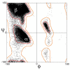
Figure 4
Ramachandran diagram [23].
Figure 5
Figure 5
Pinocytosis mechanism [27].
Scheme 2
Scheme 2
Example of Ring Closing Metathesis Stapling [28].
Scheme 3
Scheme 3
Ring closing metathesis reaction mechanism.
Scheme 4
Scheme 4
Olefinic amino acid synthesis by nucleophilic substitution.
Scheme 5
Scheme 5
Nickel-catalysed olefinic amino acid synthesis. R = -H, -Me.
Scheme 6
Scheme 6
Example of CuAAC Stapling.
Scheme 7
Scheme 7
Copper catalysed azide-alkyne cycloaddition (CuAAC) reaction mechanism.
Scheme 8
Scheme 8
Alkynyl-amino acid synthesis by nucleophilic substitution. X = -O, -S, -COO, -CONH, -CH2COO, -CH2CONH.
Scheme 9
Scheme 9
Ni-catalysed alkynyl-amino acid synthesis. R = -H, -Me.
Scheme 10
Scheme 10
Azide-amino acid synthesis by Mitsunobu reaction.
Scheme 11
Scheme 11
Azide-amino acid synthesis by Weinreb amide.
Scheme 12
Scheme 12
Azide-amino acid synthesis by Hoffmann rearrangement.
Scheme 13
Scheme 13
Azide-amino acid synthesis by Ullmann coupling.
Scheme 14
Scheme 14
Example of Lactamization Stapling [28].
Scheme 15
Scheme 15
PyBOP/HOBt coupling mechanism.
Scheme 16
Scheme 16
Example of Cysteine-Xylene Stapling [28].
Scheme 17
Scheme 17
Example of Cysteine-Perfluorobenzene Stapling [28].
Scheme 18
Scheme 18
Example of Alkyne/Alkene - Hydrothiolation Stapling [28].
Scheme 19
Scheme 19
Alkynyl-amino acid synthesis.
Scheme 20
Scheme 20
Example of Selenocysteine Stapling [54].
Scheme 21
Scheme 21
Example of Tryptophan Stapling [33].
Scheme 22
Scheme 22
Tryptophan-aldehyde coupling mechanism.
Scheme 23
Scheme 23
Example of Indole C-H activation Stapling [56].
Figure 6
Figure 6
From Huisgen, R. J. Org. Chem. 1976, 41, 403–419.
Scheme 24
Scheme 24
Example of stapling strategy with nitrones.
Scheme 25
Scheme 25
Example of Nitrile Oxide Stapling [28].
Scheme 26
Scheme 26
Nitrile oxide-amino acid synthesis from double bond oxidation.
Scheme 27
Scheme 27
Nitrile oxide-amino acid synthesis by alkyne hydroboration/oxidation.
Scheme 28
Scheme 28
Nitrile oxide-amino acid synthesis by Negishi cross-coupling.
Figure 7
Figure 7
Circular dichroism spectra of peptides measured in ddH2O (A) and in 30 mM SDS solution (B). Schematic comparison of the relative helical contents in ddH2O (C) and 30 mM SDS solution (D) (1 = unstapled; 2 = all-hydrocarbon-; 3 = lactam-; 4 = triazole-; 5 = vinyl sulfide-; 6 = xylene-; 7 = perfluoroaryl-)). The relative helicity was normalized with respect to the peptide with a lactam crosslink. n.d.: not determined as a result of additional absorption [28].
Figure 8
Figure 8
Normalized FAM intensity versus normalized retention time of FAM-labelled peptides L1b-L7b by using flow cytometry analysis in different cell lines and reversed-phase liquid chromatography. FAM intensity and retention times were normalized with respect to the linear peptide (1) (1 = unstapled; 2 = all-hydrocarbon-; 3 = lactam-; 4 = triazole-; 5 = vinyl sulfide-; 6 = xylene-; 7 = perfluoroaryl-) [28].
Figure 9
Figure 9
Flow cytometry analysis of stapled α-helical peptides with different cross-links in different cell-lines. Values are normalized with respect to Tat peptide (1 = unstapled; 2 = all-hydrocarbon-; 3 = lactam-; 4 = triazole-; 5 = vinyl sulfide-; 6 = xylene-; 7 = perfluoroaryl-) [28].
Figure 10
Figure 10
Haemolytic activity of stapled α-helical peptides with different types crosslinks [28].

References

    1. Nelson D.L., Cox M.M. Lehninger’s Principles of Biochemistry. 4th ed. W. H. Freeman and Company; New York, NY, USA: 2005.
    1. Osborne T.B. The Vegetable Protein. Longman Harlow; London, UK: 1909.
    1. Mulder G.J. Sur la composition de quelques substances animales. Bull. Sci. Phys. Nat. Neerl. 1838;1:104–119.
    1. Harold H. Origin of the Word ‘Protein’. Nature. 1951;168:244. - PubMed
    1. Kendrew J.C., Bodo G., Dintzis H.M., Parrish R.G., Wyckoff H., Phillips D.C. A three-dimensional model of the myoglobin molecule obtained by X-ray analysis. Nature. 1958;181:662–666. doi: 10.1038/181662a0. - DOI - PubMed

MeSH terms

LinkOut - more resources