. 2019 Oct 10;20(20):5021.
doi: 10.3390/ijms20205021.

Analysis of Procollagen C-Proteinase Enhancer-1/Glycosaminoglycan Binding Sites and of the Potential Role of Calcium Ions in the Interaction

Affiliations

Analysis of Procollagen C-Proteinase Enhancer-1/Glycosaminoglycan Binding Sites and of the Potential Role of Calcium Ions in the Interaction

Jan Potthoff et al. Int J Mol Sci. .

Abstract

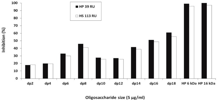
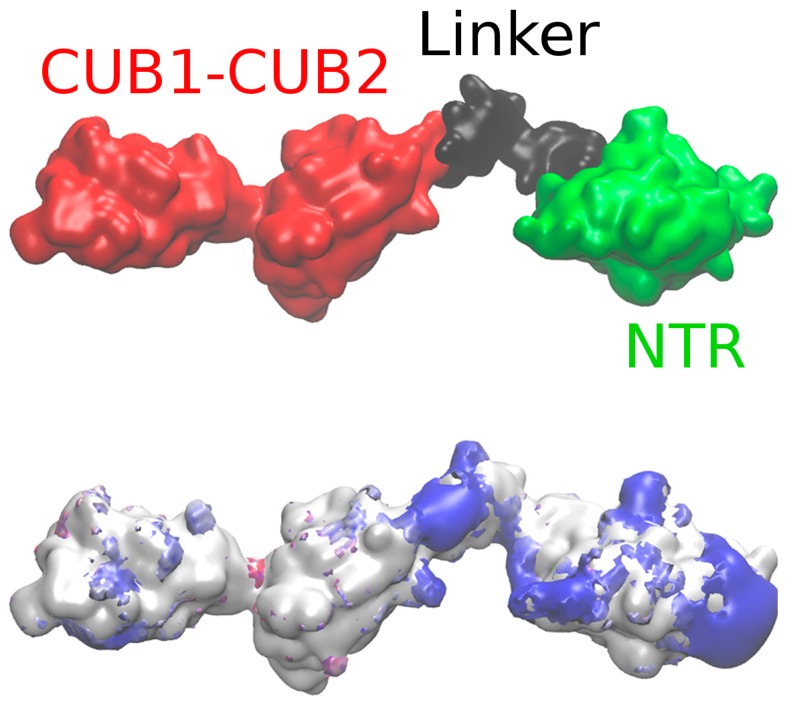
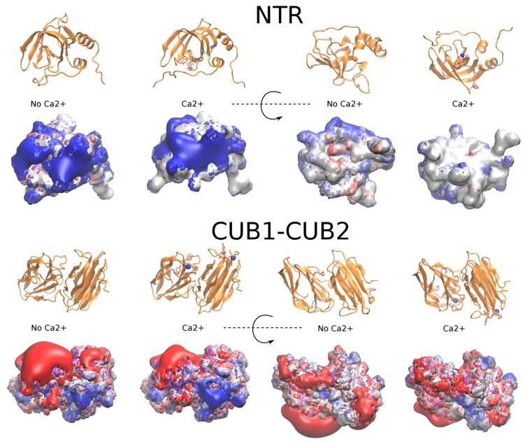
In this study, we characterize the interactions between the extracellular matrix protein, procollagen C-proteinase enhancer-1 (PCPE-1), and glycosaminoglycans (GAGs), which are linear anionic periodic polysaccharides. We applied molecular modeling approaches to build a structural model of full-length PCPE-1, which is not experimentally available, to predict GAG binding poses for various GAG lengths, types and sulfation patterns, and to determine the effect of calcium ions on the binding. The computational data are analyzed and discussed in the context of the experimental results previously obtained using surface plasmon resonance binding assays. We also provide experimental data on PCPE-1/GAG interactions obtained using inhibition assays with GAG oligosaccharides ranging from disaccharides to octadecasaccharides. Our results predict the localization of GAG-binding sites at the amino acid residue level onto PCPE-1 and is the first attempt to describe the effects of ions on protein-GAG binding using modeling approaches. In addition, this study allows us to get deeper insights into the in silico methodology challenges and limitations when applied to GAG-protein interactions.

Keywords: calcium ions; computational analysis of protein-glycosaminoglycan interactions; fragment-based docking; glycosaminoglycans; procollagen C-proteinase enhancer-1.


Conflict of interest statement

The authors declare no conflicts of interest.

Figures

Figure 1
Figure 1
Surface plasmon resonance (SPR) inhibition assays. Inhibition of the binding of recombinant human procollagen C-proteinase enhancer-1 (PCPE-1) to biotinylated heparin (HP) and heparan sulfate (HS) captured on a streptavidin sensor chip (39 and 113 resonance units (RU) respectively) by HP oligosaccharides of different degrees of polymerization (dp2‒dp18) and by HP (6 and 16 kDa) at a concentration of 5 µg/mL.
Figure 2
Figure 2
Small angle X-ray scattering (SAXS) Model (upper panel). Netrin-like (NTR) domain: green; CUB1-CUB2: red; the interdomain linker between the CUB2 and NTR domains: black. Positive electrostatic potential isosurfaces (2.0 kcal/mol · e−1) in the absence of Ca2+ ions obtained by Poisson‒Boltzmann surface area (PBSA) calculations (bottom panel).
Figure 3
Figure 3
Molecular docking and molecular mechanics-generalized born surface area (MM-GBSA) for NTR-glycosaminoglycan (GAG) complexes. The structure of the NTR domain is shown in cartoon representation at the top. For each GAG, the analyzed clusters of docking solutions are shown in blue, red, yellow and green (from the most to the less populated cluster); the top 10 residues binding to GAGs according to MM-GBSA calculations averaged per GAG are highlighted in red surface. Note that the clusters for CS6 dp6 are shown for a different protein spatial orientation to allow for a better visualization. In addition, averaging the per-residue energy for very different clusters could be misleading as shown for CS6 dp6: the residues shown in red do not overlap with the surface patches where the most representative clusters of solutions are located.
Figure 4
Figure 4
NTR amino acid residues identified in the top 10 for binding GAGs according to MM-GBSA calculations per cluster are labeled as an asterisk.
Figure 5
Figure 5
Electrostatic potential isosurfaces (blue, positive; red, negative) of NTR (–2.5 kcal/mol·e−1 and 1.0 kcal/mol·e−1) and CUB1-CUB2 (–3 kcal/mol·e−1 and 3 kcal/mol·e−1) domains in the presence and in the absence of Ca2+ ions obtained by PBSA calculations. Protein domains are shown in cartoon with the residues coordinating Ca2+ ions in licorice representation; Ca2+ ions: blue spheres.
Figure 6
Figure 6
Molecular docking results for the models of the full-length PCPE-1 SAXS Model in the absence and presence of Ca2+ ions and HP dp6. The clusters of docking solutions are shown in blue, red and yellow (from the most to the least populated clusters). NTR domain: green; CUB1-CUB2: red; the interdomain linker between the CUB2 and NTR domains: black.
Figure 7
Figure 7
Molecular docking results for the models of the full PCPE-1 SAXS Model in the absence (in blue) and the presence (in red) of Ca2+ ions and HP dp11 corresponding to the most favorable free binding energies.

References

    1. Esko J.D., Kimata K., Lindahl U. Proteoglycans and Sulfated Glycosaminoglycans. In: Varki A., Cummings R.D., Esko J.D., Freeze H.H., Stanley P., Bertozzi C.R., Hart G.W., Etzler M.E., editors. Essentials of Glycobiology. 2nd ed. Cold Spring Harbor Laboratory Press; Cold Spring Harbor, NY, USA: 2009. pp. 1–784. - PubMed
    1. Pomin V.H., Mulloy B. Glycosaminoglycans and Proteoglycans. Pharmaceuticals. 2018;11:27. doi: 10.3390/ph11010027. - DOI - PMC - PubMed
    1. Proudfoot A.E. Chemokines and Glycosaminoglycans. Front. Immunol. 2015;6:246. doi: 10.3389/fimmu.2015.00246. - DOI - PMC - PubMed
    1. Shute J. Glycosaminoglycan and chemokine/growth factor interactions. Handb. Exp. Pharmacol. 2012;207:307–324. - PubMed
    1. Iozzo R.V., Zoellerm J.J., Nyströmm A. Basement membrane proteoglycans: Modulators Par Excellence of cancer growth and angiogenesis. Mol. Cells. 2009;27:503–513. doi: 10.1007/s10059-009-0069-0. - DOI - PMC - PubMed

LinkOut - more resources