. 2019 Oct 29:8:e49058.
doi: 10.7554/eLife.49058.

Modeling the dynamics of Plasmodium falciparum gametocytes in humans during malaria infection

Affiliations

Modeling the dynamics of Plasmodium falciparum gametocytes in humans during malaria infection

Pengxing Cao et al. Elife. .

Abstract

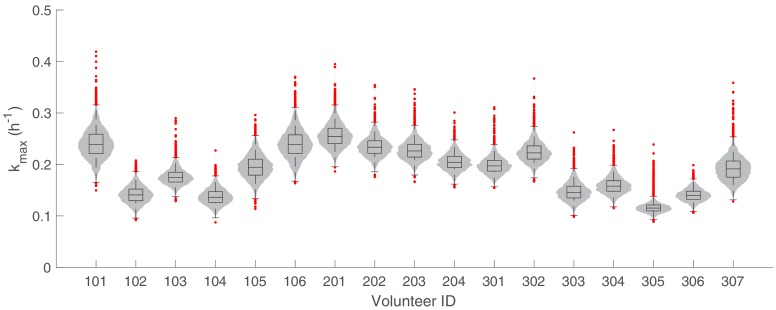
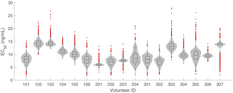
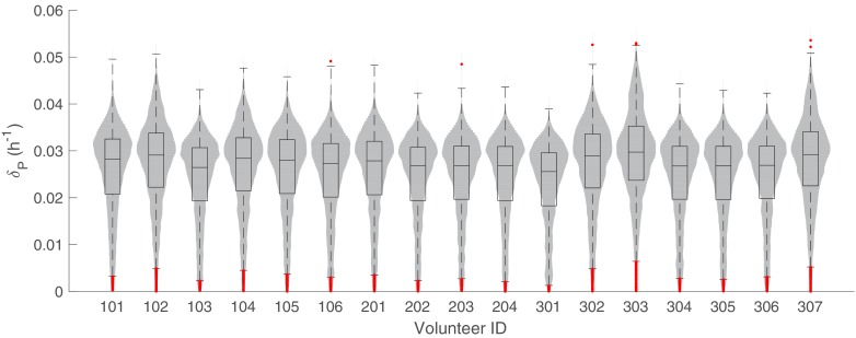
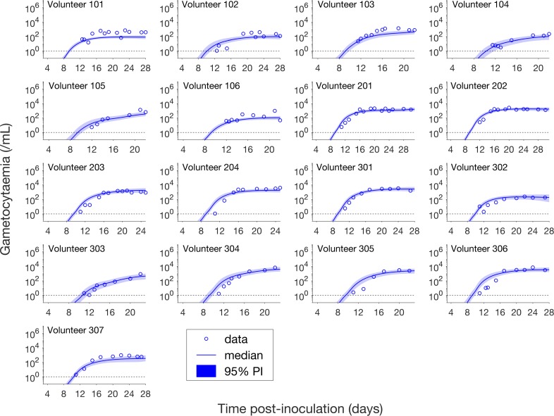
Renewed efforts to eliminate malaria have highlighted the potential to interrupt human-to-mosquito transmission - a process mediated by gametocyte kinetics in human hosts. Here we study the in vivo dynamics of Plasmodium falciparum gametocytes by establishing a framework which incorporates improved measurements of parasitemia, a novel gametocyte dynamics model and model fitting using Bayesian hierarchical inference. We found that the model provides an excellent fit to the clinical data from 17 volunteers infected with P. falciparum (3D7 strain) and reliably predicts observed gametocytemia. We estimated the sexual commitment rate and gametocyte sequestration time to be 0.54% (95% credible interval: 0.30-1.00%) per asexual replication cycle and 8.39 (6.54-10.59) days respectively. We used the data-calibrated model to investigate human-to-mosquito transmissibility, providing a method to link within-human host infection kinetics to epidemiological-scale infection and transmission patterns.

Keywords: P. falciparum; bayesian inference; computational biology; epidemiology; gametocyte dynamics; global health; mathematical modelling; systems biology.


Conflict of interest statement

PC, KC, SZ, TW, JT, JS, JM, JM No competing interests declared

Figures

Figure 1.
Figure 1.. Results of model fitting for all 17 volunteers.
Data are presented by circles. The median of posterior predictions (solid line) and 95% prediction interval (PI, shaded area) are generated by 5000 model simulations based on 5000 samples from the posterior parameter distribution as described in the Materials and methods. The histograms showing the posterior distributions of population mean and standard deviation hyperparameters are given in Figure 1—figure supplements 1 and 2. The posterior distribution of each model parameter (see the Materials and methods) for individual volunteers are given in Figure 1—figure supplements 3–14 Posterior distributions for some biological parameters are given in Figure 1—figure supplement 15, which are generated based on the posterior samples of population mean parameters (see the Materials and methods) and will be used to support the results in Table 1 shown later. The source data and computer code with instructions of implementation to generate Figure 1 and Figure 1—figure supplements 1–15 are fully publicly available at https://doi.org/10.26188/5cde4c26c8201.
Figure 1—figure supplement 1.
Figure 1—figure supplement 1.. Marginal posterior distributions for the 12 population mean parameters (hyperparameters).
5000 samples are used to generate the distributions. The dashed curves indicate the uniform prior distributions. p.i.: post-inoculation. Note that the y-axis is probability density instead of number of samples. Relevant details of the hyperparameters are provided in the Materials and methods in the main text.
Figure 1—figure supplement 2.
Figure 1—figure supplement 2.. Marginal posterior distributions for the 12 population SD parameters (hyperparameters).
5000 samples are used to generate the distributions. The dashed curves indicate the half-normal prior distributions. p.i.: post-inoculation. Note that the y-axis is probability density instead of number of samples. Relevant details of the hyperparameters are provided in the Materials and methods in the main text.
Figure 1—figure supplement 3.
Figure 1—figure supplement 3.. The marginal posterior distributions of the individual parameter of Pinit (inoculation size) for all 17 volunteers.
The violin plots (gray area) show the distributions of 5000 posterior samples. Box plots show the 25%, 50% (median) and 75% quantiles with outliers indicated by red dots. Relevant details of the individual parameter are provided in the Materials and methods in the main text.
Figure 1—figure supplement 4.
Figure 1—figure supplement 4.. The marginal posterior distributions of the individual parameter of µ (mean of the initial parasite age distribution) for all 17 volunteers.
The violin plots (gray area) show the distributions of 5000 posterior samples. Box plots show the 25%, 50% (median) and 75% quantiles with outliers indicated by red dots. Relevant details of the individual parameter are provided in the Materials and methods in the main text.
Figure 1—figure supplement 5.
Figure 1—figure supplement 5.. The marginal posterior distributions of the individual parameter of σ (Standard deviation of the initial parasite age distribution) for all 17 volunteers.
The violin plots (gray area) show the distributions of 5000 posterior samples. Box plots show the 25%, 50% (median) and 75% quantiles with outliers indicated by red dots. Relevant details of the individual parameter are provided in the Materials and methods in the main text.
Figure 1—figure supplement 6.
Figure 1—figure supplement 6.. The marginal posterior distributions of the individual parameter of rP (parasite replication rate) for all 17 volunteers.
The violin plots (gray area) show the distributions of 5000 posterior samples. Box plots show the 25%, 50% (median) and 75% quantiles with outliers indicated by red dots. Relevant details of the individual parameter are provided in the Materials and methods in the main text.
Figure 1—figure supplement 7.
Figure 1—figure supplement 7.. The marginal posterior distributions of the individual parameter of kmax (maximum rate of parasite killing by PQP) for all 17 volunteers.
The violin plots (gray area) show the distributions of 5000 posterior samples. Box plots show the 25%, 50% (median) and 75% quantiles with outliers indicated by red dots. Relevant details of the individual parameter are provided in the Materials and methods in the main text.
Figure 1—figure supplement 8.
Figure 1—figure supplement 8.. The marginal posterior distributions of the individual parameter of EC50 (half-maximum effective PQP concentration) for all 17 volunteers.
The violin plots (gray area) show the distributions of 5000 posterior samples. Box plots show the 25%, 50% (median) and 75% quantiles with outliers indicated by red dots. Relevant details of the individual parameter are provided in the Materials and methods in the main text.
Figure 1—figure supplement 9.
Figure 1—figure supplement 9.. The marginal posterior distributions of the individual parameter of γ (Hill coefficient for PQP) for all 17 volunteers.
The violin plots (gray area) show the distributions of 5000 posterior samples. Box plots show the 25%, 50% (median) and 75% quantiles with outliers indicated by red dots. Relevant details of the individual parameter are provided in the Materials and methods in the main text.
Figure 1—figure supplement 10.
Figure 1—figure supplement 10.. The marginal posterior distributions of the individual parameter of f (sexual commitment rate; not converted to percentage) for all 17 volunteers.
The violin plots (gray area) show the distributions of 5000 posterior samples. Box plots show the 25%, 50% (median) and 75% quantiles with outliers indicated by red dots. Relevant details of the individual parameter are provided in the Materials and methods in the main text.
Figure 1—figure supplement 11.
Figure 1—figure supplement 11.. The marginal posterior distributions of the individual parameter of δP (death rate of asexual and sexual parasites) for all 17 volunteers.
The violin plots (gray area) show the distributions of 5000 posterior samples. Box plots show the 25%, 50% (median) and 75% quantiles with outliers indicated by red dots. Relevant details of the individual parameter are provided in the Materials and methods in the main text.
Figure 1—figure supplement 12.
Figure 1—figure supplement 12.. The marginal posterior distributions of the individual parameter of m (maturation rate of gametocytes) for all 17 volunteers.
The violin plots (gray area) show the distributions of 5000 posterior samples. Box plots show the 25%, 50% (median) and 75% quantiles with outliers indicated by red dots. Relevant details of the individual parameter are provided in the Materials and methods in the main text.
Figure 1—figure supplement 13.
Figure 1—figure supplement 13.. The marginal posterior distributions of the individual parameter of δG (death rate of sequestered gametocytes) for all 17 volunteers.
The violin plots (gray area) show the distributions of 5000 posterior samples. Box plots show the 25%, 50% (median) and 75% quantiles with outliers indicated by red dots. Relevant details of the individual parameter are provided in the Materials and methods in the main text.
Figure 1—figure supplement 14.
Figure 1—figure supplement 14.. The marginal posterior distributions of the individual parameter of δGm (death rate of circulating gametocytes) for all 17 volunteers.
The violin plots (gray area) show the distributions of 5000 posterior samples. Box plots show the 25%, 50% (median) and 75% quantiles with outliers indicated by red dots. Relevant details of the individual parameter are provided in the Materials and methods in the main text.
Figure 1—figure supplement 15.
Figure 1—figure supplement 15.. Marginal posterior distributions of some key biological parameters.
5000 samples from the posterior parameter distribution are used to generate the figures. Full details about the definitions and expressions of those biological parameters are provided in the Materials and methods in the main text. Note that a logarithm is taken for the mean lifespan of circulating gametocyte for a better visualisation of the distribution.
Figure 2.
Figure 2.. Comparison of model predictions and clinical data for the asexual parasitemia for all 17 volunteers.
Data are presented by circles. The median of posterior predictions (solid curve) and 95% PI (shaded area) are generated by 5000 model simulations based on 5000 samples from the posterior parameter distribution as described in the Materials and methods. The data points with one parasite/mL (i.e., those points which lie on the dotted line) indicate measurements for which no parasites were detected. No data are available for Volunteer 101 and 106 to validate the model predictions. The source data and computer code with instructions of implementation to generate Figure 2 are fully publicly available at https://doi.org/10.26188/5cde4c26c8201.
Figure 3.
Figure 3.. Comparison of model predictions and clinical data for the gametocytemia for all 17 volunteers.
Data are presented by circles. The median of posterior predictions (solid curve) and 95% PI (shaded area) are generated by 5000 model simulations based on 5000 samples from the posterior parameter distribution as described in the Materials and methods. The data points with one parasite/mL (i.e. those points which lie on the dotted line) indicate measurements for which no parasites were detected. The source data and computer code with instructions of implementation to generate Figure 3 are fully publicly available at https://doi.org/10.26188/5cde4c26c8201.
Figure 4.
Figure 4.. Simulation of two scenarios predicting the dependence of human-to-mosquito transmissibility on the sexual commitment rate and gametocyte sequestration time.
(A) illustration of the first scenario: predicting the critical gametocytemia level (indicated by Gc) at the time when the total parasitemia reaches 108 parasites/mL. (B) illustration of the second scenario: predicting the non-infectious period (indicated by tc), which is defined to be time from inoculation of infected red blood cells to the time when the gametocytemia reaches 103 parasites/mL (a threshold below which human-to-mosquito transmission was not observed [Collins et al., 2018]). (C and D) Heatmaps showing the dependence of the critical gametocytemia Gc and the non-infectious period tc on the sexual commitment rate and gametocyte sequestration time. The black dots represent the value obtained by simulating the gametocyte dynamics model using the median estimates of the posterior samples of the population mean parameters as described in the Materials and methods. The red curve in C is the level curve for Gc = 103 parasites/mL. The red curve in D is the level curve for tc = 13.42 days which is the non-infectious period obtained by model simulation using the posterior estimates of the population mean parameters. The source data and computer code with instruction of implementation to generate Figure 4 are fully publicly available at https://doi.org/10.26188/5cde4c26c8201.
Figure 5.
Figure 5.. Schematic diagram showing the model compartments and transitions.
The model is comprised of three parts describing three populations of parasites: asexual parasites (Pa,t), sexually committed parasites (PGa,t) and gametocytes (Gt). P and PG are functions of asexual parasite age a and time t. Square compartments in the inner loop represent the asexual parasite population which follows a cycle of maturation and replication every aL hours. Sexual commitment occurs from age as and a fraction of asexual parasites become sexually committed (the bigger square compartments in the outer loop) and eventually enter the development of stage I–V gametocytes (G1–G5). The compartments with a dashed boundary are sequestered to tissues and thus not measurable in a blood smear. The notation for each compartment is consistent with those in the model equations and is explained in the main text.
Figure 6.
Figure 6.. The pharmacokinetic model of piperaquine (PQP).
The model is a three-compartment disposition model with two transit compartments for absorption. State D represents the dose of PQP. T1 and T2 represent the two transit compartments. C is the central compartment and PQP concentration in this compartment was measured (which are shown in Figure 6—figure supplement 1). P1 and P2 represent two peripheral compartments. kT, q1, q2 and qc are the rates of flow into or out of compartments.
Figure 6—figure supplement 1.
Figure 6—figure supplement 1.. PK data and optimized PK curves (the ‘fits’) of piperaquine (PQP) concentration for all volunteers.
The details of the optimization approach are provided in the Materials and methods in the main text and Appendix 1. Some volunteers have two peaks of PQP concentrations because they had recrudescent asexual parasitemia (see Figure 1 in the main text) and were treated with a second dose of 960 mg PQP. The source data and computer code with instructions of implementation to generate Figure 6—figure supplement 1 are fully publicly available at https://doi.org/10.26188/5cde4c26c8201.

References

    1. Adjalley SH, Johnston GL, Li T, Eastman RT, Ekland EH, Eappen AG, Richman A, Sim BK, Lee MC, Hoffman SL, Fidock DA. Quantitative assessment of Plasmodium falciparum sexual development reveals potent transmission-blocking activity by methylene blue. PNAS. 2011;108:E1214–E1223. doi: 10.1073/pnas.1112037108. - DOI - PMC - PubMed
    1. Ahn JE, Karlsson MO, Dunne A, Ludden TM. Likelihood based approaches to handling data below the quantification limit using NONMEM VI. Journal of Pharmacokinetics and Pharmacodynamics. 2008;35:401–421. doi: 10.1007/s10928-008-9094-4. - DOI - PubMed
    1. Bancells C, Llorà-Batlle O, Poran A, Nötzel C, Rovira-Graells N, Elemento O, Kafsack BFC, Cortés A. Revisiting the initial steps of sexual development in the malaria parasite Plasmodium falciparum. Nature Microbiology. 2019;4:144–154. doi: 10.1038/s41564-018-0291-7. - DOI - PMC - PubMed
    1. Bolscher JM, Koolen KMJ, van Gemert GJ, van de Vegte-Bolmer MG, Bousema T, Leroy D, Sauerwein RW, Dechering KJ. A combination of new screening assays for prioritization of transmission-blocking antimalarials reveals distinct dynamics of marketed and experimental drugs. Journal of Antimicrobial Chemotherapy. 2015;70:1357–1366. doi: 10.1093/jac/dkv003. - DOI - PubMed
    1. Bousema T, Drakeley C. Epidemiology and infectivity of Plasmodium falciparum and plasmodium vivax gametocytes in relation to malaria control and elimination. Clinical Microbiology Reviews. 2011;24:377–410. doi: 10.1128/CMR.00051-10. - DOI - PMC - PubMed

Publication types