. 2019 Oct 28;10(11):816.
doi: 10.1038/s41419-019-2055-6.

Ionizing radiation induces endothelial transdifferentiation of glioblastoma stem-like cells through the Tie2 signaling pathway

Affiliations

Ionizing radiation induces endothelial transdifferentiation of glioblastoma stem-like cells through the Tie2 signaling pathway

Pauline Deshors et al. Cell Death Dis. .

Abstract

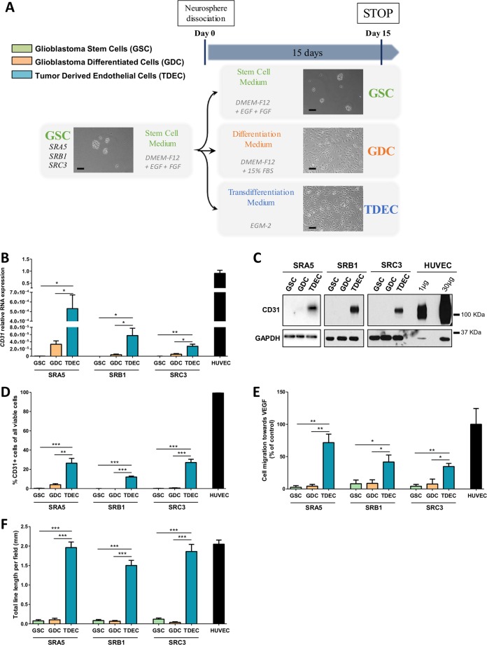
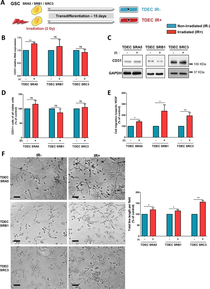
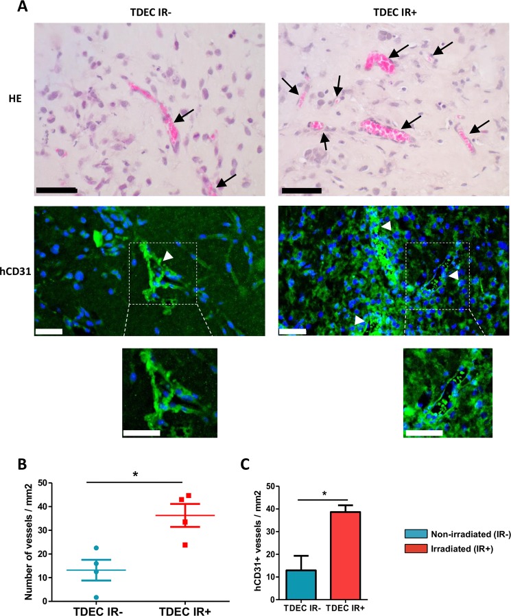
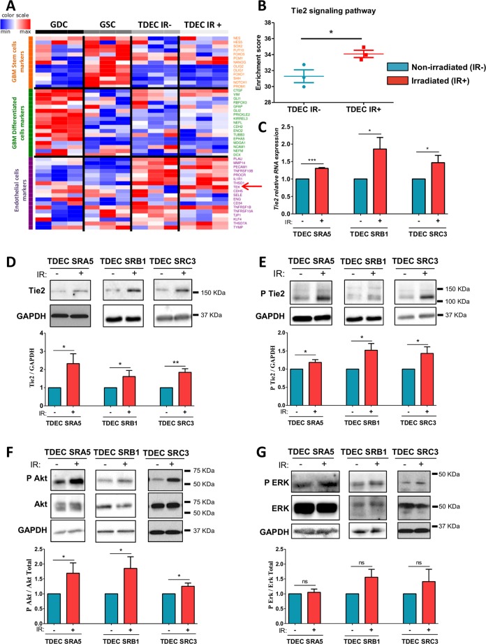
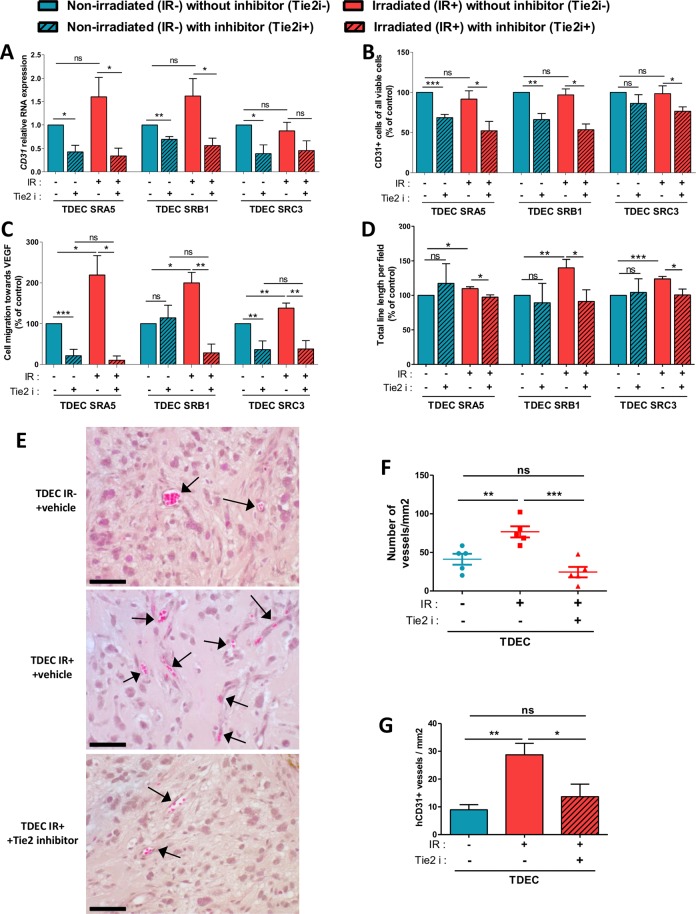
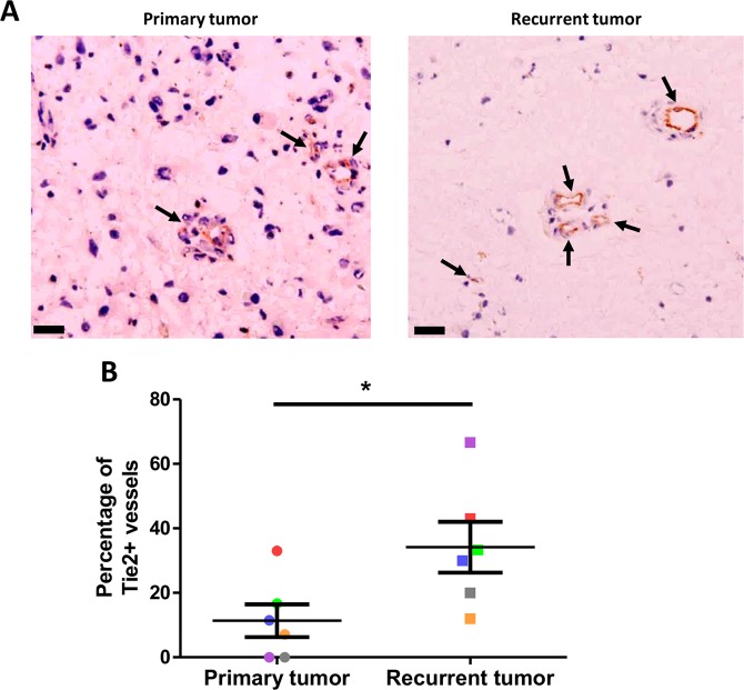
Glioblastomas (GBM) are brain tumors with a poor prognosis despite treatment that combines surgical resection and radio-chemotherapy. These tumors are characterized by abundant vascularization and significant cellular heterogeneity including GBM stem-like cells (GSC) which contribute to tumor aggressiveness, resistance, and recurrence. Recent data has demonstrated that GSC are directly involved in the formation of new vessels via their transdifferentiation into Tumor Derived Endothelial Cells (TDEC). We postulate that cellular stress such as ionizing radiation (IR) could enhance the transdifferentiation of GSC into TDEC. GSC neurospheres isolated from 3 different patients were irradiated or not and were then transdifferentiated into TDEC. In fact, TDEC obtained from irradiated GSC (TDEC IR+) migrate more towards VEGF, form more pseudotubes in MatrigelTM in vitro and develop more functional blood vessels in MatrigelTM plugs implanted in Nude mice than TDEC obtained from non-irradiated GSC. Transcriptomic analysis allows us to highlight an overexpression of Tie2 in TDEC IR+. All IR-induced effects on TDEC were abolished by using a Tie2 kinase inhibitor, which confirms the role of the Tie2 signaling pathway in this process. Finally, by analyzing Tie2 expression in patient GBMs by immunohistochemistry, we demonstrated that the number of Tie2+ vessels increases in recurrent GBM compared with matched untreated tumors. In conclusion, we demonstrate that IR potentiates proangiogenic features of TDEC through the Tie2 signaling pathway, which indicates a new pathway of treatment-induced tumor adaptation. New therapeutic strategies that associate standard treatment and a Tie2 signaling pathway inhibitor should be considered for future trials.


Conflict of interest statement

The authors declare that they have no conflict of interest.

Figures

Fig. 1
Fig. 1. GSC cultured under conditions of endothelial differentiation develop phenotypical and functional features of endothelial cells.
a Overview of the different cell culture protocols. As described in the Materials and Methods section, GSC-enriched neurospheres were isolated from patient samples and cultured in a specific stem cell medium (DMEM-F12 with EGF (epidermal growth factor) and FGF (fibroblast growth factor) growth factors). Neurospheres were then dissociated and placed for at least 15 days (i) in stem cell medium to keep GSC in culture as a control, (ii) in differentiation medium (DMEM-F12 with 15% FBS (fetal bovine serum) to obtain GDC or (iii) in transdifferentiation medium (EGM-2) to obtain TDEC. Scale bars, 100 µm. b Relative RNA expression of the endothelial marker CD31 determined by RT-qPCR in GSC, GDC, TDEC and HUVEC. Results are normalized to HUVEC expression. c Immunoblot of CD31 in GSC, GDC, TDEC, and HUVEC. Blots are representative of at least 3 independent experiments in the three patients’ GSC lines (SRA5, SRB1, and SRC3). d FACS immunofluorescence analysis of CD31 protein expression in GSC, GDC, TDEC and HUVEC. The graph represents means ± SEM of the percentage of CD31 positive cells among all viable cells of at least 3 independent experiments. e Percentage of cells that migrate towards VEGF normalized to HUVEC. f. Pseudotube formation assay. The graph represents means ± SEM of the total line length per field determined by the quantification of at least 3 fields per well
Fig. 2
Fig. 2. Ionizing radiation potentiates GSC transdifferentiation into TDEC.
a GSC isolated from tumors of 3 patients (SRA5, SRB1 and SRC3) were or were not irradiated (2 Gy) and were then cultured in EGM-2 for 15 days in order to obtain TDEC IR+ or TDEC IR−. b Relative RNA expression of the endothelial marker CD31 determined by RT-qPCR in TDEC IR− vs. TDEC IR+ from the three different GSC (SRA5, SRB1 and SRC3). The fold inductions are expressed as means ± SEM of at least three independent experiments (normalized to TDEC IR−). c Immunoblot of CD31 in TDEC IR− vs. TDEC IR+. Blots are representative of at least 3 independent experiments in the three patients’ GSC lines (SRA5, SRB1, and SRC3). d Immunofluorescence analysis by FACS of CD31 protein expression in TDEC IR− vs. TDEC IR+. The graphs represent means SEM of the percentage of CD31 positive cells among all viable cells of at least 3 independent experiments. e Cell migration towards VEGF. The graphs represent means ± SEM of the percentage of cells that migrate towards VEGF normalized to TDEC IR−. f Pseudotube formation assay. The graphs represent means ± SEM of the total line length per field determined by the quantification of at least 3 fields per well (normalized to TDEC IR−). Scale bars, 100 µm
Fig. 3
Fig. 3. Ionizing radiation potentiates TDEC vessel formation in vivo.
a MatrigelTM plug assays using TDEC IR− or TDEC IR+ obtained from SRC3 GSC stained by hematoxylin-eosin (upper panel). Immunofluorescence of plugs using a specific anti human-CD31 antibody (lower panel). Arrows indicate functional blood vessels and headed arrows indicate hCD31+ vessels. Scale bars, 50 µm. b Quantification of functional blood vessels in MatrigelTM plugs/mm2. The number of vessels containing red blood cells per mm2 was quantified by histological analysis and was expressed as means ± SEM of 4 mice. c Quantification of hCD31+ vessels in MatrigelTM plugs/mm2. The number of hCD31 vessels per mm2 was quantified and expressed as means ± SEM of 4 mice
Fig. 4
Fig. 4. Ionizing radiation increases Tie2 expression and Tie2 signaling pathway activation in TDEC.
a Transcriptome analysis using Affymetrix® chip was performed on SRC3 GSC, and on GDC, TDEC and TDEC IR+ obtained from SRC3 GSC. Because there are no gene sets for ‘GSC’ or ‘GDC’ in databases like GO, KEGG, Reactome or MSigDB, this gene list was compiled by extensive literature search (Supplementary Table. S3) b The enrichment score of Tie2 signaling pathway members in TDEC IR− versus TDEC IR+ was determined from transcriptomic analysis (n = 3). The Tie2 signaling pathway members are listed in the “Materials and Methods” section. c Relative RNA expression of Tie2 in TDEC IR− and TDEC IR+ obtained from three different GSC (normalized to TDEC IR− obtained from each GSC). dg Immunoblots of Tie2 d, P Tie2 e, P Akt and Akt f, and P ERK and ERK g in TDEC IR− and TDEC IR+ obtained from three different GSC. Blots are representative of at least 3 independent experiments. Blots were quantified and the graphs (bottom) show the protein expression ratio normalized to TDEC IR− obtained from each GSC
Fig. 5
Fig. 5. Tie2 kinase inhibitor (Tie2i) decreases the IR-induced effect on TDEC.
a Relative RNA expression of the endothelial marker CD31 determined by RT-qPCR in TDEC IR− vs. TDEC IR+ from the three different GSC (SRA5, SRB1 and SRC3) and cultured with or without Tie2i. The fold inductions are expressed as means ± SEM of at least three independent experiments (normalized to TDEC IR− obtained from each GSC and cultured without Tie2i). b Flow cytometric analysis of CD31 expression in TDEC IR− and TDEC IR+ obtained from SRA5, SRB1 and SRC3 GSC and cultured with or without Tie2i. The level of CD31 positive cells is expressed as means ± SEM normalized to TDEC IR- obtained from each GSC and cultured without Tie2i. c The percentage of cells that migrate towards VEGF normalized to TDEC IR- obtained from each GSC and cultured without Tie2i. d Pseudotube formation assay. The graph represents means ± SEM of the total line length per field determined by the quantification of at least 3 fields per well normalized to TDEC IR− without Tie2i obtained from each GSC. e MatrigelTM plug assay. Representative hematoxylin-eosin sections of MatrigelTM plugs with TDEC IR− without Tie2i, TDEC IR+ without Tie2i and TDEC IR+ with Tie2i. Black arrows indicate functional blood vessels. Scale bars, 50 µm. f Quantification of functional blood vessels in MatrigelTM plugs/mm2. The number of vessels/mm2 was expressed as means ± SEM of 5 mice for TDEC IR− without Tie2i and TDEC IR+ without Tie2i and of 4 mice for TDEC IR+ with Tie2i. g Quantification of hCD31 + vessels in MatrigelTM plugs/mm2. The number of hCD31 vessels per mm2 was quantified and expressed as means ± SEM of 5 mice for TDEC IR− without Tie2i and TDEC IR+ without Tie2i and of 4 mice for TDEC IR+ with Tie2i
Fig. 6
Fig. 6. Increase in the percentage of Tie2+ vessels in recurrent GBM.
a Representative IHC images for Tie2 expression in primary untreated tumors and post-radiation recurrent tumors from matched GBM patients. Arrows indicate functional blood vessels. Scale bars: 50 mm. b Percentage of Tie2 positive-vessels in primary untreated tumors and post-radiation recurrent tumors from matched GBM patients (n = 6). Each color is associated with one patient. *P < 0.05, by matched-pairs non-parametric Wilcoxon Signed Rank test

References

    1. Louis, D., Ohgazi, H., Wiestler, O. & Cavenee, W. Diffuse astrocytic and oligodendroglial tumours. World Health Organization Histological Classifications of Tumours of the Central Nervous System. (4th ed., Revised. Lyon: International Agency for Research Centre; 2016).
    1. Park DM, Sathornsumetee S, Rich JN. Treatment and management of malignant gliomas: medical oncology. Nat. Rev. Clin. Oncol. 2010;7:75–77. doi: 10.1038/nrclinonc.2009.221. - DOI - PubMed
    1. Stupp R, et al. Radiotherapy plus concomitant and adjuvant temozolomide for glioblastoma. New Engl. J. Med. 2005;352:987–996. doi: 10.1056/NEJMoa043330. - DOI - PubMed
    1. Stupp, R., Hegi, M. E. & Mason, W. P. Effects of radiotherapy with concomitant and adjuvant temozolomide versus radiotherapy alone on survival in glioblastoma in a randomised phase III study: 5-year analysis of the EORTC-NCIC trial. Lancet Oncol. 10, 459–466 (2009). - PubMed
    1. Tang DG. Understanding cancer stem cell heterogeneity and plasticity. Cell Res. 2012;22:457–472. doi: 10.1038/cr.2012.13. - DOI - PMC - PubMed

Publication types

MeSH terms