. 2019 Oct 28;9(1):15446.
doi: 10.1038/s41598-019-51189-7.

A murine model demonstrating reversal of structural and functional correlates of cirrhosis with progenitor cell transplantation

Affiliations

A murine model demonstrating reversal of structural and functional correlates of cirrhosis with progenitor cell transplantation

Mark D Muthiah et al. Sci Rep. .

Abstract

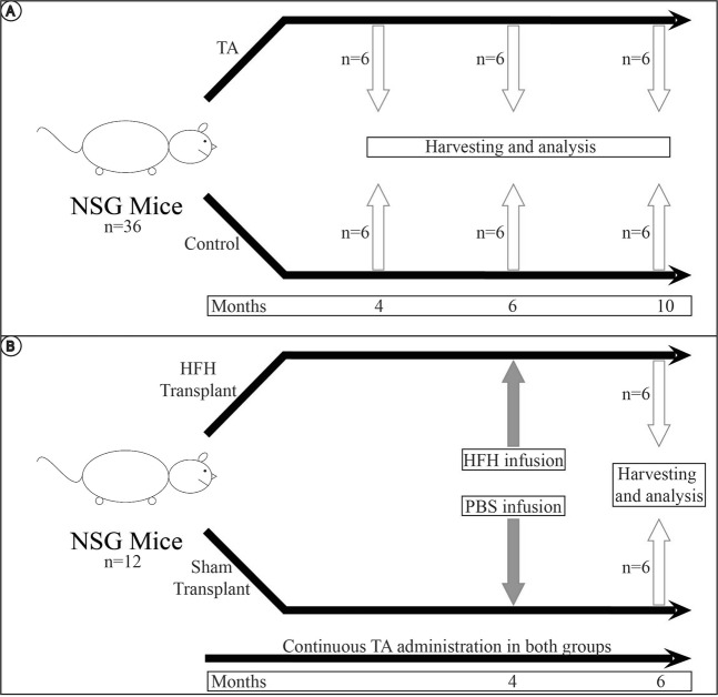
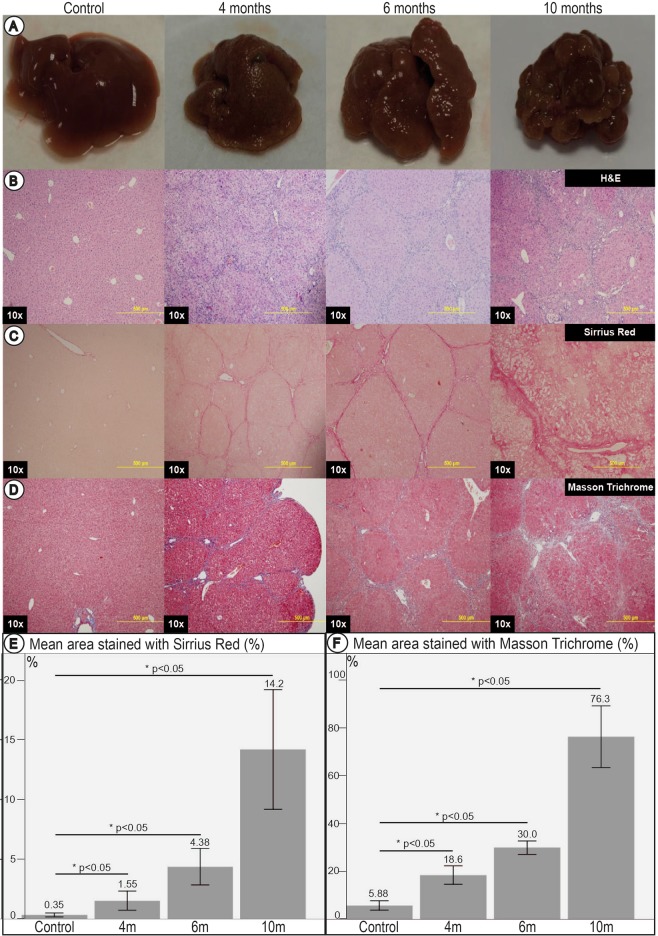
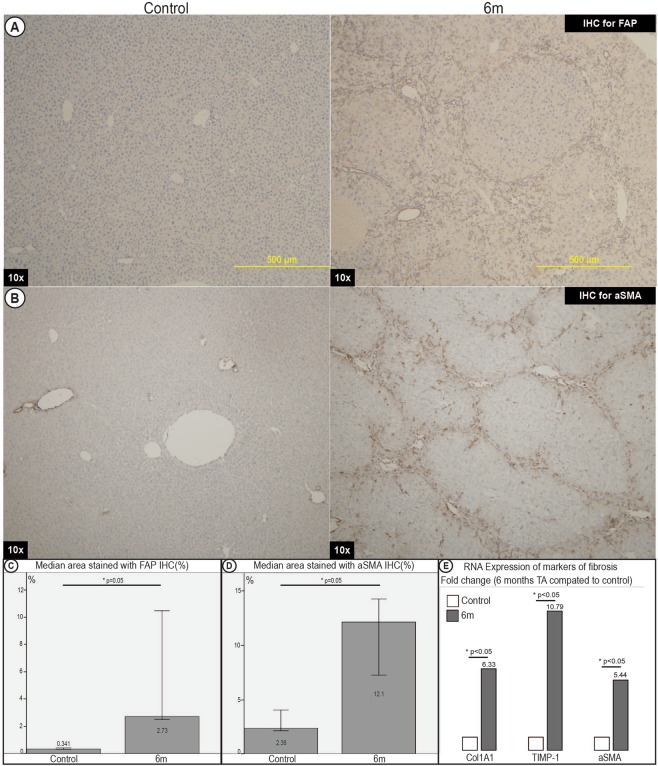
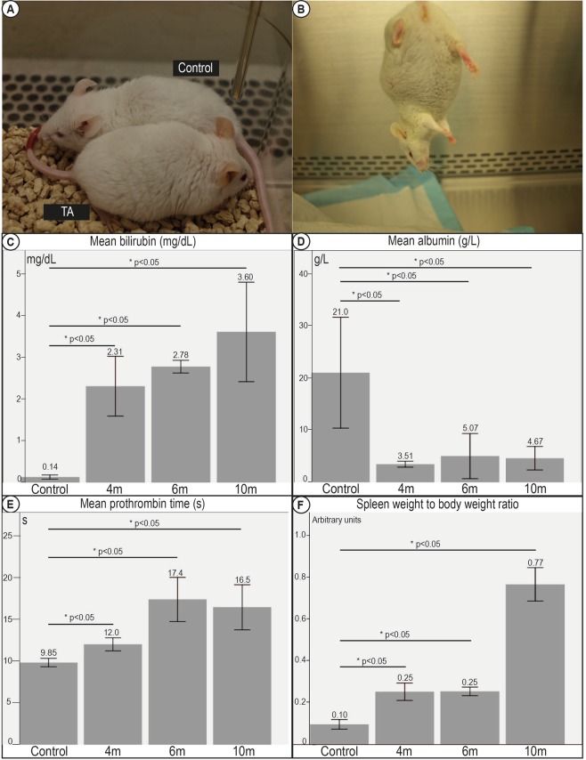
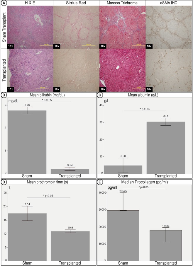
Development of cell transplantation for treating liver cirrhosis hinges critically on the availability of animal models for studying human stem cell transplantation. We report an immune-permissive murine model of liver cirrhosis with full clinical correlates of decompensated liver disease, and allows testing efficacy of stem cell transplantation. Liver cirrhosis was induced in Nod-scid gamma(NSG) mice with oral thioacetamide(TA) and compared to controls over 12 months. 4 month TA treated cirrhotic mice were then transplanted intrasplenically with 2million human fetal liver progenitor cells(HFH) and compared with cirrhotic controls 2 months after transplantation. NSG-TA mice developed shrunken and nodular livers with histological evidence of fibrosis as compared to controls. This was associated with evidence of worsening decompensated liver disease, with jaundice, hypoalbuminemia, coagulopathy, and encephalopathy in NSG-TA mice. Transplantation of HFH resulted in improvement in both fibrosis and markers of decompensated liver disease. We have demonstrated that NSG-TA mice can recapitulate the full clinical picture of structural and functional cirrhosis, both of which can be improved by transplantation of human fetal liver cells. This model serves as a valuable tool for validation of in vivo liver stem cell transplantation and opens up opportunities for studying the mechanism how stem cells reverse fibrosis.


Conflict of interest statement

The authors declare no competing interests.

Figures

Figure 1
Figure 1
Overall conduct of experiment. (A) For the first part of the experiment, 18 NSG mice were fed oral TA to induce liver cirrhosis. TA was administered, and 6 mice were sacrificed at 4 months, 6 months and 10 months respectively. They were compared to 18 control NSG mice, each given normal drinking water for the same duration. (B) For the second part of the experiment, NSG mice were given 4 months of oral TA. They were then subsequently transplanted with intrasplenic HFH, and given a further 2 months of TA, for a total duration of 6 months of TA. These mice were sacrificed at 6 months for analysis. They were compared to sham transplant mice given oral TA for 4 months, intrasplenic phosphate buffered saline (PBS), and a further 2 months of oral TA.
Figure 2
Figure 2
Livers and liver histology of control mice and at 4, 6 and 10 months of TA administration. (A) Gross livers surface of NSG mice fed with TA. Shrinkage and progressive nodularity of livers with increasing duration of TA administered was noted. By 10 months of TA administration, livers showed classical features of advanced cirrhosis. (B) Histological examination with H&E. (C) Histological examination with Sirrius Red. (D) Histological examination with Masson Trichrome. Stains revealed bridging fibrosis with thin septa at 4 months, consistent with mild cirrhosis. Nodule formation and broad septa were seen by 6 months, consistent with moderate cirrhosis. Architectural collapse with thick broad septa were seen at 10 months, consistent with severe cirrhosis. (E) Image morphometry of Sirrius Red staining. (F) Image morphometry of Masson Trichrome stain. These quantitatively demonstrated a significant increase in area stained with increasing duration of TA administered, further corroborating the findings on histology.
Figure 3
Figure 3
Immunohistochemistry and RT-qPCR for markers of fibrosis of livers of control mice and mice fed TA for 6 months. (A) Immunohistochemistry (IHC) for fibroblast activating protein (FAP). (B) IHC for alpha smooth muscle actin (aSMA). (C) Image morphometry of FAP. (D) Image morphometry of aSMA. Quantitative analysis showed a significant increase in the area stained positive for FAP and aSMA. This demonstrated a significant increase in markers consistent with fibroblast activation and myofibroblast activation respectively. (E) RT-qPCR expression compared between TA mice at 6 months and control mice for markers of fibrosis (Col1A1, TIMP-1, aSMA) demonstrated significant increase in RNA expression of markers of fibrosis.
Figure 4
Figure 4
Decompensation of clinical correlates of liver cirrhosis in mice fed TA. (A) Mice fed TA demonstrated observable jaundice. TA fed mice had Yellowing of the ears, and appeared lethargic as compared to control mice. (B) All TA fed mice developed ascites by 6 months of exposure. (C) Serum tests revealed significant hyperbilirubinemia, (D) hypoalbuminemia, (E) coagulopathy progressing with increasing duration of TA administered, and suggesting evidence of synthetic dysfunction typical of decompensated liver disease. (F) Spleen weight to body weight ratios. Increase in spleen weight to body weight ratios suggesting worsening portal hypertension was seen with increasing duration of TA administered.
Figure 5
Figure 5
Gross images and histology of nodules in TA fed mice. (A) All mice fed with TA demonstrated significant surface nodularity. 2/12 of the mice fed at least 6 months TA demonstrated dominant nodules. (B) Histology of some of these nodules revealed trabecular disarray and nuclear atypia characteristic of hepatocellular carcinoma.
Figure 6
Figure 6
Gross liver pictures and IF of transplanted mice 2 months post-transplant. (A) Gross morphology of sham transplant mice livers and HFH transplanted mice livers. Transplanted mice livers were softer in texture and less nodular than livers of sham transplant mice. (B) Co-labelling between IF of anti human albumin and anti human alpha1-antitrypsin (A1AT), (C) CYYP2A6, (D) CYP3A4, and (E) absence of co-localization between anti human albumin and AFP. This demonstrated that the engrafted hepatocytes matured, and demonstrated structural and functional viability up to 2 months post-transplant. No immature hepatocytes were seen.
Figure 7
Figure 7
Improvement in histology and clinical correlates of liver cirrhosis in transplanted mice 2 months post –transplant. (A) Histology sections of sham transplant mice livers and HFH transplanted mice livers. Histology stains with H&E, Sirrius Red and Masson Trichrome demonstrated improvement of histological markers of cirrhosis, with less bridging fibrosis, architectural distortion and nodule formation in mice transplanted with HFH. IHC for aSMA demonstrated evidence of reduced myofibroblast activation in transplanted livers. Transplanted mice demonstrated improvements in clinical correlates of cirrhosis. There was a significant improvement in (B) hyperbiliruminemia, (C) hypoalbuminemia, and (D) coagulopathy. (E) Elisa for serum pro-collagen was significantly lower after transplant, demonstrating improvement in serum markers of fibrosis.

References

    1. Friedman SL. Liver fibrosis–from bench to bedside. J Hepatol. 2003;38(Suppl 1):S38–53. doi: 10.1016/S0168-8278(02)00429-4. - DOI - PubMed
    1. Gines P, Cardenas A, Arroyo V, Rodes J. Management of cirrhosis and ascites. The New England journal of medicine. 2004;350:1646–1654. doi: 10.1056/NEJMra035021. - DOI - PubMed
    1. Schuppan D, Afdhal NH. Liver cirrhosis. Lancet. 2008;371:838–851. doi: 10.1016/S0140-6736(08)60383-9. - DOI - PMC - PubMed
    1. Tapper EB, Parikh ND. Mortality due to cirrhosis and liver cancer in the United States, 1999–2016: observational study. Bmj. 2018;362:k2817. doi: 10.1136/bmj.k2817. - DOI - PMC - PubMed
    1. Perera MTPR, Mirza DF, Elias E. Liver transplantation: Issues for the next 20 years. J Gastroenterol Hepatol. 2009;24(Suppl 3):124–131. doi: 10.1111/j.1440-1746.2009.06081.x. - DOI - PubMed

Publication types