. 2019 Dec;11(12):1113-1123.
doi: 10.1038/s41557-019-0351-5. Epub 2019 Oct 28.

Expedited mapping of the ligandable proteome using fully functionalized enantiomeric probe pairs

Affiliations

Expedited mapping of the ligandable proteome using fully functionalized enantiomeric probe pairs

Yujia Wang et al. Nat Chem. 2019 Dec.

Abstract

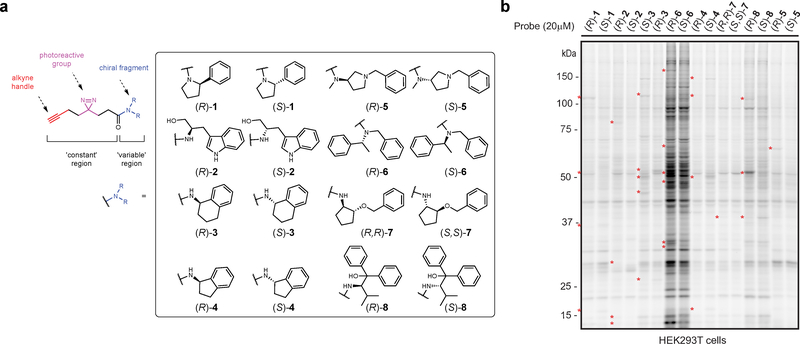
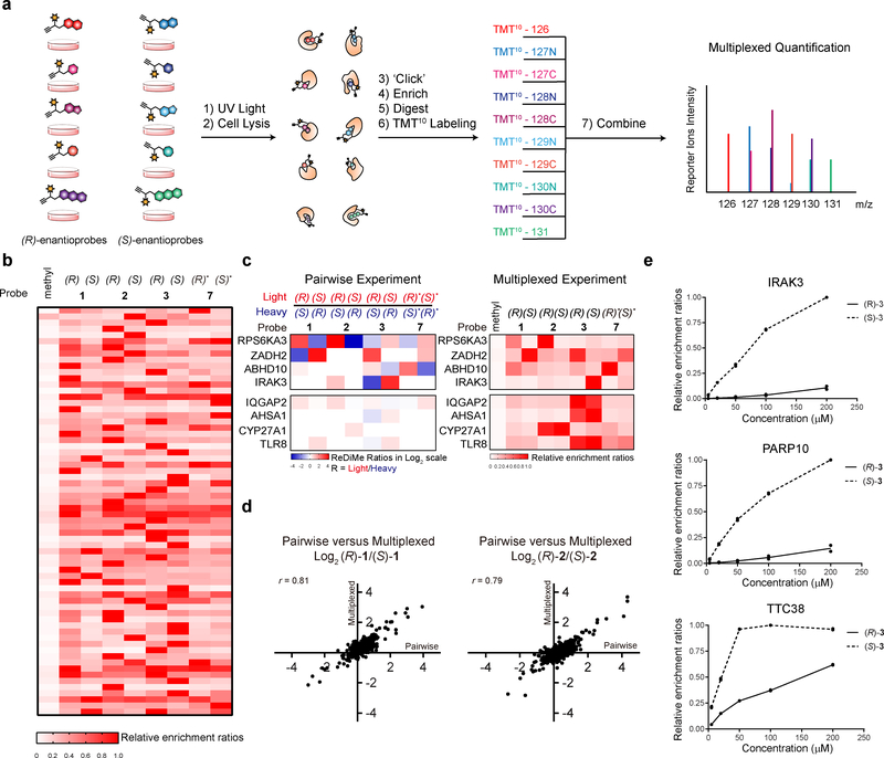
A fundamental challenge in chemical biology and medicine is to understand and expand the fraction of the human proteome that can be targeted by small molecules. We recently described a strategy that integrates fragment-based ligand discovery with chemical proteomics to furnish global portraits of reversible small-molecule/protein interactions in human cells. Excavating clear structure-activity relationships from these 'ligandability' maps, however, was confounded by the distinct physicochemical properties and corresponding overall protein-binding potential of individual fragments. Here, we describe a compelling solution to this problem by introducing a next-generation set of fully functionalized fragments differing only in absolute stereochemistry. Using these enantiomeric probe pairs, or 'enantioprobes', we identify numerous stereoselective protein-fragment interactions in cells and show that these interactions occur at functional sites on proteins from diverse classes. Our findings thus indicate that incorporating chirality into fully functionalized fragment libraries provides a robust and streamlined method to discover ligandable proteins in cells.


Conflict of interest statement

Competing Interests

The authors declare competing financial interests. B.F.C. is a founder and advisor to Vividion Therapeutics, a biotechnology company interested in using chemical proteomic methods to develop small-molecule drugs to treat human disease. C.G.P. serves as a consultant for Vividion Therapeutics.

Figures

Figure 1.
Figure 1.. Enantioprobes for mapping stereoselective protein-small molecule fragment interactions in human cells.
a, Structures of enantioprobes, which consist of a “variable” element of stereopure fragment pairs (enclosed box) and a “constant” region containing a diazirine photoreactive group and a clickable alkyne handle. b, Gel-based profiling of enantioprobe-protein interactions in human cells. HEK293T cells were treated with enantioprobes (20 μM) for 30 min, photocrosslinked, lysed, and proteomes conjugated to an azide-rhodamine tag using CuAAC chemistry and analyzed by SDS-PAGE and in-gel fluorescent scanning. Red asterisks mark representative stereoselective enantioprobe-protein interactions. Gel image reflects representative results from two independently performed experiments.
Figure 2.
Figure 2.. MS-based profiling of enantioprobe-protein interactions in human cells.
a, Schematic workflow for identifying stereoselective enantioprobe-protein interactions in human cells. b, Heatmap showing relative protein enrichment ratios for pairwise comparisons of (R) and (S) enantioprobes (200 μM each) in both isotopic directions in human peripheral blood mononuclear cells (PBMCs). White signals in the heatmap either correspond to proteins with ratio values of ~ 1 or proteins that were not enriched and quantified with the indicated enantioprobe pair. (R)* and (S)*- represent (R,R) and (S,S)- for enantioprobe 7. c, Representative scatter plot showing protein enrichment ratios for (R)-1 versus (S)-1 in PBMCs. Proteins enriched > 2.5-fold by one enantiomer over the other are considered stereoselective targets. Red and blue protein targets show stereoselective interactions with (R)-1 and (S)-1, respectively. Data reflect an average of at least two independently performed experiments for each isotopic direction that provided similar results (see Supplementary Dataset 2). d, Similar stereoselective interactions are observed in different cell types. Plot depicts Log2 values of protein enrichment ratios for (R)-1/(S)-1 in HEK293T cells (x-axis) versus PBMCs (y-axis). The graph contains 812 total quantified proteins. r values are Pearson correlation coefficients. Data reflect an average of two independently performed experiments that provided similar results (see Supplementary Dataset 2). e, Number of stereoselective protein interactions found for each enantioprobe pair in PBMCs. f, Number of proteins showing stereoselective interactions with the indicated number of enantioprobe pairs in PBMCs. g, Quantity of aggregate spectral counts for PYGB (left graph) and ABRAXAS2 (right graph) enriched by each enantioprobe in PBMCs.
Figure 3.
Figure 3.. Characterization of stereoselective protein targets of enantioprobes.
a, Functional classes of stereoselective protein targets of enantioprobes in PBMCs and HEK293T cells. b, Fraction of stereoselective protein targets of enantioprobes showing evidence of ligandability with cysteine and/or lysine-reactive fragments, as determined previously. The left graph includes all stereoselective targets; the right graph shows only those stereoselective targets with quantified cysteines and/or lysines in previous studies. c-f, Top: Confirmation of stereoselective enantioprobe-protein interactions with recombinantly expressed proteins. RPS6KA3 (c), PACSIN2 (d), SMYD3 (e), and UNC119B (f) were recombinantly expressed with FLAG epitope tags by transient transfection in HEK293T cells, and transfected cells were then treated with the indicated concentrations of enantioprobes, photocrosslinked, lysed, and proteomes conjugated to an azide-rhodamine tag by CuAAC chemistry and analyze by SDS-PAGE and in-gel fluorescence scanning. Gel images reflect representative results from two independently experiments. Bottom left: Extracted MS1 chromatograms of representative tryptic peptides for endogenous forms of the protein targets in HEK293T cells or PBMCs treated with indicated enantioprobes (200 μM). Bottom right: quantification of protein labeling by the indicated enantioprobes derived from gel-based profiles show in Top section. Data reflect two independently performed experiments. Confirmation of additional stereoselective interactions shown in Supplementary Fig 3.
Figure 4.
Figure 4.. Stereoselective interactions occur at functional and druggable sites on protein targets of enantioprobes.
a-b, Left: Structure of competitor ligands (a) EPZ031686; (b) Squarunkin A. Middle: Waterfall plots of competitive blockade of enantioprobe (200 μM) interactions with endogenous protein targets for corresponding ligands (20 μM) in HEK293T cells. Data reflect average values from two independently performed experiments that provided similar results (see Supplementary Dataset 2). Right: Gel-based profiles of competitive blockade of enantioprobe interactions with recombinantly expressed protein targets for corresponding ligands in transfected HEK293T cells. Gel images reflect representative results from two independently performed experiments. c, Structure of SMYD3 in complex with EPZ031686 (shown as stick model; PDB 5CCM) highlighting (R)-1-modified tryptic peptide (aa 255–265, light red; predicted probe-modified residues D255-Y257, dark red). d, Predicted binding modes of enantioprobe (R)-1 (top, green sticks) or (S)-1 (bottom, cyan sticks) to SMYD3, as determined by docking simulations, superimposed on the co-crystallized EPZ030456 inhibitor-SMYD3 complex (brown sticks). Predicted hydrogen bonds between (R)-1 or (S)-1 and T184 of SMYD3 are depicted as red dashed lines. e, Structure of UNC119A in complex with a myristoylated peptide from NPHP3 (yellow; PDB 5L7K) highlighting (R)-1-modified tryptic peptide (aa 227–235, light red, predicted probe-modified residues S227-Y230, dark red).
Figure 5.
Figure 5.. Multiplexed MS-based quantification for expedited discovery of stereoselective protein-enantioprobe interactions.
a, Schematic of TMT-based workflow for mapping enantioprobe-protein interactions in a multi (10)-plex format. b, Heatmap depicting TMT quantification of stereoselective protein targets in PBMCs. Relative enrichment ratios are calculated as a percent of maximum signal per protein. c, Similar profiles are found for stereoselective protein targets of enantioprobes in pairwise (ReDiMe) versus multiplexed (TMT) experiments (top panels). Multiplexed experiments also enable the identification of proteins that interact with enantioprobes in a chemotype-selective manner (bottom panels). White signals in the heatmap either correspond to proteins with ratio values of ~ 1 or proteins that were not enriched and quantified with the indicated enantioprobe pair. d, Representative scatter plots showing the correlation between pairwise (x-axis) and multiplexed (y-axis) experiments performed with enantioprobes (R/S)-1 and (R/S)-2. Left graph contains 1095 total quantified proteins; right graph contains 1005 total quantified proteins. r values are Pearson correlation coefficients. Data reflect an average of two independently performed experiments that provided similar results (see Supplementary Dataset 2 and Supplementary Dataset 3). e, Concentration-dependent profiles for representative stereoselective enantioprobe-protein interactions as determined by multiplexed experiments of PBMCs treated with 0, 5, 20, 50, 100 and 200 μM of the indicated enantioprobe pair. Data reflect two independently performed experiments.

Comment in

  • Ligandable proteome mapping.
    Singh A. Singh A. Nat Methods. 2019 Dec;16(12):1204. doi: 10.1038/s41592-019-0673-7. Nat Methods. 2019. PMID: 31780831 No abstract available.

References

    1. Knight ZA & Shokat KM Chemical genetics: where genetics and pharmacology meet. Cell 128, 425–430, doi:10.1016/j.cell.2007.01.021 (2007). - DOI - PubMed
    1. Belshaw PJ, Ho SN, Crabtree GR & Schreiber SL Controlling protein association and subcellular localization with a synthetic ligand that induces heterodimerization of proteins. P Natl Acad Sci USA 93, 4604–4607, doi:DOI 10.1073/pnas.93.10.4604 (1996). - DOI - PMC - PubMed
    1. Lai AC & Crews CM Induced protein degradation: an emerging drug discovery paradigm. Nat Rev Drug Discov 16, 101–114, doi:10.1038/nrd.2016.211 (2017). - DOI - PMC - PubMed
    1. Lu G et al. The myeloma drug lenalidomide promotes the cereblon-dependent destruction of Ikaros proteins. Science 343, 305–309, doi:10.1126/science.1244917 (2014). - DOI - PMC - PubMed
    1. Schreiber S A chemical biology view of bioactive small molecules and a binder-based approach to connect biology to precision medicines. bioRxiv (2018). - PMC - PubMed

Publication types