. 2019 Dec;94(6):2101-2137.
doi: 10.1111/brv.12550.

Fungal evolution: diversity, taxonomy and phylogeny of the Fungi

Affiliations

Fungal evolution: diversity, taxonomy and phylogeny of the Fungi

Miguel A Naranjo-Ortiz et al. Biol Rev Camb Philos Soc. 2019 Dec.

Abstract

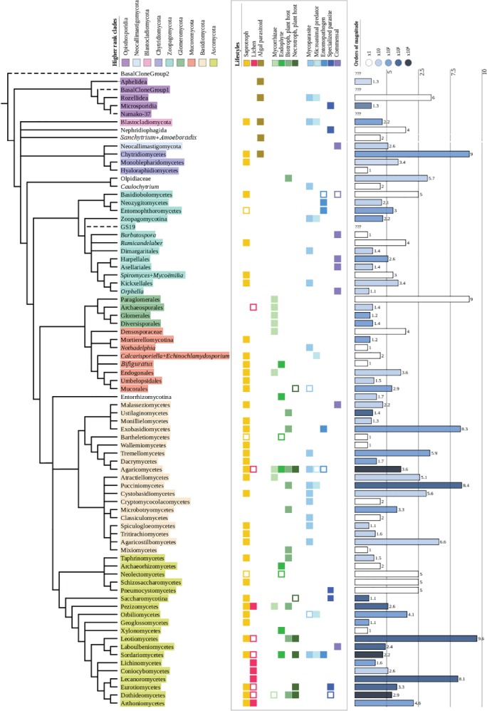
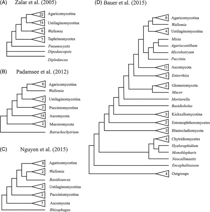
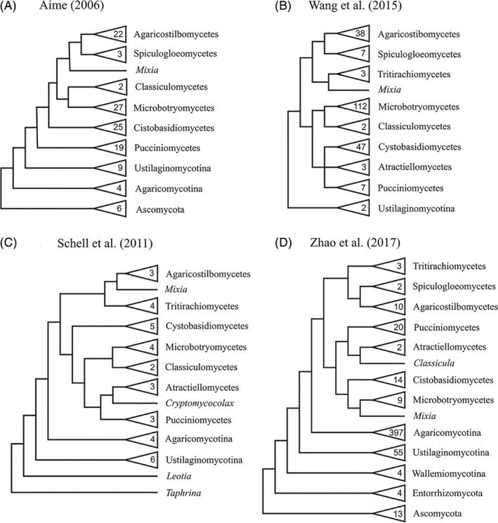
The fungal kingdom comprises a hyperdiverse clade of heterotrophic eukaryotes characterized by the presence of a chitinous cell wall, the loss of phagotrophic capabilities and cell organizations that range from completely unicellular monopolar organisms to highly complex syncitial filaments that may form macroscopic structures. Fungi emerged as a 'Third Kingdom', embracing organisms that were outside the classical dichotomy of animals versus vegetals. The taxonomy of this group has a turbulent history that is only now starting to be settled with the advent of genomics and phylogenomics. We here review the current status of the phylogeny and taxonomy of fungi, providing an overview of the main defined groups. Based on current knowledge, nine phylum-level clades can be defined: Opisthosporidia, Chytridiomycota, Neocallimastigomycota, Blastocladiomycota, Zoopagomycota, Mucoromycota, Glomeromycota, Basidiomycota and Ascomycota. For each group, we discuss their main traits and their diversity, focusing on the evolutionary relationships among the main fungal clades. We also explore the diversity and phylogeny of several groups of uncertain affinities and the main phylogenetic and taxonomical controversies and hypotheses in the field.

Keywords: Fungi; diversity; phylogenomics; phylogeny; taxonomy.


Figures

Figure 1
Figure 1
The fungal tree of life. Tree showcasing currently described groups within the Kingdom Fungi up to the class level, as well as incertae sedis lineages that cannot be assigned to any other class. In the case of zygomycetous fungi, due to historical reasons, we have included lineages up to order level. The first column uses colours to cluster clades in corresponding phyla. The second column compiles the lifestyles present in each group. Empty squares indicate that the given lifestyle is anecdotic or hypothetical. The third column shows the number of described species in each group according to the Catalogue of Life (Bisby & Roskov, 2010) or, for certain groups that are not represented in this database, Wikispecies (Wikimedia, 2011). Since the number of species might vary by several orders of magnitude, species number bars are coded using different colours. Tree generated using the interactive Tree of Life (iTOL) server (Letunic & Bork, 2016).
Figure 2
Figure 2
Diversity of zoosporic Fungi. (A) Transmission electron micrograph of a sporoblast of Fibrillanosema crangonycis (Microsporidia). The nucleus is clearly visible in the image and a series of concentrical structures with a highly electrodense core that appear tightly packed around the perimeter of the cell. This peculiar structure corresponds to a coiled polar tube, an infective harpoon‐like structure characteristic of Microsporidia. Original photograph taken by Leon White, CC BY‐SA 3.0 license. (B) Mature zoosporangia of Rozella allomycis (Rozellidea) during the last stages of infection of a mycelium of Allomyces sp. (Blastocladiomycota). Like many zoosporic fungi, Rozella is a parasitoid that invades and consumes the host cytoplasm, after which it produces sporangia. Original photograph by Timothy Y. James, CC BY‐SA 3.0 license. (C) Zoosporangium of Rhizophidium keratinophylum (Chytridiomycetes), appearing as a globular structure, growing on a human hair (fibrous brown structure). Beyond their parasitic roles, many chytrids have important roles in aquatic environments as saprotrophs specialized for degrading highly recalcitrant organic matter, such as pollen grains, arthropod exuviae or keratin. Original photograph by Wikipedia user TelosCricket, CC BY‐SA 4.0 license. (D) Micrograph of a group of oogonia from Gonapodya polymorpha (Monoblepharidomycetes, Chytridiomycota). The Monoblepharidomycetes are the only group of Fungi that present morphologically distinct gametes (i.e. anisogamy). They are also the only group within Chytridiomycota that have developed true hyphae, which evolved independently from those of terrestrial Fungi. Original photograph by Marilyn R. N. Mollicone. All rights reserved. (E) Mature sporangia of Catenaria anguillulae (Blastocladiomycota) growing inside a nematode alongside a true mycelium. Despite its relatively low number of species, Blastocladiomycota is a highly diverse group in terms of ecology, including saprotrophs, plant pathogens, algal parasitoids and even animal parasites. Catenaria, in particular, has been studied for its potential use as a pest‐control agent in agriculture. Original photograph by George Barron. Licensed for non‐commercial academic and research use only. (F) Microscopic preparation of a monocentric thallus from Neocallimastix frontalis (Neocallimastigomycota) isolated from deer faeces. The thallus possess a bulbous structure that corresponds with the zoosporangia and a series of root‐like protrusions, the rhizoids. The Neocallimastigomycota are a group of Fungi almost exclusively associated with the gut of herbivorous mammals. They have lost their mitochondria and present a highly expanded repertoire of cellulolytic enzymes. Original photograph from Atanasova‐Pancevska & Kungulovski (2017), CC BY‐NC 4.0 license.
Figure 3
Figure 3
Phylogenetic position of Neocallimastigomycota in different studies. Simplified topology from several phylogenetic studies covering the phylogenetic position of Neocallimastigomycota. Numbers inside triangles represent the number of sampled species within the clade. (A) Topology obtained from James et al. (2006b). Phylogeny constructed from a concatenation of 18S rRNA, 28S rRNA and 5.8S rRNA, using Bayesian inference. (B) Topology obtained from James et al. (2006a). Phylogeny constructed from a concatenation of 18S rRNA, 28S rRNA, 5.8S rRNA, EF1‐α, RPB1 and RPB2, using Bayesian inference. (C) Topology obtained from Sekimoto et al. (2011). Phylogeny constructed from RPB1, RPB2, EF1‐α, rRNA and actin genes, using a maximum‐likelihood approach. (D) Topology obtained from Ebersberger et al. (2012). Phylogeny reconstructed from a supermatrix of 46 single‐copy genes, using a maximum‐likelihood approach.
Figure 4
Figure 4
Diversity of zygomycetous Fungi. (A) Zygospore from Rhizopus stolonifer (Mucorales, Mucoromycotina). Zygospores are naked sexual spores formed in the intersection of two mating hyphae in both Zoopagomycota and Mucoromycota. Original photograph by George Barron. Licensed for non‐commercial academic and research use only. (B) Mycelium and multinucleated spores from Rhizophagus intrarradices (Glomerales, Glomeromycotina) growing in association with a plant root, appearing as a foamy structure in the lower part of the picture. The spores, appearing as dark brown globular structures, contain multiple nuclei that are thought to form a chimeric population (heterokaryon). Original photograph by Banco de Glomeromycota in vitro, CC BY‐NC‐ND 2.5 AR license. (C) Entomophthora muscae (Entomophthorales, Entomophthoromycotina) growing in a fly. The Entomophthorales include mostly entomopathogenic species that form an unwalled coenocytic mycelium that invades the host body before killing it. Original photograph by Hans Hillewaert, CC BY‐SA 4.0 license. (D) Hyphae from Zoophagus insidians (Zoopagales, Zoopagomycotina) attacking a group of rotifers. Zoopagales is a group of parasitic fungi that mostly infect other fungi, protozoans and microinvertebrates. Original photograph by George Barron. Licensed for non‐commercial academic and research use only. (E) Pin mould [probably Rhizopus stolonifer (Mucorales, Mucoromycotina)] growing on a tomato. Most members of the mucorales are fast‐growing saprotrophs that present very large sporangia, appearing here as dark globose structures at the end of long aerial hyphae. Original photograph by Wikipedia user Calimo, CC BY‐SA 3.0 license. (F) Scanning electron micrograph of a Mortierella hyalina (Mortierellales, Mortierellomycotina) sporangium. Members of the Mortierellomycotina have similar ecologies to Mucorales, but they can be easily differentiated by the absence of an inflated base to their sporangia (columella). Original photograph by flickr user ZygoLife Research Consortium, CC BY‐SA 2.0 license.
Figure 5
Figure 5
Diversity of Basidiomycota. (A) Basidia from Coprinus (Agaricomycetes, Agaricomycotina). Basidia are reproductive structures formed by a cell attached to the (typically four) derived spores produced by meiosis, appearing here as dark structures. Original photograph by Wikipedia user Jon Houseman, CC BY‐SA 3.0 license. (B) Puccinia recondita (Pucciniomycetes, Pucciniomycotina) growing on the back of a leaf. Pucciniomycetes are a diverse class of biotrophic plant pathogens within the Pucciniomycotina. Original photograph by flickr user Line Sabroe, CC BY 2.0 license. (C) Micrograph of a skin cell infected by Malassezia furfur (Malasseziomycetes, Ustilaginomycotina). Although most Ustilaginomycotina are plant pathogens, the genus Malassezia is commonly found in the skin of mammals. Original photograph in the public domain. (D) Fruiting bodies of Amanita muscaria (Agaricomycetes, Agaricomycotina), a poisonous mushroom famous for its bright white and red colour and its hallucinogenic properties. Original photograph in the public domain. (E) Micrograph of Wallemia ichthyophaga (Wallemiomycetes, Wallemiomycotina), appearing as a rounded mass. Wallemiomycetes contains a few species of highly extremotolerant fungi. W. ichthyophaga in particular requires high salinity to grow, as can be seen from the presence of cubic salt crystals in the picture. Photograph by Wikipedia user Anticicklon, CC BY‐SA 3.0 license. (F) A Ginkgo biloba leaf covered by clustersof black Bartheletia paradoxa telia. B. paradoxa represents a divergent lineage that has probably co‐evolved with Ginkgopsida, an ancient plant lineage of which there is only one extant species. Original photograph by flicker user AJC1, CC BY‐SA 2.0 license.
Figure 6
Figure 6
Diversity of Ascomycota. (A) Asci from Sordaria fimicola (Sordariomycetes, Pezizomycotina). Asci are reproductive structures that enclose (typically four or eight) spores produced by meiosis, appearing here as dark structures. Original photograph by Wikipedia user CarmelitaLevin CC BY‐SA 4.0 license. (B) Fruiting bodies of Neolecta vitellina (Neolectomycetes, Taphrinomycotina). Taphrinomycotina includes several lineages with a wide range of body plans, ranging from intracellular parasites to complex multicellular fungi. Original photograph by Mushroom Observer user gillow2e, CC BY‐SA 3.0 license. (C) Mating cells (Shmoo) of Saccharomyces cerevisiae. Under the right conditions haploid cells enter the shmoo mating state and fuse with a mating cell of the opposite mating type, producing a diploid cell. The diploid cell can enter meiosis, producing an ascus with four spores, from which haploid cells germinate. Original photograph by Wikipedia user Pilarbini, CC BY‐SA 4.0 license. (D) Micrograph of a group of conidia from Penicillium spinulosum (Eurotiomycetes, Pezizomycotina). Penicillium is a genus of cosmopolitan moulds that mostly propagate by producing high numbers of asexual conidiospores. Original photograph by Wikipedia user Medmyco, CC BY‐SA 4.0 license. (E) Photograph of a ladybird infected with Hesperomyces virescens (Laboulbeniomycetes, Pezizomycotina), appearing here as light‐coloured digitiform structures (see arrow). Laboulbeniales are a diverse order of fungi associated with arthropod surfaces that present determinate growth and separate sexes. Original photograph by flickr user Gilles San Martin, CC BY‐SA 2.0 license. (F) Xanthoria parietina, a lichen, growing on a branch. In the picture, disk‐like structures can be observed sprouting prefentially in the centre of the formation. These correspond with the apothecia, support tissues containing the asci. Original photograph by Wikipedia user Marianne Perdomo, CC BY‐SA 2.0 license.
Figure 7
Figure 7
Phylogenetic position of Wallemia in different studies. Simplified topology from several phylogenetic studies covering the phylogenetic position of Wallemiomycetes. Numbers inside triangles represent the number of sampled species within the clade. (A) Topology extracted from Zalar et al. (2005). Phylogeny constructed using a maximum‐parsimony approach. (B) Topology extracted from Padamsee et al. (2012). Phylogeny constructed from a data set of 71 protein‐coding genes, using a Bayesian inference approach. (C) Topology extracted from Nguyen et al. (2015). Pylogeny constructed from a data set of 35 single‐copy protein‐coding genes, using a Bayesian inference approach. (D) Topology extracted from Bauer et al. (2015). Phylogeny constructed from a concatenation of 18S rRNA, 28S rRNA, 5.8S rRNA, RPB1 and RPB2, using a combination of Bayesian inference, maximum‐likelihood and maximum‐parsimony approaches.
Figure 8
Figure 8
Phylogenetic relationships among the different clades within Pucciniomycotina in different studies. Numbers inside triangles represent the number of sampled species within the clade. (A) Topology extracted from Aime et al. (2006). Phylogeny reconstructed from LSU rRNA and SSU rRNA genes, using a maximum‐parsimony approach. (B) Topology extracted from Wang et al. (2015c). Phylogeny reconstructed from a concatenation of SSU rRNA and LSU rRNA D1/D2, using a maximum‐likelihood approach. (C) Topology extracted from Schell et al. (2011). Phylogeny constructed from a concatenation of EF1‐α, LSU rRNA and SSU rRNA genes, using a maximum‐parsimony approach. (D) Topology extracted from Zhao et al. (2017). Phylogeny reconstructed from a concatenation of LSU rRNA, SSU rRNA, 5.8S rRNA, TEF1, RPB1 and RPB2, using a maximum‐likelihood approach.

References

    1. Adl, S. M. , Simpson, A. G. , Lane, C. E. , Lukeš, J. , Bass, D. , Bowser, S. S. , Brown, M. , Burki, F. , Dunthorn, M. , Hampl, V. , Heiss, A. , Hoppenrath, M. , Lara, E. , Le Gall, L. , Lynn, D. H. , et al. (2012). The revised classification of eukaryotes. Journal of Eukaryotic Microbiology 59, 1–45. - PMC - PubMed
    1. Adl, S. M. , Bass, D. , Lane, C. E. , Lukeš, J. , Schoch, C. L. , Smirnov, A. , Agatha, S. , Berney, C. , Brown, M. W. , Burki, F. , Cárdenas, P. , Čepička, I. , Chistyakova, L. , del Campo, J. , Dunthorn, M. , et al. (2018). Revisions to the classification, nomenclature, and diversity of eukaryotes. Journal of Eukaryotic Microbiology 66, 4–119. - PMC - PubMed
    1. Ahrendt, S. R. , Quandt, C. A. , Ciobanu, D. , Clum, A. , Salamov, A. , Andreopoulos, B. , Cheng, J.‐F. , Woyke, T. , Pelin, A. , Henrissat, B. , Reynolds, N. K. , Benny, G. L. , Smith, M. E. , James, T. Y. , Grigoriev, I. V. , et al. (2018). Leveraging single‐cell genomics to expand the fungal tree of life. Nature Microbiology 3, 1417–1428. - PMC - PubMed
    1. Aime, M. C. , Matheny, P. B. , Henk, D. A. , Frieders, E. M. , Nilsson, R. H. , Piepenbring, M. , Mclaughlin, D. J. , Szabo, L. J. , Begerow, D. , Sampaio, J. P. , Bauer, R. , Weiß, M. , Oberwinkler, F. & Hibbett, D. (2006). An overview of the higher level classification of Pucciniomycotina based on combined analyses of nuclear large and small subunit rDNA sequences. Mycologia 98, 896–905. - PubMed
    1. Aime, M. C. , Toome, M. & McLaughlin, D. J. (2014). 10 Pucciniomycotina In Systematics and Evolution (eds Karl Esser), pp. 271–294. Springer, Berlin Heidelberg.

Publication types