

Measurement of percentage dose at the surface for a 6 MV photon beam
- PMID: 31660051
- PMCID: PMC6807031
- DOI: 10.1016/j.rpor.2019.09.007
Measurement of percentage dose at the surface for a 6 MV photon beam
Abstract
Aim: To evaluate if a radiochromic film (RF) Gafchromic EBT3 is suitable for surface dose measurements of radiotherapy treatments performed with a 6 MV linear accelerator. Two aspects of RF were analyzed, beam energy dependence and surface dose determination.
Background: The measurements done at the surface or near the radiation source are done without charged electronic equilibrium and also have contribution of electron contamination. The detectors used for these measurements should not alter the dose to the target. To counteract these dosimetric problems it is proposed to do the measurements with radiochromic films which are thin detectors and have tissue equivalent properties.
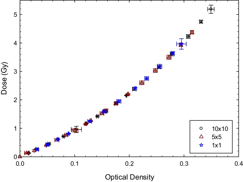
Materials and methods: The measurements were done using a Novalis linear accelerator (LINAC) with nominal energy of 6 MV. To determine the surface dose, the total scatter factors (TSF) of three different field sizes were measured in a water phantom at 5 cm depth. Energy dependence of EBT3 was studied at three different depths, using a solid water phantom. The surface measurements were done with the RF for the same field sizes of the TSF measurements. The value of the percentage depth dose was calculated normalizing the doses measured in the RF with the LINAC output, at 5 cm depth, and the TSF.
Results: The radiochromic films showed almost energy independence, the differences between the curves are 1.7% and 1.8% for the 1.5 cm and 10 cm depth, respectively. The percentage depth doses values at the surface measured for the 10 cm × 10 cm, 5 cm × 5 cm and 1 cm × 1 cm were 26.1 ± 1.3%, 21.3 ± 2.4% and 20.2 ± 2.6%, respectively.
Conclusions: The RF-EBT3 seems to be a detector suitable for measurements of the dose at the surface. This suggests that RF-EBT3 films might be good candidates as detectors for in vivo dosimetry.
Keywords: Energy independence; Radiochromic film EBT3; Surface dose.
© 2019 Greater Poland Cancer Centre. Published by Elsevier B.V. All rights reserved.
Figures


References
-
- Devic S., Seuntjens J., Abdel-Rahman W. Accurate skin dose measurements using radiochromic film in clinical applications. Med Phys. 2006;33(4):1116. - PubMed
-
- Saini A.S. 2007. In-vivo radiation diode dosimetry for therapeutic photon beams.
-
- Mijnheer B., Beddar S., Izewska J., Reft C. In vivo dosimetry in external beam radiotherapy. Med Phys. 2013;40(7) - PubMed
-
- Leunens G., Van Dam J., Dutreix A., van der Schueren E. Quality assurance in radiotherapy by in vivo dosimetry. 2. Determination of the target absorbed dose. Radiother Oncol. 1990 - PubMed
LinkOut - more resources
Full Text Sources
