

doi: 10.1093/ofid/ofz342.
eCollection 2019 Oct.
Rescue of Replication-Competent ZIKV Hidden in Placenta-Derived Mesenchymal Cells Long After the Resolution of the Infection
Affiliations
- PMID: 31660333
- PMCID: PMC6785694
- DOI: 10.1093/ofid/ofz342
Item in Clipboard
Rescue of Replication-Competent ZIKV Hidden in Placenta-Derived Mesenchymal Cells Long After the Resolution of the Infection
Open Forum Infect Dis.
.
Abstract
The Zika virus (ZIKV) genome, its negative-strand viral proteins, and virus-like particles were detected in placenta-derived mesenchymal cells (MSCs), indicating that ZIKV persists after virus clearance from maternal blood and can be rescued by in vitro cultivation. We report for the first time the presence of replication-competent ZIKV in MSCs from an asymptomatic woman who acquired infection during pregnancy.
Keywords: Zika virus; mesenchymal cells; placenta; vertical transmission; viral reservoir.
© The Author(s) 2019. Published by Oxford University Press on behalf of Infectious Diseases Society of America.
Figures
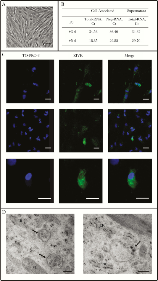
Characterization of Zika virus (ZIKV) infection of decidua-derived mesenchymal cells (dd-MSCs). A, Morphology of dd-MSCs (P0, 21 days post expansion (DPE)) observed by light microscope. Scale bar = 500 μm. B, ZIKV total-RNA and neg-RNA quantification in dd-MSCs cells and culture supernatants sampled at different days of P0 culture. C, Immunolocalization by confocal microscopy of the ZIKV glycoprotein, detected by anti-pan-flavi-specific IgG antibody (ZIKV, green) on dd-MSCs (day 3 of P0). Left panels: nuclear staining; middle panels: ZIKV-specific staining; right panels: merged images. The 2 higher rows show different microscopy fields; the lower row shows a higher magnification of the central row pictures. A dotted immunolabeling can be observed in the cytoplasm of positive cells. Scale bar = 100 μm. D, Ultrastructural analysis of dd-MSCs (day 3 of P0), assessed by transmission electron microscopy. The cultured cells show virus-like structures measuring approximately 40–50 nm in diameter in cytoplasmic vesicles close to the rough endoplasmic reticulum (arrows). The morphologic characteristics of the virus-like structures are consistent with viral particles of the Flaviviridae family. Scale bars = 200 μm. Abbreviations: ER, endoplasmic reticulum; M, mitochondrion.
References
-
- Wikan N, Smith DR. Zika virus: history of a newly emerging arbovirus. Lancet Infect Dis 2016; 16:e119–26. - PubMed
-
- Ministério da Saúde. Boletins epidemiológicos—Secretaria de Vigilância em Saúde Available at: http://portalarquivos2.saude.gov.br/images/pdf/2018/janeiro/10/2017-046-.... Accessed 2 November 2018.
-
- de Araújo TVB, Ximenes RAA, Miranda-Filho DB, et al. ; investigators from the Microcephaly Epidemic Research Group; Brazilian Ministry of Health; Pan American Health Organization; Instituto de Medicina Integral Professor Fernando Figueira; State Health Department of Pernambuco Association between microcephaly, Zika virus infection, and other risk factors in Brazil: final report of a case-control study. Lancet Infect Dis 2018; 18:328–36. - PMC - PubMed
-
- Martines RB, Bhatnagar J, de Oliveira Ramos AM, et al. . Pathology of congenital Zika syndrome in Brazil: a case series. Lancet 2016; 388:898–904. - PubMed
-
- Schaub B, Monthieux A, Najihoullah F, et al. . Late miscarriage: another Zika concern? Eur J Obstet Gynecol Reprod Biol 2016; 207:240–1. - PubMed
