. 2020 Apr;13(4):e201900167.
doi: 10.1002/jbio.201900167. Epub 2019 Nov 25.

Photoacoustic imaging of the human placental vasculature

Affiliations

Photoacoustic imaging of the human placental vasculature

Efthymios Maneas et al. J Biophotonics. 2020 Apr.

Abstract

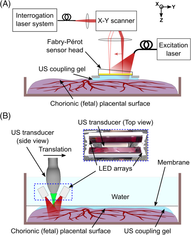
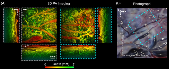
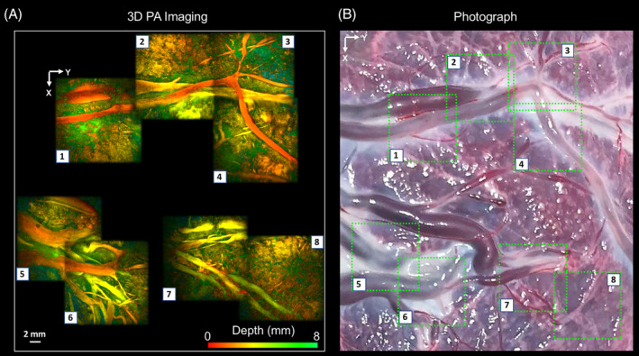
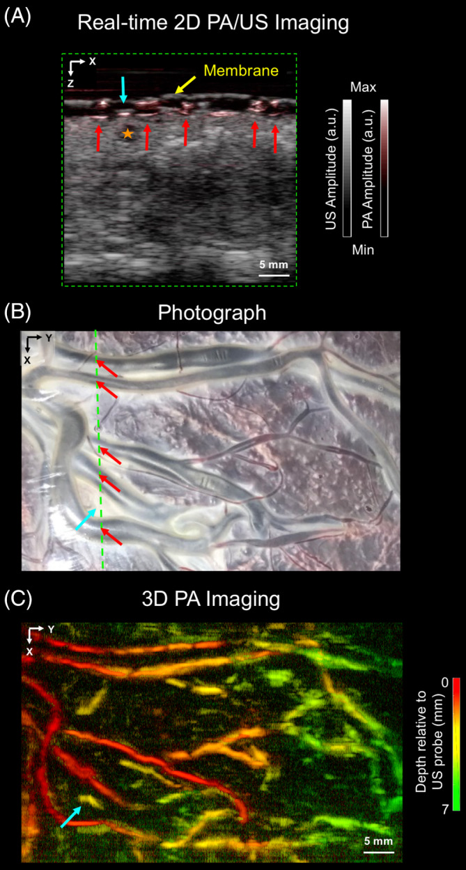
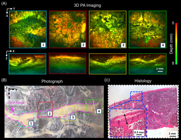
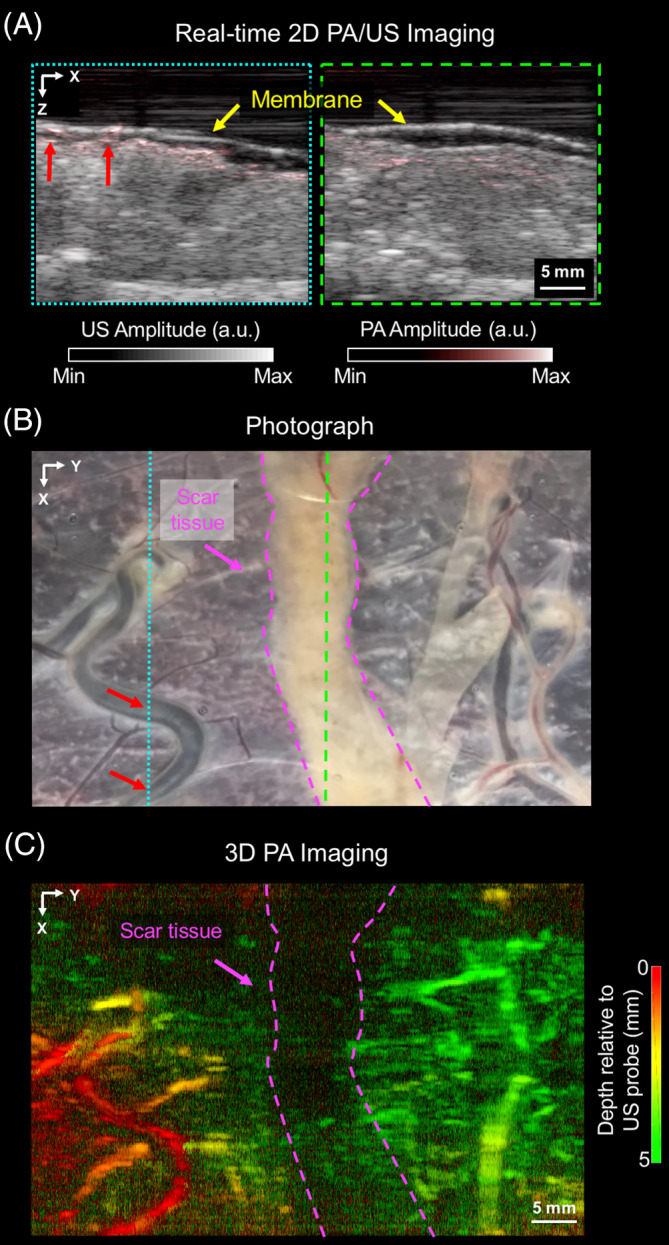
Minimally invasive fetal interventions require accurate imaging from inside the uterine cavity. Twin-to-twin transfusion syndrome (TTTS), a condition considered in this study, occurs from abnormal vascular anastomoses in the placenta that allow blood to flow unevenly between the fetuses. Currently, TTTS is treated fetoscopically by identifying the anastomosing vessels, and then performing laser photocoagulation. However, white light fetoscopy provides limited visibility of placental vasculature, which can lead to missed anastomoses or incomplete photocoagulation. Photoacoustic (PA) imaging is an alternative imaging method that provides contrast for hemoglobin, and in this study, two PA systems were used to visualize chorionic (fetal) superficial and subsurface vasculature in human placentas. The first system comprised an optical parametric oscillator for PA excitation and a 2D Fabry-Pérot cavity ultrasound sensor; the second, light emitting diode arrays and a 1D clinical linear-array ultrasound imaging probe. Volumetric photoacoustic images were acquired from ex vivo normal term and TTTS-treated placentas. It was shown that superficial and subsurface branching blood vessels could be visualized to depths of approximately 7 mm, and that ablated tissue yielded negative image contrast. This study demonstrated the strong potential of PA imaging to guide minimally invasive fetal therapies.

Keywords: fetal therapy; human placenta imaging; photoacoustic imaging; twin-to-twin-transfusion syndrome.


Figures

Figure 1
Figure 1
Photoacoustic imaging experimental setups. A, Fabry‐Pérot‐based planar sensor PA imaging system. The sensor head gently touched the placenta from the chorionic (fetal) surface, with ultrasound (US) gel placed in between the sensor and the tissue for acoustic coupling. B, Clinical linear‐array ultrasound probe‐based PA imaging system. The US imaging probe with light emitting diode (LED) arrays was translated across the placenta using a linear motorized stage. The placenta was coated with US gel, covered with a plastic membrane (cling film) and placed inside a water‐filled container. The plastic membrane minimized leakage of blood from the placenta to the water above it
Figure 2
Figure 2
Photoacoustic (PA) images of the chorionic placental vasculature of a normal term placenta. The 3D PA images were obtained from two locations (white and cyan dashed squares) using the Fabry‐Pérot‐based planar sensor PA imaging system. They are displayed as maximum intensity projections of the reconstructed 3D image volume. The scale bar applies to all PA images. Superficial chorionic blood vessels are apparent in both xy PA images, A, and the corresponding photograph, B. Several subsurface structures that could be attributed to blood vessels are visible down to a depth of approximately 7 mm from the chorionic fetal surface of the placenta
Figure 3
Figure 3
Photoacoustic (PA) images of the chorionic placental vasculature of an identical twin placenta treated for TTTS using laser photocoagulation of the placental vascular equator. The 3D PA images were obtained using a Fabry‐Pérot‐based PA scanner and are displayed as maximum intensity projections of the reconstructed 3D image volume. The images are arranged as a mosaic to capture a wider area of the placenta. Superficial blood vessels are clearly resolved, A, which correspond well to those apparent in the photograph, B
Figure 4
Figure 4
Wide‐field field photoacoustic (PA) and ultrasound (US) images of the chorionic placental vasculature in an untreated part of a TTTS photocoagulated placenta. The PA and US images were acquired using the clinical linear‐array US probe‐based PA imaging system. A, a 2D PA and a 2D US image, acquired from a region corresponding to the purple line in, B, are merged and displayed in a grayscale and a color‐scale, respectively. Several superficial blood vessels (red arrows) are visible in this 2D PA image but not clearly visible in the 2D US image; they correspond well to those apparent in the photograph, B. A prominent signal from the 2D PA image (solid blue arrow) was not apparent in the photograph, B. Additionally, a fluid‐filled cavity (orange star; 2D US image) was not visible in the 2D PA image. The placenta was covered with a cling film membrane (yellow arrow). The images are displayed on logarithmic scales. C, 3D PA image displayed as a maximum intensity projection of the reconstructed image volume, which was acquired by linear translation of the US probe. Superficial chorionic structures are clearly resolved, with good correspondence to the vessels apparent in the photograph, B
Figure 5
Figure 5
Photoacoustic (PA) images of the laser photocoagulated (scar) tissue of a TTTS‐treated placenta. A, The 3D PA images were obtained using a Fabry‐Pérot based PA scanner and are displayed on a logarithmic scale as maximum intensity projections (MIPs) of the reconstructed 3D image volume. The scar tissue is a weak absorber for excitation light and thereby provides negative contrast to the image. The photocoagulation depth is visible in the xz MIPs. The scale bar applies to all images and axes orientations. B, Corresponding photograph that includes the locations that were imaged in A. C, An H&E‐stained section was obtained from within imaging location 4 (dashed orange line). The photocoagulated tissue depth on histological examination is approximately 1 mm (double black arrow), which is consistent with that observed from the PA image acquired at the same location in A
Figure 6
Figure 6
Wide‐field photoacoustic (PA) and ultrasound (US) images of tissue of a placenta that was treated for TTTS. The PA and US images were acquired with the PA/US imaging system based on a clinical linear array. A, Two frames of merged 2D PA and US images that were acquired from two locations: non‐treated tissue (dashed blue box in A corresponding to the dashed blue line in B) and scar tissue from photocoagulation treatment (dashed green lines). Superficial blood vessels (red arrows) are visible in the left PA image, which correspond well with those in the photograph, B. The placenta was covered with a cling film membrane (yellow arrow). The images are displayed on logarithmic scales. B, 3D PA image volume, which was acquired by linear translation of the US probe and displayed as a maximum intensity projection. Superficial chorionic structures are visualized, with good correspondence to the photograph, B

Similar articles

Cited by

References

    1. Burton G. J., Fowden A. L., Philos. Trans. R. Soc. B Biol. Sci. 2015, 370, 20140066. - PMC - PubMed
    1. Baschat A. A., Hecher K., Semin. Perinatol. 2004, 28, 67. - PubMed
    1. Lewi L., Deprest J., Hecher K., Am. J. Obstet. Gynecol. 2013, 208, 19. - PubMed
    1. Bamberg C., Hecher K., Best Pract. Res. Clin. Obstet. Gynaecol. 2019, 58, 55. - PubMed
    1. Senat M.‐V., Deprest J., Boulvain M., Paupe A., Winer N., Ville Y., N. Engl. J. Med. 2004, 351, 136. - PubMed

Publication types

LinkOut - more resources