. 2019 Oct 28;20(21):5358.
doi: 10.3390/ijms20215358.

Fatty Acid-Binding Protein 3 is Critical for α-Synuclein Uptake and MPP+-Induced Mitochondrial Dysfunction in Cultured Dopaminergic Neurons

Affiliations

Fatty Acid-Binding Protein 3 is Critical for α-Synuclein Uptake and MPP+-Induced Mitochondrial Dysfunction in Cultured Dopaminergic Neurons

Ichiro Kawahata et al. Int J Mol Sci. .

Abstract

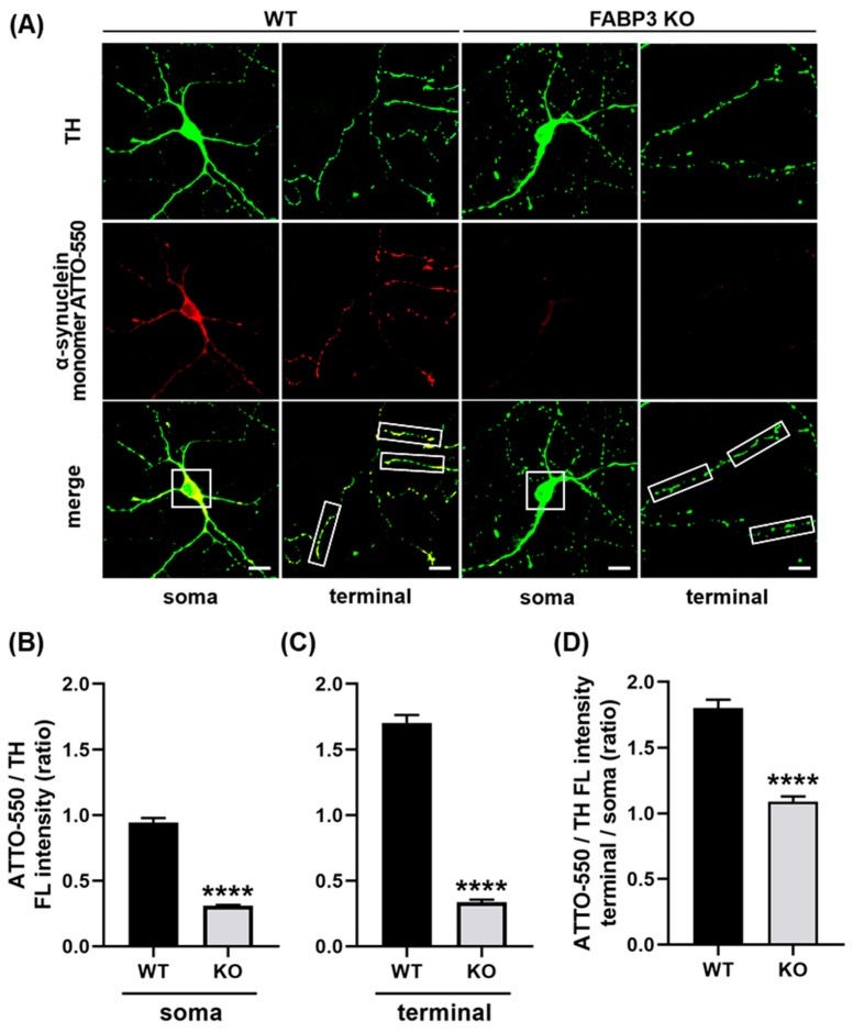
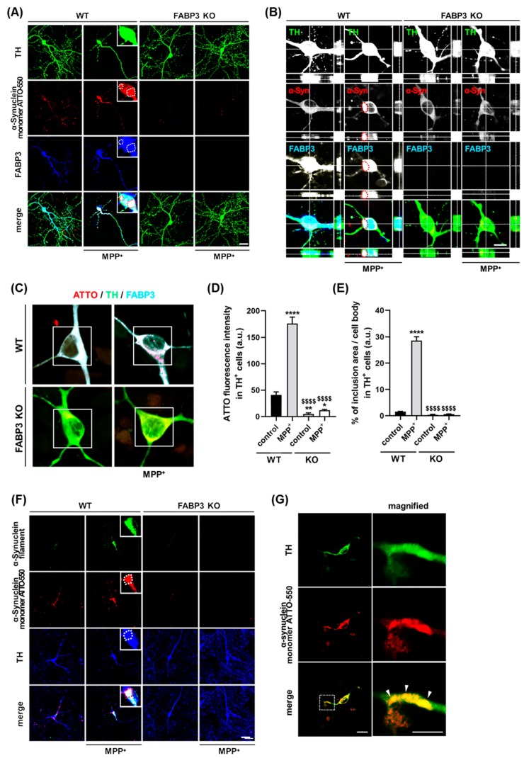
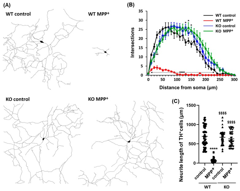
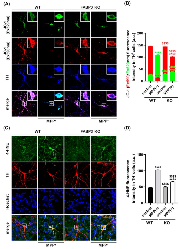
α-Synuclein is an abundant neuronal protein that accumulates in insoluble inclusions in Parkinson's disease and other synucleinopathies. Fatty acids partially regulate α-Synuclein accumulation, and mesencephalic dopaminergic neurons highly express fatty acid-binding protein 3 (FABP3). We previously demonstrated that FABP3 knockout mice show decreased α-Synuclein oligomerization and neuronal degeneration of tyrosine hydroxylase (TH)-positive neurons in vivo. In this study, we newly investigated the importance of FABP3 in α-Synuclein uptake, 1-methyl-4-phenylpyridinium (MPP+)-induced axodendritic retraction, and mitochondrial dysfunction. To disclose the issues, we employed cultured mesencephalic neurons derived from wild type or FABP3-/- C57BL6 mice and performed immunocytochemical analysis. We demonstrated that TH+ neurons from FABP3+/+ mice take up α-Synuclein monomers while FABP3-/- TH+ neurons do not. The formation of filamentous α-Synuclein inclusions following treatment with MPP+ was observed only in FABP3+/+, and not in FABP3-/- neurons. Notably, detailed morphological analysis revealed that FABP-/- neurons did not exhibit MPP+-induced axodendritic retraction. Moreover, FABP3 was also critical for MPP+-induced reduction of mitochondrial activity and the production of reactive oxygen species. These data indicate that FABP3 is critical for α-Synuclein uptake in dopaminergic neurons, thereby preventing synucleinopathies, including Parkinson's disease.

Keywords: 1-methyl-4-phenylpyridinium (MPP+); Parkinson’s disease; fatty acid-binding protein 3; mitochondria; synucleinopathy; α-Synuclein.


Conflict of interest statement

The authors declare no conflict of interest.

Figures

Figure 1
Figure 1
Cultured primary dopaminergic neurons require fatty acid-binding protein 3 (FABP3) to take up α-Synuclein. (A) Representative images of TH+ mesencephalic neurons at days in vitro (DIV) 12 derived from wild type (WT) or FABP3−/− C57BL6 mice. Neurons were exposed to 1 μM ATTO-550-labeled α-Synuclein monomer for 48 h and stained with antibody against tyrosine hydroxylase (TH, green). Scale bar: 10 μm. (B) Quantitative analysis of the ratio of ATTO-550-labeled α-Synuclein fluorescence intensity (FL) to TH immunoreactivity in the region of interest (ROI)-selected soma of individual TH+ neurons shown in A (white square 15 × 15 μm). **** p < 0.0001 in wild type (WT) versus FABP3−/− (KO), n > 20. (C) Ratio of ATTO-550 fluorescence intensity to TH immunoreactivity in ROI-selected neuronal processes shown in A (white square 10 × 30 μm). **** p < 0.0001 in WT versus KO, n > 60. (D) The calculated ratio of ATTO-550 to TH in the individual terminal (C) was divided by the value in the soma (B) to represent the superiority on the α-Synuclein uptake in the axonal processes compared to the uptake in the soma. **** p < 0.0001 in WT versus KO, n > 20.
Figure 2
Figure 2
FABP3 is required for the formation of α-Synuclein inclusions in 1-methyl-4-phenylpyridinium (MPP+)-treated dopaminergic neurons. (A) Representative images of TH+ mesencephalic neurons at DIV 12 derived from wild type (WT) and FABP3−/− C57BL6 mice. Neurons were exposed to 1 μM ATTO-550-labeled α-Synuclein monomer with or without 10 μM MPP+ for 48 h. The cells are stained with antibodies against TH (green) and FABP3 (blue). The dotted circles indicate α-Synuclein and FABP3-positive aggregates. Scale bar: 20 μm. (B) Confocal microscopy reveals ATTO-labelled α-Synuclein within the intracellular compartment of dopaminergic neurons. Image taken from an optical section approximately in the center of the z-axis of a dopaminergic neuron stained with anti-TH and anti-FABP3 antibodies. The red dotted circles indicate α-Synuclein and FABP3-positive aggregates. Scale bar: 10 μm. (C) Representative images of TH+ mesencephalic neurons either treated or not treated with MPP+. The images were acquired in photon counting mode using Leica TCS SP8 with a higher magnification (×63 objective). Scale bar: 10 μm. (D) Quantitative analysis of ATTO-550-labeled α-Synuclein monomer fluorescence intensity in ROI-selected soma of individual TH+ neurons shown in C (white square). **** p < 0.0001, ** p < 0.01, * p < 0.05 versus WT control; $$$$ p < 0.0001 versus WT MPP+; n > 60. (E) Percentage of ATTO-550-labeled α-Synuclein-positive inclusions (defined as dot-like signals 3 standard deviations (SD) above the background levels) in the total cell area [31]. **** p < 0.0001 versus WT control; $$$$ p < 0.0001 versus WT MPP+; n > 60. (F) Representative images of TH+ mesencephalic neurons in the same treatment conditions as the neurons in A. The cells are stained with antibodies against α-Synuclein filament (green) and TH (blue). Scale bar: 20 μm. (G) Representative images of a TH+ mesencephalic neuronal process stained with an antibody against TH (green). The magnified images of the dotted box are shown in the right, and the white arrowheads indicate Lewy neurite-like structures. Scale bar: 10 μm.
Figure 3
Figure 3
Representative dendritic morphology of TH+ dopaminergic neurons derived from wild type or FABP3−/ mice. (A) Representative traced dendritic patterns of wild type (WT) or FABP3−/ (KO) dopaminergic neurons either treated or not treated with 10 μM MPP+ for 48 h. (B) Sholl analysis of WT or FABP3 KO dopaminergic neurons either treated or not treated with MPP+. **** p < 0.0001, ** p < 0.01, * p < 0.05 versus WT control, n > 20. (C) Average neurite length of WT or FABP3−/ dopaminergic neurons. **** p < 0.0001 versus WT control; $$$$ p < 0.0001 versus WT MPP+, n > 20.
Figure 4
Figure 4
FABP3 is involved in MPP+-induced reduction of mitochondrial activity and ROS production in dopaminergic neurons. (A) Representative images of JC-1-stained TH+ neurons. The white boxes indicate regions of interest (ROIs), which are magnified in the upper right in each image and quantified to measure the fluorescence intensity. Scale bar: 10 μm. (B) Quantitative analysis of Ex590 (red) and Ex529 (green) fluorescence intensity in JC-1-stained TH+ neurons. **** p < 0.0001 versus wild type (WT) control; $$$$ p < 0.0001 versus WT MPP+, n > 20. (C) Representative images of TH+ neurons (red) stained with anti-4HNE antibody (green). The white boxes show ROIs to measure 4-HNE fluorescence intensity. Scale bar: 10 μm. (D) Quantitative analysis of 4-HNE immunoreactivity in TH+ cells. **** p < 0.0001 versus WT control; $$$$ p < 0.0001 versus WT MPP+, n > 20.

Similar articles

Cited by

References

    1. Parkinson J. An essay on the shaking palsy. 1817. J. Neuropsychiatry Clin. Neurosci. 2002;14:223–236. doi: 10.1176/jnp.14.2.223. discussion 222. - DOI - PubMed
    1. Van Den Eeden S.K., Tanner C.M., Bernstein A.L., Fross R.D., Leimpeter A., Bloch D.A., Nelson L.M. Incidence of parkinson’s disease: Variation by age, gender, and race/ethnicity. Am. J. Epidemiol. 2003;157:1015–1022. doi: 10.1093/aje/kwg068. - DOI - PubMed
    1. De Lau L.M., Breteler M.M. Epidemiology of parkinson’s disease. Lancet Neurol. 2006;5:525–535. doi: 10.1016/S1474-4422(06)70471-9. - DOI - PubMed
    1. Dauer W., Przedborski S. Parkinson’s disease: Mechanisms and models. Neuron. 2003;39:889–909. doi: 10.1016/S0896-6273(03)00568-3. - DOI - PubMed
    1. Beyer K., Domingo-Sabat M., Ariza A. Molecular pathology of lewy body diseases. Int. J. Mol. Sci. 2009;10:724–745. doi: 10.3390/ijms10030724. - DOI - PMC - PubMed

MeSH terms

LinkOut - more resources