Review
. 2019 Oct 28;20(21):5364.
doi: 10.3390/ijms20215364.

Molecular Links between Central Obesity and Breast Cancer

Affiliations
Review

Molecular Links between Central Obesity and Breast Cancer

Alina-Andreea Zimta et al. Int J Mol Sci. .

Abstract

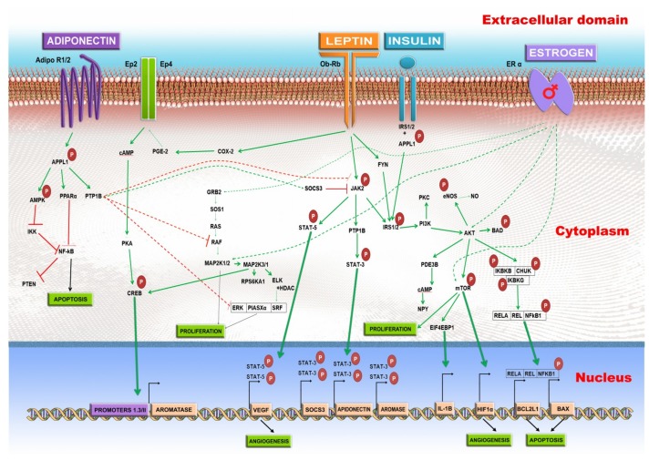
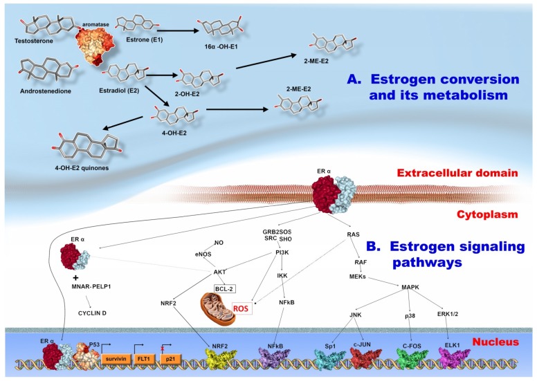
: Worldwide, breast cancer (BC) is the most common malignancy in women, in regard to incidence and mortality. In recent years, the negative role of obesity during BC development and progression has been made abundantly clear in several studies. However, the distribution of body fat may be more important to analyze than the overall body weight. In our review of literature, we reported some key findings regarding the role of obesity in BC development, but focused more on central adiposity. Firstly, the adipose microenvironment in obese people bears many similarities with the tumor microenvironment, in respect to associated cellular composition, chronic low-grade inflammation, and high ratio of reactive oxygen species to antioxidants. Secondly, the adipose tissue functions as an endocrine organ, which in obese people produces a high level of tumor-promoting hormones, such as leptin and estrogen, and a low level of the tumor suppressor hormone, adiponectin. As follows, in BC this leads to the activation of oncogenic signaling pathways: NFκB, JAK, STAT3, AKT. Moreover, overall obesity, but especially central obesity, promotes a systemic and local low grade chronic inflammation that further stimulates the increase of tumor-promoting oxidative stress. Lastly, there is a constant exchange of information between BC cells and adipocytes, mediated especially by extracellular vesicles, and which changes the transcription profile of both cell types to an oncogenic one with the help of regulatory non-coding RNAs.

Keywords: abdominal fat; adiponectin; breast cancer; exosomes; hormone dependency; leptin; menopause; miRNA; obesity.


Conflict of interest statement

The authors declare no conflict of interest. The funders had no role in the design of the study; in the collection, analyses, or interpretation of data; in the writing of the manuscript, or in the decision to publish the results.

Figures

Figure 1
Figure 1
The pathways activated by several dysregulated hormones in central obesity. The green arrow stands for activation and the red line for repression. Leptin binds to the Ob-Rb receptor, and activates several signaling pathways. The JAK2-STAT3 pathway is activated; which will result in the stimulated transcription of the SOCS3, and ultimately AROMASE (CYP19A) genes. The JAK-STAT5 pathway results in the increased expression of vascular endothelial growth factor (VEGF), which leads to angiogenesis. The IRS1/2-PI3K-AKT is also stimulated by Ob-Rb receptor which increases the NO level, and activates BCL2 associated agonist of cell death - BAD. The IRS1/2-PI3K-AKT stimulation also leads to the activation of mTOR which increases the proliferation, and stimulates the expression of HIF-1α, respectively, it initiates the angiogenesis process. The AKT interaction with the IKBKB/CHUK/IKBKG complex is followed by the activation of RELA/REL/NFκB1 which will lead to the increased expression of BCL2 Like 1- BCL2L1, and BCL2-associated X, apoptosis regulator-BAX, two genes which form a complex involved in apoptosis. The AKT activation phosphorylates the mTOR pathway, leading to the stimulated transcription of HIF1α, IL-1β, and increased proliferation. The leptin – Ob-Rb interaction causes the activation of COX-2 – PGE-2, and the prostaglandin and in EP2/4 receptor. This is followed by the cAMP-PKA-CREB activation. CREB will bind to the Promoter 1.3/II of aromatase and it will stimulate the AROMASE (CYP19A) gene transcription. The adiponectin interaction with one of its two receptors, AdipoR1 or AdipoR2 leads to the adaptor protein, phosphotyrosine, interacting with PH domain and leucine zipper 1 APPL1 phosphorylation, which will activate protein tyrosine phosphatase non-receptor Type 1-PTP1B. The PTP1B represses Janus kinase-JAK by direct interaction or through the activation of suppressor of cytokine signaling 3 - SOCS3. The tyrosine-protein phosphatase non-receptor type 1, also known as protein-tyrosine phosphatase 1B (PTP1B), also leads to the repression of RAF, and the ERK/PIASX alpha/SRF complex, thus inhibiting proliferation. The APPL1-PPARα or APPL1/AMPK-IKK causes the NFκB inhibition, leading to apoptosis. The overstimulation of the ER leads to the activation of GRB2, AKT, and mTOR.
Figure 2
Figure 2
(A) Estrogen biogenesis, and its metabolism. The male hormones, androstenedione, and testosterone are converted by aromatase to estrone (E1) and estradiol (E2). Estrone is converted to 16α-hidroxy-estrone. The estradiol is converted to 2-hidroxy-estradiol (2-OH-E2) or to 4-hydroxy-estradiol (4-OH-E2). The 2-OH-E2 can be further metabolized to 2-methyl-estradiol. The 4-OH-E3 is also converted to 4-methyl-estradiol (4-ME-E2). The 4-OH-E2 can also interact with quinone, giving rise to the 4-hidroxy-quinones. (B) The signaling pathways initiated by estrogen interaction with its receptor. The ERα interaction with estrogen leads to the activation of several pathways. The RAS-RAF-MEK-MAPK pathway activates: JNK, p38, and ERK1/2 pathways. The JNK activates the Sp1, and c-JUN transcription factors. P38 leads to the activation of c-FOS and ERK1/2 to activation of ELK1. The estrogen receptor also interacts with GRB2/SOS/SRC/SHO, leading to the activation of PI3K-IKK, and the NFκB transcription factor. The PI3K activates AKT leading to the activation of the anti-apoptotic factor BCL-2, and the production of nitric oxide. The ERα can translocate to the cytoplasm or to the nucleus. In the cytoplasm, it interacts with MNAR-PELP1, and stimulates the activation of cyclin D. In the nucleus, ERα interacts with p53 and the DNA, leading to the up-regulation of survivin gene, Fms related tyrosine kinase 1-FLT1, and the down-regulation of p21. The 3D structure of proteins was taken from the RCSB-PDB database (https://www.rcsb.org/).

References

    1. Shah R., Rosso K., Nathanson S.D. Pathogenesis, prevention, diagnosis and treatment of breast cancer. World J. Clin. Oncol. 2014;5:283–298. doi: 10.5306/wjco.v5.i3.283. - DOI - PMC - PubMed
    1. Jurj A., Braicu C., Pop L.-A., Tomuleasa C., Gherman C.D., Berindan-Neagoe I. The new era of nanotechnology, an alternative to change cancer treatment. Drug Des. Dev. Ther. 2017;11:2871–2890. doi: 10.2147/DDDT.S142337. - DOI - PMC - PubMed
    1. Tomuleasa C., Braicu C., Irimie A., Craciun L., Berindan-Neagoe I. Nanopharmacology in translational hematology and oncology. Int. J. Nanomed. 2014;9:3465–3479. doi: 10.2147/ijn.s60488. - DOI - PMC - PubMed
    1. Braicu C., Catana C., Calin G.A., Berindan-Neagoe I. NCRNA combined therapy as future treatment option for cancer. Curr. Pharm. Des. 2014;20:6565–6574. doi: 10.2174/1381612820666140826153529. - DOI - PubMed
    1. Onitilo A.A., Engel J.M., Greenlee R.T., Mukesh B.N. Breast cancer subtypes based on ER/PR and Her2 expression: Comparison of clinicopathologic features and survival. Clin. Med. Res. 2009;7:4–13. doi: 10.3121/cmr.2008.825. - DOI - PMC - PubMed

MeSH terms