. 2020 Jan 1;80(1):44-56.
doi: 10.1158/0008-5472.CAN-19-1310. Epub 2019 Oct 29.

Phosphorylation of RAB7 by TBK1/IKKε Regulates Innate Immune Signaling in Triple-Negative Breast Cancer

Affiliations

Phosphorylation of RAB7 by TBK1/IKKε Regulates Innate Immune Signaling in Triple-Negative Breast Cancer

Jessica L Ritter et al. Cancer Res. .

Abstract

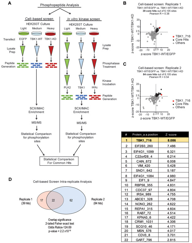
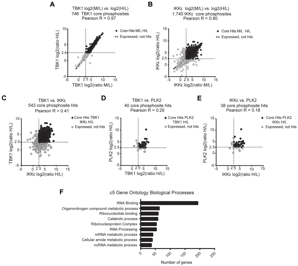
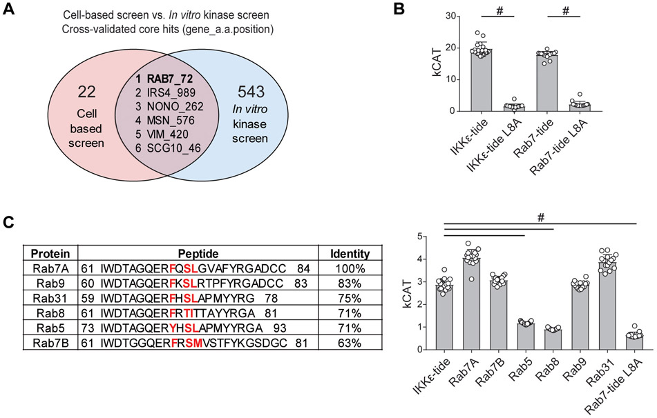
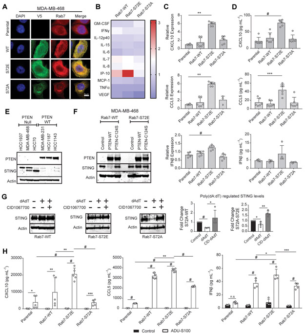
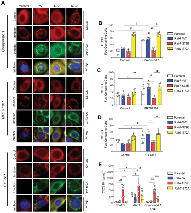
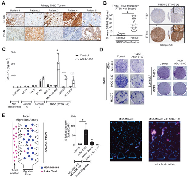
Triple-negative breast cancer (TNBC) is a heterogeneous disease enriched for mutations in PTEN and dysregulation of innate immune signaling. Here, we demonstrate that Rab7, a recently identified substrate of PTEN phosphatase activity, is also a substrate of the innate immune signaling kinases TANK-binding kinase 1 (TBK1)/IκB kinase ε (IKKε) on the same serine-72 (S72) site. An unbiased search for novel TBK1/IKKε substrates using stable isotope labeling with amino acids in cell culture phosphoproteomic analysis identified Rab7-S72 as a top hit. PTEN-null TNBC cells expressing a phosphomimetic version of Rab7-S72 exhibited diffuse cytosolic Rab7 localization and enhanced innate immune signaling, in contrast to a kinase-resistant version, which localized to active puncta that promote lysosomal-mediated stimulator of interferon genes (STING) degradation. Thus, convergence of PTEN loss and TBK1/IKKε activation on Rab7-S72 phosphorylation limited STING turnover and increased downstream production of IRF3 targets including CXCL10, CCL5, and IFNβ. Consistent with this data, PTEN-null TNBC tumors expressed higher levels of STING, and PTEN-null TNBC cell lines were hyperresponsive to STING agonists. Together, these findings begin to uncover how innate immune signaling is dysregulated downstream of TBK1/IKKε in a subset of TNBCs and reveals previously unrecognized cross-talk with STING recycling that may have implications for STING agonism in the clinic. SIGNIFICANCE: These findings identify Rab7 as a substrate for TBK1 for regulation of innate immune signaling, thereby providing important insight for strategies aimed at manipulating the immune response to enhance therapeutic efficacy in TNBC.


Conflict of interest statement

Conflict of Interest Statement. T.U.B. is a consultant for N-of-One/Qiagen. D.A.B. is consultant for N-of-One/Qiagen, Tango Therapeutics, and MADALON consulting. D.A.B. is cofounder of Xsphera Biosciences. D.A.B has received research funding from Novartis and Bristol Myers Squibb.

Figures

Fig. 1
Fig. 1
Stable isotope labeling with amino acids in cell culture (SILAC) determination of novel TBK1 substrates. A, Schematic of SILAC screen analysis for the determination of TBK1 substrates. Left panel: cell-based screen where HEK 293T cells were cultured in the presence of light, medium, or heavy isotopes then transfected with three plasmids containing the following proteins for expression: EGFP, TBK1-WT, or TBK1-KD. Right panel: in vitro kinase screen where following isotope culturing, HEK 293T lysates were generated and dephosphorylated. Light labeled lysates were left untreated while heavy and medium-labeled lysates were incubated in one of the following recombinant kinases: PLK2, TBK1 or IKKε. Each screen was then evaluated for kinase specific phosphorylation sites by tandem mass spectrometry. B and C, Graphical representation of the phosphopeptides common in TBK1-WT/EGFR and TBK1-WT/TBK1-KD analyses from cell-based screen experimental replicate 1 (B) and replicate 2 (C). D, Venn diagram of the comparison of phosphopeptides from replicate 1 and replicate 2 for common core hits. List of phosphopeptides that were significant if their respective z-scores were >2.5 in both conditions.
Fig. 2
Fig. 2
In vitro SILAC screen analysis of phosphopeptides for the determination of novel TBK1 substrates. Enzyme phosphopeptide ratios were determined from peptides identified from medium or heavy labeled lysates incubated with either IKKε, TBK1, or PLK2 over no enzyme light-labeled lysates. A and B, Intra replicate concordance for TBK1(A) and IKKε (B). C-E, Cross enzyme concordance between TBK1 and IKKε (C), TBK1 and PLK2 (D), and IKKε and PLK2 (E). In all phosphopeptide analyses, hits were considered positive if z-scores were >2.5 in both conditions. F, Pathway analysis for phosphopeptides from TBK1/IKKε core hits from (C) utilizing Molecular Signature Database C5 Gene Ontology Biological Processes Collection.
Fig. 3
Fig. 3
Determination and confirmation of Rab7-S72 as a TBK1/IKKε substrate target. A, Venn diagram showing the six common phosphorylation sites resulting from analysis of cell-based screen and in vitro kinase screen core hits (sites were selected based on z-score >2.5). B, Short peptide sequences were generated for the positive control TBK1 substrate, IKKε-tide and surrounding the predicted substrate, Rab7-S72. Peptide sequences were unaltered (IKKε-tide or Rab7-tide) or contained a negative control +1 mutation, where leucine was mutated to alanine (IKKε-Tide L8A or Rab7-Tide L8A). Synthetic peptides were incubated with recombinant TBK1 kinase and ATP, and kCat was calculated based upon the ADP generated (bars, n=16, #p<0.0001 One-way ANOVA with Tukey post-test). C, Selected Rab GTPase family members containing the conserved TBK1/IKKε motif, identified by BLAST analysis, and used for TBK1 enzymatic analysis. Small peptides were generated for Rab proteins identified and analyzed by biochemical assay as in B (bars, n=16, #p<0.0001 One-way ANOVA with Tukey post-test).
Fig. 4
Fig. 4
Regulation of STING turnover and innate immune signaling by Rab7-S72 phosphorylation. A, Representative immunofluorescent image showing localization of Rab7 or V5-Rab7 in parental and mutant MDA-MB-468 cells under basal conditions (n=3, scale=6μm). B, Multiplexed cytokine analysis of conditioned medium from Rab7 mutant cell lines cultured under basal conditions for 24 h. (n=6). C, qRT-PCR measurement of CXCL10, CCL5 and IFNβ levels in MDA-MB-468 Rab7 mutant cell lines under basal conditions at 24 h (CXCL10) or 72 h (CCL5 and IFNβ) (bars, n=6; **p<0.01, #p<0.0001 by One-way ANOVA with Tukey post-test). D, Cytokine expression of CXCL10, CCL5, and IFNβ in supernatants from cells cultured for 24 h (CXCL10) or 72 h (CCL5 and IFNβ) and analyzed by ELISA (bars, n=6; ***p<0.001, #p<0.0001 by One-way ANOVA with Tukey post-test). E, Immunoblot of STING and PTEN expression in a panel of TNBC cell lines under basal conditions. F, Immunoblot of STING and PTEN expression in MDA-MB-468 cells expressing the indicated PTEN and Rab7 constructs. G, Immunoblots of STING levels in MDA-MB-468 Rab7 cell lines stimulated with poly(dA:dT) (1μg mL−1) +/− 100nM CID1067700 for 6 h. Densitometry quantification of enhanced poly(dA:dT) STING-degradation by Rab7-S72A and sensitivity to CID106770 (bars, n=4; *p<0.5, **p<0.01, #p<0.0001 by unpaired t-test). H, Expression of CXCL10, CCL5, and IFNβ by MDA-MB-468 Rab7 cell lines cultured for either 24 h (CXCL10) or 72 h (CCL5 and IFNβ) in the presence or absence of 10μM ADU-S100 (bars, n=6; *p<0.05, **p<0.01, ***p<0.001, #p<0.0001 by Two-way ANOVA with Tukey post-test).
Fig. 5
Fig. 5
Rab7 S72E is resistant to TBK1/IKKε inhibition on STING localization and immune signaling in Rab7 mutants. A, Immunofluorescence imaging of parental and Rab7 cell lines: WT, S72E, and S72A, treated with either 1μM Compound 1 (6 h), 5μM MRT67307 (8 h) or 5μM CYT387 (3 h) alone (representative, n=2; scale=10μm). B, Quantification of STING foci containing cells treated with Compound 1 as in A (bars, n=6; #p<0.0001 by Two-way ANOVA with Tukey post-test). C, Quantification of STING foci in containing cells treated with MRT67307 as in A (bars, n=6; *p<0.05, ***p<0.001, #p<0.0001 by One-way ANOVA with a Tukey post-test). D, Quantification of images from CYT387 treated cells A (bars, n=6; **p<0.01, ***p<0.001, #p<0.0001 by One-way ANOVA with a Tukey post-test). E, Analysis of conditioned media from Rab7 expressing cells following 30 min pretreatment with 1μM Compound 1, then stimulated for 48 h with poly(dA:dT) (Compound 1 dA:dT) or poly(dA:dT) alone (dA:dT) for CXCL10 expression by ELISA (bars, n=6; *p<0.05, **p<0.01, ***p<0.001, #p<0.0001 by Two-way ANOVA with Tukey post-test).
Fig. 6
Fig. 6
PTEN loss increases TNBC STING expression in vivo and enhances STING agonist response. A, Patient-derived TNBC histology sections stained for PTEN or STING by immunohistochemistry. B, Commercially available tissue microarray (TMA) of breast cancer carcinomas were stained for STING and PTEN. Left panel: Quantitative analysis of percent STING staining of PTEN null TNBC cores (bar, n=26; #p<0.001 by unpaired t-test). Right panel: representative images of PTEN negative and STING positive staining from duplicate TNBC cores. C, Expression of CXCL10 in supernatants from either non-transformed (MCF-10A), Luminal A (MCF7 and T-47D), Luminal B (ZR-751 and SKBr3) or TNBC cell lines (MDA-MB-468, HCC1937, and HCC70) following 10μM ADU-S100 treatment for 24 h (bars, n=6; ***p<0.001, #p<0.0001 by Two-way ANOVA with Tukey post-test). D, Breast cancer cell line growth inhibition following twice weekly treatment with 10μM ADU-S100 for 6 weeks. Colony formation was visualized with crystal violet staining. E, First panel: Schematic of T-cell migration assay, utilizing a 3D microfluidic device with MDA-MB-468 spheroids embedded in a central collagen-filled channel, co-cultured with CXCR3 expressing Jurkat T-cells. Second panel: Quantification of Jurkat T-cell migration towards MDA-MB-468 cells −/+ ADU-S100 treatment (bars, n=3; #p<0.0001 by One-way ANOVA with Tukey post-test). Third and Fourth Panels: Representative images of T-cell migration towards MDA-MB-468 cells −/+ ADU-S100 treatment.

Similar articles

Cited by

References

    1. Carey LA, Dees EC, Sawyer L, Gatti L, Moore DT, Collichio F, et al. The triple negative paradox: primary tumor chemosensitivity of breast cancer subtypes. Clin Cancer Res 2007;13:2329–34 - PubMed
    1. Liedtke C, Mazouni C, Hess KR, Andre F, Tordai A, Mejia JA, et al. Response to neoadjuvant therapy and long-term survival in patients with triple-negative breast cancer. J Clin Oncol 2008;26:1275–81 - PubMed
    1. Lin NU, Vanderplas A, Hughes ME, Theriault RL, Edge SB, Wong YN, et al. Clinicopathologic features, patterns of recurrence, and survival among women with triple-negative breast cancer in the National Comprehensive Cancer Network. Cancer 2012;118:5463–72 - PMC - PubMed
    1. Lehmann BD, Bauer JA, Chen X, Sanders ME, Chakravarthy AB, Shyr Y, et al. Identification of human triple-negative breast cancer subtypes and preclinical models for selection of targeted therapies. J Clin Invest 2011;121:2750–67 - PMC - PubMed
    1. Phuah SY, Looi LM, Hassan N, Rhodes A, Dean S, Taib NA, et al. Triple-negative breast cancer and PTEN (phosphatase and tensin homologue) loss are predictors of BRCA1 germline mutations in women with early-onset and familial breast cancer, but not in women with isolated late-onset breast cancer. Breast Cancer Res 2012;14:R142. - PMC - PubMed

Publication types

MeSH terms