. 2019 Dec 13;294(50):19365-19380.
doi: 10.1074/jbc.RA119.010193. Epub 2019 Oct 29.

The ribosome assembly factor Nop53 controls association of the RNA exosome with pre-60S particles in yeast

Affiliations

The ribosome assembly factor Nop53 controls association of the RNA exosome with pre-60S particles in yeast

Leidy Paola P Cepeda et al. J Biol Chem. .

Abstract

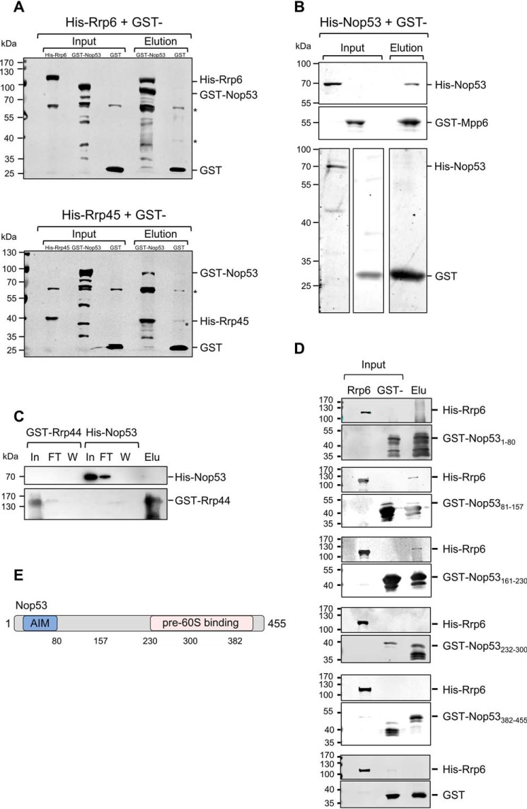
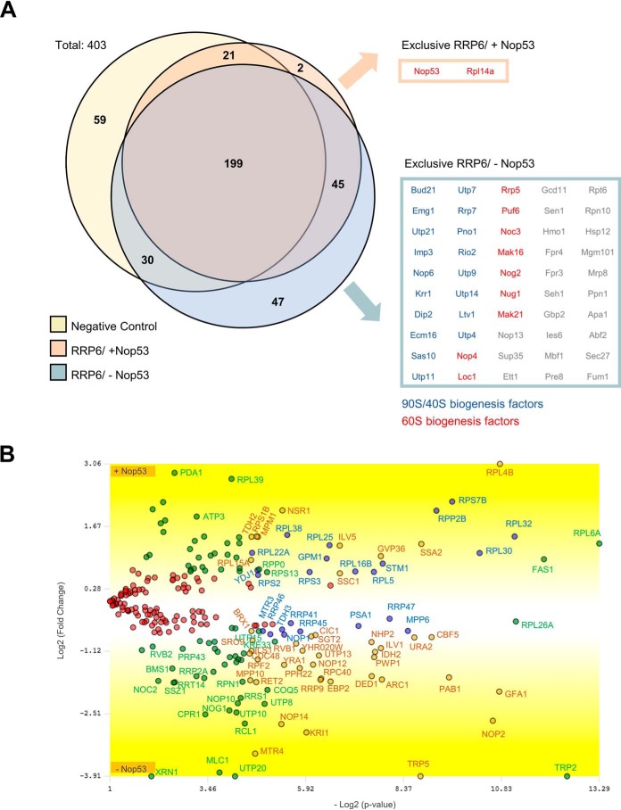
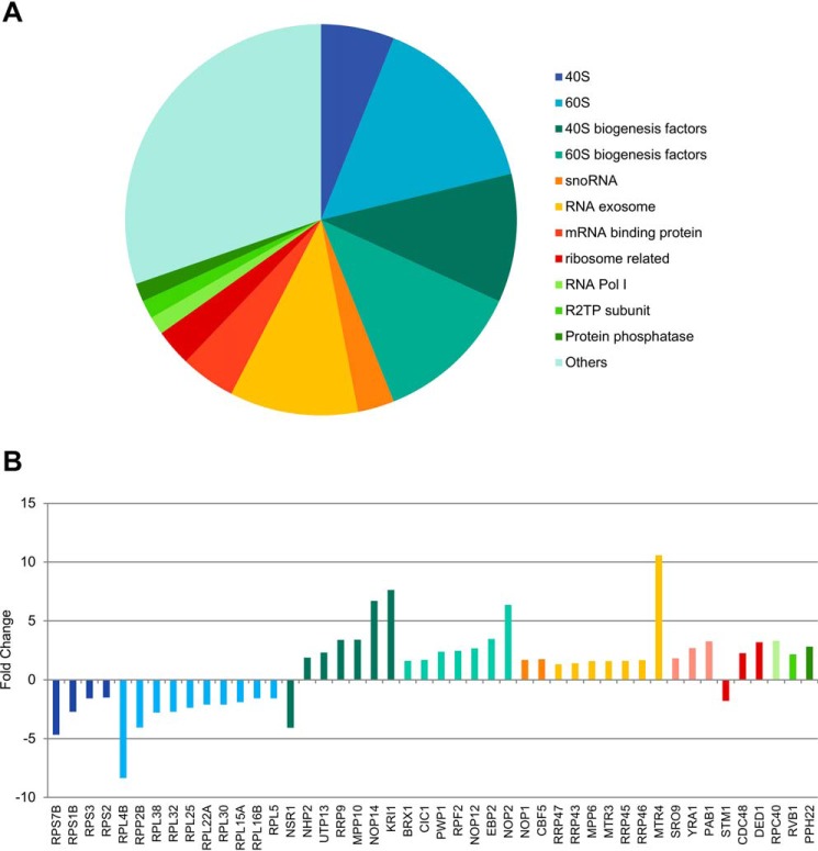
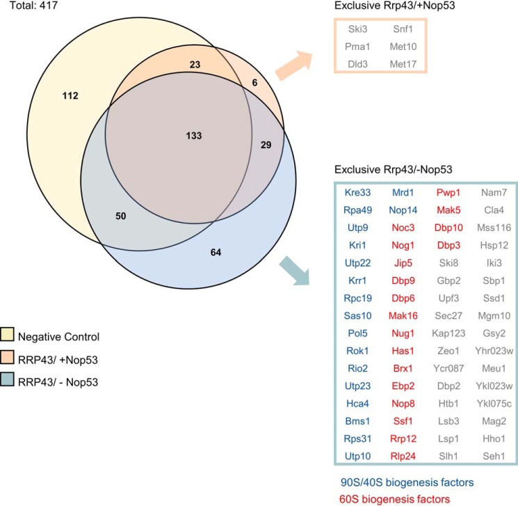
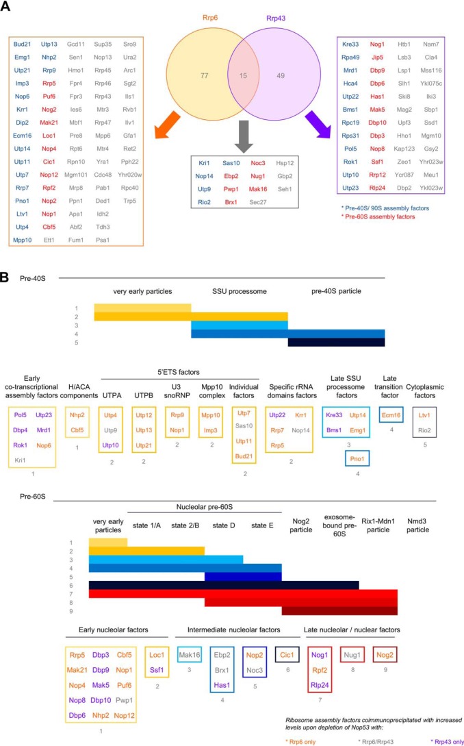
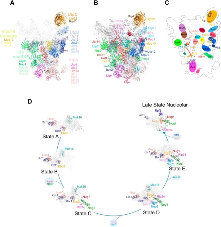
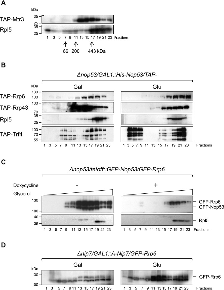
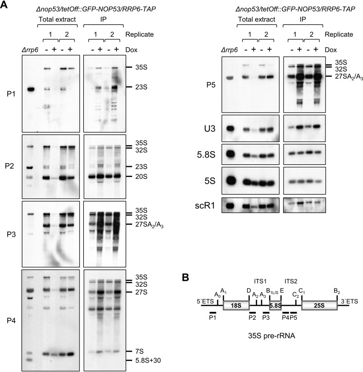
Eukaryotic ribosomal biogenesis is a high-energy-demanding and complex process that requires hundreds of trans-acting factors to dynamically build the highly-organized 40S and 60S subunits. Each ribonucleoprotein complex comprises specific rRNAs and ribosomal proteins that are organized into functional domains. The RNA exosome complex plays a crucial role as one of the pre-60S-processing factors, because it is the RNase responsible for processing the 7S pre-rRNA to the mature 5.8S rRNA. The yeast pre-60S assembly factor Nop53 has previously been shown to associate with the nucleoplasmic pre-60S in a region containing the "foot" structure assembled around the 3' end of the 7S pre-rRNA. Nop53 interacts with 25S rRNA and with several 60S assembly factors, including the RNA exosome, specifically, with its catalytic subunit Rrp6 and with the exosome-associated RNA helicase Mtr4. Nop53 is therefore considered the adaptor responsible for recruiting the exosome complex for 7S processing. Here, using proteomics-based approaches in budding yeast to analyze the effects of Nop53 on the exosome interactome, we found that the exosome binds pre-ribosomal complexes early during the ribosome maturation pathway. We also identified interactions through which Nop53 modulates exosome activity in the context of 60S maturation and provide evidence that in addition to recruiting the exosome, Nop53 may also be important for positioning the exosome during 7S processing. On the basis of these findings, we propose that the exosome is recruited much earlier during ribosome assembly than previously thought, suggesting the existence of additional interactions that remain to be described.

Keywords: exosome complex; pre-60S particles; precursor ribosomal RNA (pre-rRNA); protein–protein interaction; rRNA processing; ribosome assembly.


Figures

Figure 1.
Figure 1.
Nop53 interacts with the exosome and with exosome cofactors. GST pulldown assays were performed to test the interaction between Nop53 and the exosome components Rrp44 and Rrp45 and with the exosome cofactor Mpp6. A, GST–Nop53 immobilized in GSH-Sepharose beads pulled down His–Rrp6 and His–Rrp45. GST was used as negative control of interaction. *, E. coli proteins nonspecifically recognized by anti-GST antibody. B, GST–Mpp6, but not GST, also pulled down His–Nop53, whereas GST–Rrp44 did not (C). D, Rrp6 interacts with the N-terminal half of Nop53. The interaction between different GST-fused Nop53 truncation mutants was tested against His–Rrp6 by GST pulldown assay indicating that only the N-terminal region of Nop53 is able to interact with the exosome catalytic subunit Rrp6. GST was used as a negative control. Total extract of cells expressing His–Rrp6 was incubated either with purified GST or GST–Nop53 mutants. In, input; FT, flow-through; W, wash; Elu, elution. E, schematic representation of Nop53 with the relative positions of the amino acids indicated in the truncation mutants.
Figure 2.
Figure 2.
Depletion of Nop53 affects the Rrp6 interactome. To evaluate how Nop53 modulates the interaction of Rrp6 with other factors, a coimmunoprecipitation assay was performed both in the presence (−doxycycline) and upon depletion (+doxycycline) of Nop53 using the conditional strain Δnop53/tetOff::GFP-NOP53 carrying the endogenous Rrp6–TAP fusion. As a negative control, the same strain carrying only the TAP tag was employed. For each condition, the elution of biological triplicates was subjected to label-free quantitative analysis. Using the PatternLab software, the identified proteins in at least two biological replicates were grouped in a Venn diagram (A), showing that among a total of 403 proteins, two were exclusively identified in the presence of Nop53, and 47 uniquely upon depletion of Nop53. Three additional proteins were included in this latter group because, despite being present in the negative control, they were significantly enriched upon depletion of Nop53 (Fig. S3). Separately, the exclusively identified proteins in each condition are listed in boxes, highlighting the over-representation of pre-60S (red) and pre-90S/pre-40S (blue) assembly factors. B, proteins that were coimmunoprecipitated with Rrp6 both in the presence and absence of Nop53 (identified in at least two replicates per group) were compared through TFold analysis (benjamini-hochberg q-value; F-stringency (fold-change stringency parameter); L-stringency (stringency parameter for lowly-abundant proteins)) using PatternLab software (52). The top and bottom of the volcano plot display proteins, respectively, depleted and enriched upon depletion of Nop53. Among the 244 commonly identified proteins, 104 did not show a statistically significant change upon depletion of Nop53 (red dots), 74 met the fold change criteria but were not statistically significant (green dots), 43 are low abundance proteins that met both the fold change and statistical criteria (orange dots), and 23 proteins met both the fold change and statistical criteria (blue dots).
Figure 3.
Figure 3.
Rrp6 remains associated with the core exosome complex and is retained in immature pre-ribosomal subunits upon depletion of Nop53. A, proteins that showed a statistically significant enrichment or depletion (both orange and blue dots in Fig. 4B) in the absence of Nop53 were categorized according to their biological function in a pie chart. Among the most enriched classes, both ribosome biogenesis and exosome complex stood out. B, same proteins, significantly affected by Nop53 depletion, are depicted with their respective fold changes. Positive and negative fold change values indicate, respectively, enrichment and decrease upon Nop53 depletion. Of note, several early pre-60S and pre-40S transacting factors were found coimmunoprecipitated with increased levels in the absence of Nop53. The exosome subunits and cofactors were slightly enriched, but not as much as the RNA helicase Mtr4.
Figure 4.
Figure 4.
Depletion of Nop53 also affects the Rrp43 interactome. To analyze how Nop53 modulates the interaction of Rrp43 with other factors, a coimmunoprecipitation assay was performed both in the presence (−doxycycline) and upon depletion (+doxycycline) of Nop53 using the conditional strain Δnop53/tetOff::GFP-NOP53 containing RRP43-TAP fusion in a plasmid. As a negative control, the same strain carrying only the TAP tag was used. For each condition, the elution of biological triplicates was subjected to label-free quantitative analysis. Using the PatternLab software, the identified proteins in at least two biological replicates were grouped in a Venn diagram, showing that among a total of 417 proteins, six were exclusively identified in the presence of Nop53, and 64 uniquely upon depletion of Nop53. The exclusively identified proteins in each condition are listed in boxes, highlighting the over-representation of pre-60S (red) and pre-90S/pre-40S (blue) assembly factors.
Figure 5.
Figure 5.
Comparison of the proteins copurified with Rrp6 and Rrp43 that showed increased levels upon depletion of Nop53. A, Venn diagram depicting the identified proteins in at least two replicates coimmunoprecipitated with Rrp6 and Rrp43, whose levels were increased upon depletion of Nop53. B, schematics of pre-40S and pre-60S maturation pathways, depicting stages at which the identified proteins copurified with Rrp6 or Rrp43 associate with pre-ribosomes. Classification was based on Ref. . Proteins not classified are as follows: pre-40S proteins Rpa49 and Rpc19; pre-60S proteins Jip5 and Rrp12. No common proteins had their levels reduced upon depletion of Nop53.
Figure 6.
Figure 6.
Proteins coimmunoprecipitated with the exosome in higher levels in the absence of Nop53 participate in different phases of ribosomal maturation. A, structural representation of 90S pre-ribosomes with the identified protein complexes are depicted in different colors as follows: pink, UTP-A; blue, UTP-B; orange, UTP-C; green, U3 snoRNP; yellow, Mpp10 complex. Proteins in bold letters are those indicated in Fig. 5B. The remaining parts of the particle are represented in light gray. B, individual proteins from the subcomplexes indicated in (A) are highlighted in different colors. C, schematics of the positions of the proteins in the 90S particle and their interactions within the particle. Note that all proteins interacting more efficiently with the exosome in the absence of Nop53 are exposed on the same face of 90S. Structure of the 90S particle was based on Ref. (PDB 5WLC). D, representation of pre-60S maturation pathway, with the factors identified here highlighted in different colors. The exosome associates with various pre-60S intermediates in the absence of Nop53. Schematics show the interactions between the proteins identified here within the pre-60S. Structures of pre-60S particles were based on Refs. , . State A (PDB 6EM3), state B (PDB 6EM4), state C (PDB 6EM1), state D (PDB 6EM5), state E (PDB 6ELZ), and late nuclear states (PDB 3JCT) are shown.
Figure 7.
Figure 7.
Nop53 affects exosome association with pre-60S. The conditional strain Δnop53/GAL::NOP53 was transformed with plasmids expressing either the exosome subunits Rrp43 and Rrp6 or the TRAMP subunit Tfr4 fused to the TAP tag to monitor the association of these complexes to the pre-60S particle in the presence or absence of Nop53. A, cell extract from a strain expressing TAP–Mtr3 was subjected to centrifugation through the glycerol gradient for separation of soluble proteins from ribosomal particles. Rpl5 was used as a control for the 60S and pre-60S ribosomal subunit. Molecular mass markers were centrifuged in parallel, and their fractionation is indicated by arrows. B, fractionation of Rrp43, Rrp6, and Trf4 in the presence (Gal) or absence (Glu) of Nop53 shows their concentration in lower fractions of the gradient after depletion of Nop53. C, similar experiments with the conditional strain Δnop53/tetOff::GFP-NOP53-expressing GFP–Rrp6 show the same concentration of this exosome subunit in lower fractions after depletion of Nop53 (+doxycycline). Numbers of fractions from the gradient are indicated. D, conditional strain Δnip7/GAL::A-NIP7 expressing GFP-Rrp6 was used as a control for a pre-60S maturation factor and shows that its depletion (Glu) does not affect Rrp6 fractionation.
Figure 8.
Figure 8.
Rrp6–TAP co-purifies early precursor rRNAs. A, Δnop53/tetOff::GFP-NOP53/RRP6-TAP strain was used for RNA coimmunoprecipitation with Rrp6–TAP in the presence (−Dox (doxycycline )), or absence (+Dox) of Nop53. Total RNA extracted from Δrrp6 strain was used as a control of precursor rRNAs accumulating in the absence of Rrp6. Left panels show two biological replicates of RNAs extracted from aliquots of total cell extracts in the indicated conditions used in the coimmunoprecipitations as shown on the right panels. Precursor rRNAs are indicated on the right. 5S rRNA and scR1 were used as controls for nonspecific binding to the resin. Total RNA extracted from replicate 1 in the absence of Nop53 (+Dox) was lost during loading on the gel, but it was maintained in the figure because it clearly shows the accumulation of 7S pre-rRNA under this condition. B, schematic representation of the yeast 35S pre-rRNA indicating the hybridizing positions of the different probes.

References

    1. de la Cruz J., Karbstein K., and Woolford J. L. Jr. (2015) Functions of ribosomal proteins in assembly of eukaryotic ribosomes in vivo. Annu. Rev. Biochem. 84, 93–129 10.1146/annurev-biochem-060614-033917 - DOI - PMC - PubMed
    1. Kressler D., Hurt E., and Bassler J. (2017) A puzzle of life: crafting ribosomal subunits. Trends Biochem. Sci. 42, 640–654 10.1016/j.tibs.2017.05.005 - DOI - PubMed
    1. Fatica A., and Tollervey D. (2002) Making ribosomes. Curr. Opin Cell Biol. 14, 313–318 10.1016/S0955-0674(02)00336-8 - DOI - PubMed
    1. Allmang C., Kufel J., Chanfreau G., Mitchell P., Petfalski E., and Tollervey D. (1999) Functions of the exosome in rRNA, snoRNA and snRNA synthesis. EMBO J. 18, 5399–5410 10.1093/emboj/18.19.5399 - DOI - PMC - PubMed
    1. Mitchell P., Petfalski E., Shevchenko A., Mann M., and Tollervey D. (1997) The exosome: a conserved eukaryotic RNA processing complex containing multiple 3′ → 5′ exoribonucleases. Cell 91, 457–466 10.1016/S0092-8674(00)80432-8 - DOI - PubMed

Publication types

MeSH terms

Substances