. 2019 Oct 29;10(5):e02376-19.
doi: 10.1128/mBio.02376-19.

Shielding the Next Generation: Symbiotic Bacteria from a Reproductive Organ Protect Bobtail Squid Eggs from Fungal Fouling

Affiliations

Shielding the Next Generation: Symbiotic Bacteria from a Reproductive Organ Protect Bobtail Squid Eggs from Fungal Fouling

Allison H Kerwin et al. mBio. .

Abstract

The importance of defensive symbioses, whereby microbes protect hosts through the production of specific compounds, is becoming increasingly evident. Although defining the partners in these associations has become easier, assigning function to these relationships often presents a significant challenge. Here, we describe a functional role for a bacterial consortium in a female reproductive organ in the Hawaiian bobtail squid, Euprymna scolopes Bacteria from the accessory nidamental gland (ANG) are deposited into the egg jelly coat (JC), where they are hypothesized to play a defensive role during embryogenesis. Eggs treated with an antibiotic cocktail developed a microbial biomass primarily composed of the pathogenic fungus Fusarium keratoplasticum that infiltrated the JC, resulting in severely reduced hatch rates. Experimental manipulation of the eggs demonstrated that the JC was protective against this fungal fouling. A large proportion of the bacterial strains isolated from the ANG or JC inhibited F. keratoplasticum in culture (87.5%), while a similar proportion of extracts from these strains also exhibited antifungal activity against F. keratoplasticum and/or the human-pathogenic yeast Candida albicans (72.7%). Mass spectral network analyses of active extracts from bacterial isolates and egg clutches revealed compounds that may be involved in preventing microbial overgrowth. Several secondary metabolites were identified from ANG/JC bacteria and egg clutches, including the known antimicrobial lincomycin as well as a suite of glycerophosphocholines and mycinamicin-like compounds. These results shed light on a widely distributed but poorly understood symbiosis in cephalopods and offer a new source for exploring bacterial secondary metabolites with antimicrobial activity.IMPORTANCE Organisms must have strategies to ensure successful reproduction. Some animals that deposit eggs protect their embryos from fouling/disease with the help of microorganisms. Although beneficial bacteria are hypothesized to contribute to egg defense in some organisms, the mechanisms of this protection remain largely unknown, with the exception of a few recently described systems. Using both experimental and analytical approaches, we demonstrate that symbiotic bacteria associated with a cephalopod reproductive gland and eggs inhibit fungi. Chemical analyses suggest that these bacteria produce antimicrobial compounds that may prevent overgrowth from fungi and other microorganisms. Given the distribution of these symbiotic glands among many cephalopods, similar defensive relationships may be more common in aquatic environments than previously realized. Such defensive symbioses may also be a rich source for the discovery of new antimicrobial compounds.

Keywords: Euprymna scolopes; chemical defense; defensive symbiosis; host-microbe symbioses; interaction-driven molecular networking.


Figures

FIG 1
FIG 1
Antibiotic treatment of eggs leads to fungal fouling. (A) Ventral dissection of a female E. scolopes squid showing locations of reproductive system organs relative to the light organ and eye of the squid. (B and C) Detailed images and diagrams of female reproductive tract (B) and egg (C). Treatment of clutches with an antibiotic cocktail led to the formation of fungal/bacterial fouling. (D to I) Clutches were either left untreated (D to F) or were treated with antibiotics (G to I; n = 17). (J and K) The fungal biomass was composed of hyphae interspersed with bacterial cells (white arrows). (F) Electron micrographs of JCs from eggs at day 21 of embryogenesis show that untreated egg JCs contained both single bacterial cells and small clusters of cells (white arrows). (I) The antibiotic-treated egg JCs contained numerous fungal conidia and hyphae (insets). (L) After 3 days of treatment, a 98% reduction in bacterial load was observed (t8 = 1.685, P = 0.131, n = 5 clutches). The bacterial load of both the untreated and the antibiotic-treated clutch segments increased over embryogenesis, but a significant reduction in bacteria was found both at day 10 (t8 = 5.011, P = 0.001) and at day 15 (t8 = 2.511) (P = 0.036; 98% and 97%, respectively; data in graph presented as means ± standard errors of the means [SEM]). NG, nidamental gland; ANG, accessory nidamental gland; JC, jelly coat; LO, light organ.
FIG 2
FIG 2
Treatment of eggs with an antibiotic cocktail and subsequent development of fungal fouling significantly reduced hatch of juveniles. Eggs treated with an antibiotic cocktail developed a fungal biomass between days 8 and 14 of embryogenesis and had a reduced hatch rate (t4 = 3.572, P = 0.023). Hatching rates were unaffected when eggs were treated with the antibiotic cocktail and maintained in a laminar flow hood (t2 = 1.289, P = 0.326) or at low temperatures (15 to 20°C, t3 = 0.738, P = 0.514) to prevent fungal growth. The antibiotic cocktail included penicillin G, kanamycin, spectinomycin, streptomycin, and gentamicin, each at a concentration of 25 μg/ml. Matching data point shapes represent eggs taken from the same initial clutch.
FIG 3
FIG 3
Embryos lacking jelly coats are more susceptible to fungal infection. Eggs either were left untreated (A, C, and E) or were challenged with F. keratoplasticum FSSC-2g at 104 conidia/ml for 18 days (B, D, and F). Eggs left intact (A and B) or that were decapsulated, leaving only the jelly coat and developing embryos (C and D), were observed with few hyphae when challenged (B and D; yellow arrows), while eggs that were decapsulated and for which the jelly coat was removed, leaving only the embryo (E and F), were covered with fungal hyphae when challenged (F). Hyphae were stained with Syto9 nucleic acid stain and visualized with a confocal microscope (F, inset). (n = 4 trials, 10 eggs/treatment). Shown are representative images from a single trial.
FIG 4
FIG 4
Bacterial secondary metabolites from the ANG and JC isolates demonstrate differential activity against fungal pathogens. (A) Many extracts from both ANG and JC bacterial isolates exhibited antifungal activity against one or more strains of Fusarium keratoplasticum and the human pathogen Candida albicans. Amphotericin B (4 μg/ml) was used as a positive control. Data shown are based on average percent growth values of technical triplicates from at least two experimental replicates. (B) Over 51% of ANG and JC bacterial extracts strongly or moderately inhibited the egg-isolated F. keratoplasticum strains, FSSC-2i and FSSC-2g, compared to the clinical F. keratoplasticum FSSC-2d isolate. (C) Venn diagram showing extracts that were active against each fusarial strain. Overall, six ANG and JC extracts exhibited antifungal activity against all three F. keratoplasticum strains, with other extracts showing activity against just one or two of the strains. Highlighted boxes indicate strain numbers listed in panel A.
FIG 5
FIG 5
Comparative metabolomics identifies antimicrobial bacteria-derived secondary metabolites in egg clutches and active ANG and JC bacterial extracts. (A) LC-MS/MS molecular network of challenged and control clutches (see Fig. 6) with antifusarium active bacterial extracts (yellow circles) and inactive bacterial extracts (blue circles) resulted in a large, complex network (enlarged image of network provided in Fig. S7). Active bacterial extracts included only those with strong inhibition (0% to 25% fungal growth, n = 8), while inactive bacterial extracts included only those with minimal to no inhibition (≥76% fungal growth, n = 13). Parent masses are represented within each node, and the thickness of the edge is based on the cosine similarity score. (B) Ten MS features (circled) were prioritized for further investigation, including seven from challenged clutches that overlapped active bacterial extracts (pink squares) and three from control clutches that overlapped active bacterial extracts (teal squares). (C) Lincomycin B ([M + H]+ m/z 393.1480), a structural analogue of the FDA-approved antimicrobial drug lincomycin A, was identified in both the control clutches and the active extract, Labrenzia sp. ANG18 (identification confirmed via comparison with purchased standards). Lincomycin A was also found in the control clutch but was not detectable in the Labrenzia sp. ANG18 isolate.
FIG 6
FIG 6
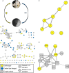
Comparative metabolomics identified specialized metabolite production by egg clutch-associated bacteria induced in the presence of Fusarium keratoplasticum. (A) The experimental design involved splitting egg clutches, challenging one portion with F. keratoplasticum FSSC-2g, and leaving one portion as an unchallenged control. No antibacterial treatment was added, leaving JC-associated bacteria potentially able to produce defensive metabolites. (B) LC-MS/MS molecular networking revealed numerous metabolites present only in challenged clutches (yellow circles), including two clusters with metabolites found only in challenged clutches (C). Metabolites were deprioritized if found only in control clutches (blue circles) and/or in F. keratoplasticum FSSC-2g extract (gray circles), with features found in two or more extracts shown in gray squares or diamonds. Parent masses are represented within each node, and the thickness of the edge is based upon the cosine similarity score. (D) Structurally distinct glycerophosphocholines (see the text) were also present in another cluster in this network, found in challenged clutches, control clutches, and/or F. keratoplasticum FSSC-2g. Several of the metabolites in this cluster were found only in the challenged clutches (yellow circles) or the F. keratoplasticum FSSC-2g extract (gray circles), while other metabolites in this cluster were found in both challenged and control clutches (gray squares) or in all three samples (gray diamonds) (see Table S3a).

Similar articles

Cited by

References

    1. Gil-Turnes MS, Fenical W. 1992. Embryos of Homarus americanus are protected by epibiotic bacteria. Biol Bull 182:105–108. doi:10.2307/1542184. - DOI - PubMed
    1. Gil-Turnes MS, Hay ME, Fenical W. 1989. Symbiotic marine bacteria chemically defend crustacean embryos from a pathogenic fungus. Science 246:116–118. doi:10.1126/science.2781297. - DOI - PubMed
    1. Fraune S, Anton-Erxleben F, Augustin R, Franzenburg S, Knop M, Schröder K, Willoweit-Ohl D, Bosch T. 2015. Bacteria-bacteria interactions within the microbiota of the ancestral metazoan Hydra contribute to fungal resistance. ISME J 9:1543–1556. doi:10.1038/ismej.2014.239. - DOI - PMC - PubMed
    1. Caldera E, Poulsen M, Suen G, Currie C. 2009. Insect symbioses: a case study of past, present, and future fungus-growing ant research. Environ Entomol 38:78–92. doi:10.1603/022.038.0110. - DOI - PubMed
    1. Lopanik N. 2014. Chemical defensive symbioses in the marine environment. Funct Ecol 28:328–340. doi:10.1111/1365-2435.12160. - DOI

Publication types

MeSH terms