. 2019 Nov-Dec;33(6):1773-1784.
doi: 10.21873/invivo.11668.

Ionizing Radiation Promotes Epithelial-to-Mesenchymal Transition in Lung Epithelial Cells by TGF-β-producing M2 Macrophages

Affiliations

Ionizing Radiation Promotes Epithelial-to-Mesenchymal Transition in Lung Epithelial Cells by TGF-β-producing M2 Macrophages

Hae-Ran Park et al. In Vivo. 2019 Nov-Dec.

Abstract

Background/aim: Ionizing radiation induces pulmonary fibrosis, which is a common dose-limiting complication in patients receiving radiotherapy. Fibrosis occurs through the accumulation of large amounts of ECM components, synthesized by myofibroblasts in damaged lung tissue. Epithelial cells serve as one of the cellular sources of myofibroblasts via the epithelial-to-mesenchymal transition (EMT) process. In this study, we investigated the role of TGF-β-secreting M2 macrophages in association with ionizing radiation-induced EMT.

Materials and methods: The lung epithelial cell line MLE12, was irradiated and the expression of EMT markers and chemokines was examined. Moreover, the mouse lung macrophage MH-S cell line was cultured with conditioned media from irradiated MLE12 cells, to examine the effects of the secreted factors on the migration ability of macrophages. For the murine pulmonary fibrosis model, mice were locally irradiated and the levels of M1 or M2 macrophage-related markers and cytokines were measured in bronchoalvelolar lavage (BAL) fluid and lung tissue.

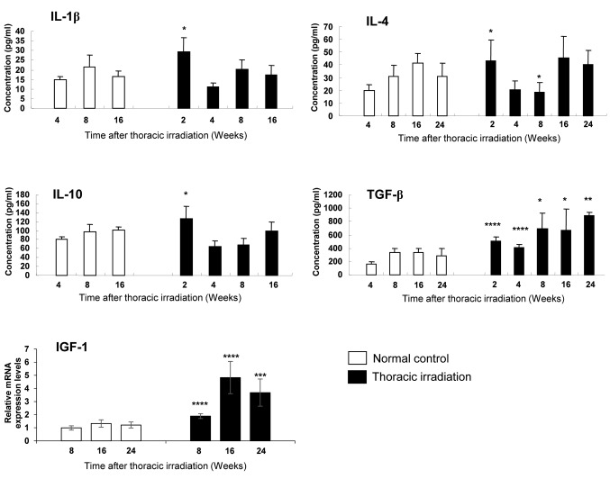
Results: In MLE12 cells, irradiation directly induced expression of EMT-related markers and secretion of various chemokines, which lead to macrophage migration. Interestingly, the sub-population of macrophages recruited in the lung of mice after thoracic irradiation was M2 macrophages that expressed Arg-1 and CD206. M2 macrophages induced the MLE12 to undergo phenotypic conversion to form fibroblast-like cells, which leads to a down-regulation of epithelial markers and an up-regulation of new EMT-related markers. In thoracic irradiated mice, pro-inflammatory cytokines such as IL-1β, IL-4 and IL-10 were increased at 2 weeks, but returned to normal levels from 16 weeks or 24 weeks after irradiation. However, thoracic irradiation led to a rapid increase of TGF-β and IGF-1 levels, which lasted up to 24 weeks. It was confirmed that M2 macrophages secreted the high levels of TGF-β. Moreover, the elimination of TGF-β from M2 macrophages attenuated mesenchymal transition of MLE12.

Conclusion: TGF-β-secreting M2 macrophages play an important regulatory role in mesenchymal transition of epithelial cells in the lung of irradiated mice, thus contributing to radiation-induced pulmonary fibrosis.

Keywords: Ionizing radiation; TGF-β; alternatively activated macrophages; epithelial to mesenchymal transition; fibrosis; myofibroblast.


Conflict of interest statement

The Authors declare that they have no competing interests.

Figures

Figure 1
Figure 1. Ionizing radiation induced a mesenchymal phenotype and increased epithelial–to–mesenchymal transition (EMT) markers in MLE12 in vitro. MLE12 cells were irradiated at a dose of 5Gy or 10Gy and then analysis was conducted at 7 days after irradiation. Representative cell morphology images of MLE12 after single-dose irradiation are shown (A). Scale bar, 50 μm. Expression levels of EMT-related markers in MLE12 after single-dose irradiation were examined by western blot (B). The experiment was repeated three times independently and the bars indicate the means±SD.
Figure 2
Figure 2. Irradiation induced the expression of various chemokines in epithelial cells and promoted macrophage migration. A representative of the mRNA levels of various chemokines in MLE12 cells at indicated time points after irradiation (10 Gy) is shown (A). After irradiation at a dose of 10 Gy, CCL2 levels in culture supernatants of MLE12 cells were analyzed by ELISA (B). CCL2 levels in the BAL fluid at 2, 4, 8, 16, and 24 weeks after thoracic irradiation were tested using ELISA. Each group consisted of five mice and the bars indicate the means±SD (C). Representative images (upper) and statistical column plots (lower) demonstrated increased potential of migration of MH-S cells cultured in conditioned media from irradiated MLE12 cells, compared to MH-S cells cultured in control media (from non-irradiated MLE12 cells) (D). C, Control; *p<0.05, **p<0.01, ***p<0.001.
Figure 3
Figure 3. Thoracic irradiation increased the expression of Arg-1 and CD206 in the lung. At 1, 2, 4, and 6 months after irradiation, the RNA expression levels of CD32, Arg-1, and CD206 in the lung tissue were measured through quantitative real-time PCR. There were five or six mice in each group. The bars indicate the means±SD. *p<0.05, **p<0.005, ***p<0.001 compared to the age-matched control mice. NC, Normal control; TI, thoracic irradiation.
Figure 4
Figure 4. Soluble protein of M2 macrophages induced a mesenchymal phenotype and increased epithelial–to–mesenchymal transition (EMT) markers in MLE12 cells, in vitro. MLE12 cells were cultured with conditioned media of M1 polarized, M2 polarized, or non-stimulated macrophages, for 14 days. Representative cell morphology images of MLE12 cells cultured in conditioned media of M1-polarized or M2-polarized macrophages are shown (A). Scale bar, 50 μm. Expression of EMT-related markers in MLE12 cells cultured in conditioned media obtained from non-treated (NTCM), M1 (M1-CM), or M2 macrophages (M2-CM) (B). Experiment was repeated three times independently and the bars indicate the means±SD.
Figure 5
Figure 5. Thoracic irradiation increased pro-inflammatory and pro-fibrotic cytokines in the lung of mice. At 2, 4, 8, 16, and 24 weeks after irradiation, interleukin (IL)-1β, IL-4, IL-10, and transforming growth factor (TGF)-β in bronchoalveolar lavage (BAL) fluid of mice were measured through ELISA, and relative mRNA expression levels of insulin-like growth factor (IGF)-1 in the lung tissue were examined through quantitative real-time PCR. There were four or five mice in each group. The bars indicate the means±SD. *p<0.05, **p<0.01, ***p<0.005, and ****p<0.001 compared to the age-matched control mice.
Figure 6
Figure 6. Inhibition of transforming growth factor (TGF-β) derived from M2 macrophages attenuated epithelial–to–mesenchymal transition (EMT) in MLE12 cells. The levels of interleukin (IL)-10 and TGF-β from M1-polarized or M2-polarized MH-S macrophages are presented (A). The bars indicate the means ± SD. *p<0.01 and **p<0.005 compared the control macrophages that were cultured without stimulants. Protein levels of EMTrelated markers in MLE12 cells after treatment with interferon (IFN)-γ, IL-4/IL-13, TGF-β, on untreated (Con) were evaluated by western blot (B). Protein levels of E-cadherin, N-cadherin, α-SMA, and Slug in MLE12 cells treated with anti-TGF-β antibody or anti-IL-10 antibody under M1 or M2-conditioned culture media were evaluated by western blot (C). The bars indicate the means±SD. *p<0.05, **p<0.005 and ***p<0.0005 (compared the control MLE12 cells). NT, Non-treatment (cultured in RPMI 1640 media supplemented with 10% FBS).

References

    1. Thannickal VJ, Toews GB, White ES, Lynch III JP, Martinez FJ. Mechanisms of pulmonary fibrosis. Ann Rev Med. 2004;55:395–417. PMID: 14746528. DOI: 10.1146/annurev.med. 55.091902.103810. - PubMed
    1. Wynn TA. Integrating mechanisms of pulmonary fibrosis. J Exp Med. 2011;208:1339–1350. PMID: 21727191. DOI: 10.1084/jem.20110551. - PMC - PubMed
    1. Lee K, Nelson CM. New insights into the regulation of epithelial-mesenchymal transition and tissue fibrosis. Int Rev Cell Mol Biol. 2012;294:171–221. PMID: 22364874. DOI: 10.1016/B978-0-12-394305-7.00004-5. - PubMed
    1. Zavadil J, Bottinger EP. TGF-beta and epithelial-to-mesenchymal transition. Oncogene. 2005;24:5764–5774. PMID: 16123809. DOI: 10.1038/sj.onc.1208927. - PubMed
    1. Willis BC, duBois RM, Borok Z. Epithelial origin of myofibroblasts during fibrosis in the lung. Proc Am Thorac Soc. 2006;3:377–382. PMID: 16738204. DOI: 10.1513/pats.200601-004TK. - PMC - PubMed

MeSH terms