. 2019 Sep 30:2019:4185942.
doi: 10.1155/2019/4185942. eCollection 2019.

Healing of Bone Defects in Pig's Femur Using Mesenchymal Cells Originated from the Sinus Membrane with Different Scaffolds

Affiliations

Healing of Bone Defects in Pig's Femur Using Mesenchymal Cells Originated from the Sinus Membrane with Different Scaffolds

Rita Bou Assaf et al. Stem Cells Int. .

Abstract

Objective: Repairing bone defects, especially in older individuals with limited regenerative capacity, is still a big challenge. The use of biomimetic materials that can enhance the restoration of bone structure represents a promising clinical approach. In this study, we evaluated ectopic bone formation after the transplantation of human maxillary Schneiderian sinus membrane- (hMSSM-) derived cells embedded within various scaffolds in the femur of pigs.

Methods: The scaffolds used were collagen, gelatin, and hydroxyapatite/tricalcium phosphate (HA/βTCP) where fibrin/thrombin was used as a control. Histological analysis was performed for the new bone formation. Quantitative real-time PCR (qRT-PCR) and immunohistochemistry (IHC) were used to assess mRNA and protein levels of specific osteoblastic markers, respectively.

Results: Histological analysis showed that the three scaffolds we used can support new bone formation with a more pronounced effect observed in the case of the gelatin scaffold. In addition, mRNA levels of the different tested osteoblastic markers Runt-Related Transcription Factor 2 (RUNX-2), osteonectin (ON), osteocalcin (OCN), osteopontin (OPN), alkaline phosphatase (ALP), and type 1 collagen (COL1) were higher, after 2 and 4 weeks, in cell-embedded scaffolds than in control cells seeded within the fibrin/thrombin scaffold. Moreover, there was a very clear and differential expression of RUNX-2, OCN, and vimentin in osteocytes, osteoblasts, hMSSM-derived cells, and bone matrix. Interestingly, the osteogenic markers were more abundant, at both time points, in cell-embedded gelatin scaffold than in other scaffolds (collagen, HA/βTCP, fibrin/thrombin).

Conclusions: These results hold promise for the development of successful bone regeneration techniques using different scaffolds embedded with hMSSM-derived cells. This trial is registered with NCT02676921.


Conflict of interest statement

The authors declare that there is no conflict of interest regarding the publication of this article.

Figures

Figure 1
Figure 1
The eight defects were divided into two groups depending on the type of scaffolds and cells that they have received. a1: mesenchymal sinus membrane cell with collagen. b1: mesenchymal sinus membrane cell with gelatin (hemostatic sponge). c1: mesenchymal sinus membrane cell with βTCP and HA. d1: mesenchymal sinus membrane cell with fibrin and thrombin. a2: collagen without stem cells. b2: gelatin without stem cells. c2: βTCP and HA without stem cells. d2: fibrin and thrombin.
Figure 2
Figure 2
Hematoxylin and eosin staining of histological micrographs from paraffin-embedded scaffolds implanted in the femur of pigs at 2 and 4 weeks. Asterisks () represent the new bone formation and F corresponds to the fibroblastic reaction while I represents the inflammatory reactions. New bone formation was detected in groups implanted with scaffolds along with the hMSSM cells, in comparison with the control group with scaffolds alone. Magnification is 40x.
Figure 3
Figure 3
Hematoxylin and eosin staining of histological micrographs from paraffin-embedded scaffolds implanted in the femur of pigs at 6 and 8 weeks. Asterisks () represent the mature bone in the inner and outer areas of the scaffolds. Note the presence of a chronic inflammatory exudate within the sections. Magnification is 40x.
Figure 4
Figure 4
Quantitative real-time PCR (qRT-PCR) of different osteoblastic markers. (a) RUNX-2, (b) ON, (c) OCN, (d) OPN, (e) ALP, and (f) COL1 mRNA levels in cell-embedded scaffolds from different implants at 2 or 4 weeks. The expression levels are relative to those obtained in cells+fibrin-thrombin (control). Data were normalized to GAPDH levels. Each value represents a mean ± SEM for three independent experiments (n = 3). p < 0.05, ∗∗p < 0.01, and ∗∗∗p < 0.001vs. cells with gelatin scaffold (Student's t-test).
Figure 5
Figure 5
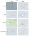
Immunohistochemistry (IHC) of RUNX-2 osteoblastic marker. Expression of RUNX-2 protein in scaffold-embedded hMSSM-derived cells from different implants after 2 or 4 weeks. Asterisks () represent the new bone formation while solid arrows (→) correspond to the positively stained osteoblast cells.
Figure 6
Figure 6
Immunohistochemistry (IHC) of OCN osteoblastic marker. Expression of OCN protein in scaffold-embedded hMSSM-derived cells from different implants after 2 or 4 weeks. Staining showed a very clear differential expression of OCN in osteocytes, osteoblasts, hMSSM-derived cells, and bone matrix. Interestingly, OCN was more abundant, at both time points, in gelatin scaffold-embedded cells than the other scaffolds (collagen, HA/βTCP, and fibrin/thrombin). Asterisks () represent the new bone matrix formation and regular arrows (→) correspond to the positively stained osteoblast cells while bold arrows (•→) correspond to the positively stained osteocytes.
Figure 7
Figure 7
Immunohistochemistry (IHC) of vimentin osteoblastic marker. Expression of vimentin protein in scaffold-embedded hMSSM-derived cells from different implants after 2 or 4 weeks. Staining showed a very clear differential expression of vimentin in osteocytes, osteoblasts, hMSSM-derived cells, and bone matrix. Interestingly, vimentin was more abundant, at both time points, in gelatin scaffold-embedded cells than the other scaffolds (collagen, HA/βTCP, and fibrin/thrombin). Asterisks () represent the new bone matrix formation and solid short arrows correspond to the positively stained osteoblast cells while solid long arrows correspond to the positively stained osteocytes.

References

    1. Myeroff C., Archdeacon M. Autogenous bone graft: donor sites and techniques. The Journal of Bone and Joint Surgery-American Volume. 2011;93(23):2227–2236. doi: 10.2106/JBJS.J.01513. - DOI - PubMed
    1. Ebraheim N. A., Elgafy H., Xu R. Bone-graft harvesting from iliac and fibular donor sites: techniques and complications. Journal of the American Academy of Orthopaedic Surgeons. 2001;9(3):210–218. doi: 10.5435/00124635-200105000-00007. - DOI - PubMed
    1. Joyce M. J. Safety and FDA regulations for musculoskeletal allografts: perspective of an orthopaedic surgeon. Clinical Orthopaedics and Related Research. 2005;435:22–30. doi: 10.1097/01.blo.0000165849.32661.5e. - DOI - PubMed
    1. Amini A. R., Laurencin C. T., Nukavarapu S. P. Bone tissue engineering: recent advances and challenges. Critical Reviews in Biomedical Engineering. 2012;40(5):363–408. doi: 10.1615/CritRevBiomedEng.v40.i5.10. - DOI - PMC - PubMed
    1. Roseti L., Parisi V., Petretta M., et al. Scaffolds for bone tissue engineering: state of the art and new perspectives. Materials Science and Engineering: C. 2017;78:1246–1262. doi: 10.1016/J.MSEC.2017.05.017. - DOI - PubMed

Associated data