. 2019 Sep 29:2019:2160642.
doi: 10.1155/2019/2160642. eCollection 2019.

SOD2 Mediates Curcumin-Induced Protection against Oxygen-Glucose Deprivation/Reoxygenation Injury in HT22 Cells

Affiliations

SOD2 Mediates Curcumin-Induced Protection against Oxygen-Glucose Deprivation/Reoxygenation Injury in HT22 Cells

Yuqing Wang et al. Evid Based Complement Alternat Med. .

Abstract

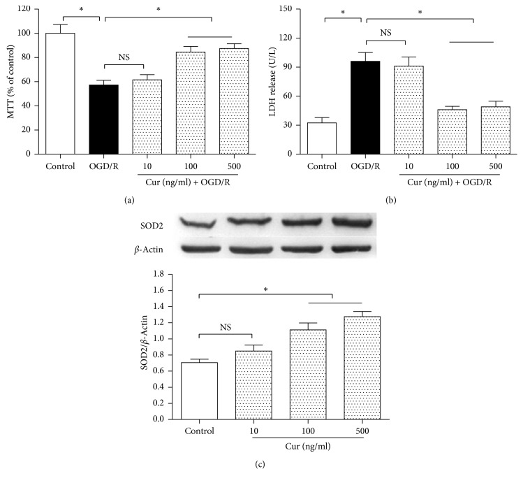
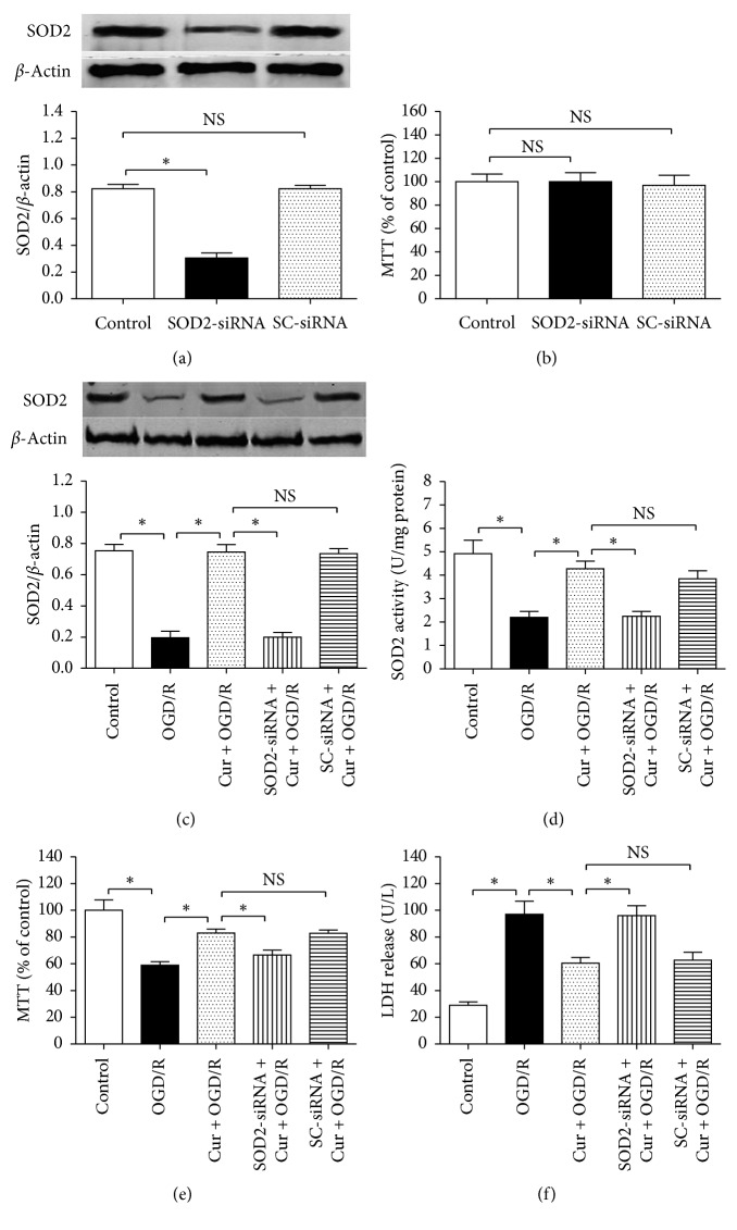
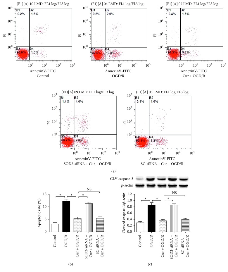
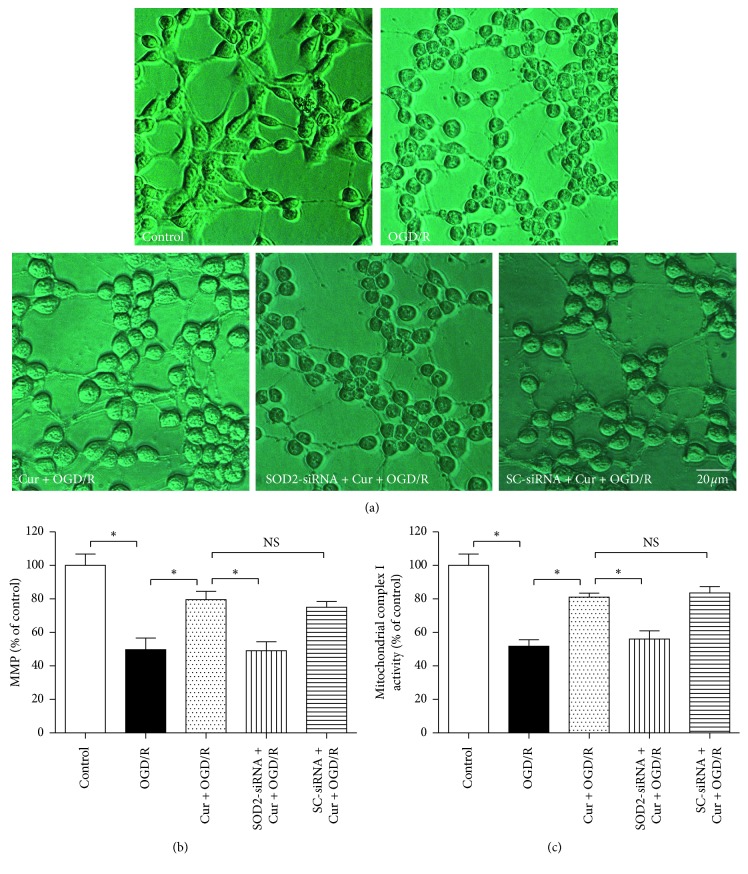
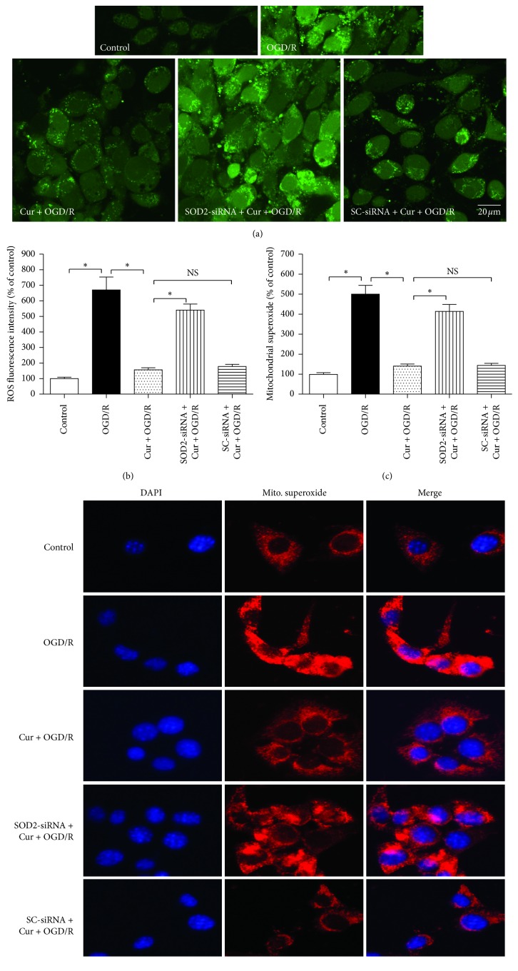
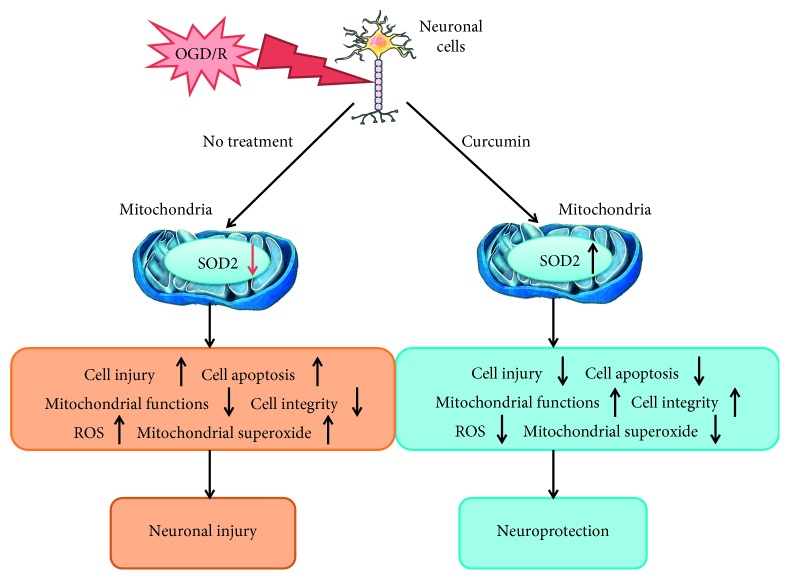
Curcumin (Cur) induces neuroprotection against brain ischemic injury; however, the mechanism is still obscure. The aim of this study is to explore the potential neuroprotective mechanism of curcumin against oxygen-glucose deprivation/reoxygenation (OGD/R) injury in HT22 cells and investigate whether type-2 superoxide dismutase (SOD2) is involved in the curcumin-induced protection. In the present study, HT22 neuronal cells were treated with 3 h OGD plus 24 h reoxygenation to mimic ischemia/reperfusion injury. Compared with the normal cultured control group, OGD/R treatment reduced cell viability and SOD2 expression, decreased mitochondrial membrane potential (MMP) and mitochondrial complex I activity, damaged cell morphology, and increased lactic dehydrogenase (LDH) release, cell apoptosis, intracellular reactive oxygen species (ROS), and mitochondrial superoxide (P < 0.05). Meanwhile, coadministration of 100 ng/ml curcumin reduced the cell injury and apoptosis, inhibited intracellular ROS and mitochondrial superoxide accumulation, and ameliorated intracellular SOD2, cell morphology, MMP, and mitochondrial complex I activity. Downregulating the SOD2 expression by using siRNA, however, significantly reversed the curcumin-induced cytoprotection (P < 0.05). These findings indicated that curcumin induces protection against OGD/R injury in HT22 cells, and SOD2 protein may mediate the protection.


Conflict of interest statement

The authors declare no conflicts of interest.

Figures

Figure 1
Figure 1
Experimental protocol diagram. (a) Searching a suitable curcumin concentration. The cells were divided into 5 groups, including the normal cultured control group, oxygen glucose 3 h OGD plus 24 reoxygenation treatment group (OGD/R), and 3 concentrations of curcumin treatment groups. After the treatments, cell viability and LDH release were assessed. (b) Observing curcumin-induced effect on SOD2 protein expression. The cells were divided into 4 groups, including normal cultured control group and 3 concentrations of curcumin treatment groups; after 3 h treatment, SOD2 expression was assessed by using western blot analysis. (c) Evaluating interfering effect of SOD2-siRNA. The cells were divided into 3 groups, including control group, SOD2-siRNA treatment group, and scrambled (SC)-siRNA treatment group. After the treatments, SOD2 protein expression was evaluated by using western blot analysis. (d) Exploring the role of SOD2 in curcumin-induced protection in HT22 cells. The cells were divided into 5 groups, including control, OGD/R treatment group, Cur + OGD/R group, SOD2-siRNA + Cur + OGD/R group, and SC-siRNA + Cur + OGD/R group; after the treatments, cell injury, apoptosis, SOD2 expression, cell morphology, intracellular ROS, mitochondrial functions, and superoxide were assessed.
Figure 2
Figure 2
Curcumin decreased cell injury in HT22 cells exposed to OGD/R and upregulated SOD2 expression in normal condition. The HT22 cells were divided into 5 groups, including control, OGD/R, and 3 concentrations (10 ng/ml, 100 ng/ml, and 500 ng/ml) of curcumin plus OGD/R groups. After the treatments, cell viability and LDH release were measured by using the MTT method and reagent kit, respectively. Then, the cells were divided into 4 groups, including control and 3 concentrations (10 ng/ml, 100 ng/ml, and 500 ng/ml) of curcumin treatment groups; after 3 h exposure, western blot was performed to assess SOD2 expression. (a) Curcumin restored cell viability (n = 8). (b) Curcumin reduced LDH release (n = 8). (c) Curcumin increased SOD2 expression (n = 4). Results are expressed as means ± SD. P < 0.05; NS: no significance.
Figure 3
Figure 3
SOD2-siRNA reversed curcumin-induced cytoprotection and SOD2 upregulation in HT22 cells exposed to OGD/R. The cells were divided into 3 groups, including control, SOD2-siRNA, and scrambled (SC)-siRNA; after 6 h incubation, western blot and MTT assay were taken to assess SOD2 expression and cell viability, respectively. Then, the cells were divided into 5 groups, including control, OGD/R, Cur + OGD/R, SOD2-siRNA + Cur + OGD/R, and SC-siRNA + Cur + OGD/R; after 3 h OGD plus 24 h reoxygenation, SOD2 expression and activity, cell viability, and lactic dehydrogenase (LDH) were assessed. (a) SOD2-siRNA inhibited SOD2 protein expression (n = 4). (b) Either SOD2-siRNA or SC-siRNA induced no obvious cytotoxicity (n = 8). (c, d) SOD2-siRNA reversed curcumin-induced effects on SOD2 expression (n = 4) and activity (n = 8). (e) SOD2-siRNA reversed curcumin-induced cell viability restoration (n = 8). (f) SOD2-siRNA reversed curcumin-induced LDH release decrease (n = 8). Results are expressed as means ± SD. P < 0.05; NS: no significance.
Figure 4
Figure 4
SOD2-siRNA reversed curcumin-induced antiapoptotic effects in HT22 cells exposed to OGD/R. The cells were divided into 5 groups, including control, OGD/R, Cur + OGD/R, SOD2-siRNA + Cur + OGD/R, and SC-siRNA + Cur + OGD/R; after 3 h OGD and 24 h reoxygenation, cell apoptotic rate and cleaved caspase-3 expression were evaluated by using flow cytometry and western blot, respectively. (a) Flow cytometry results of cells. (b) SOD2-siRNA reversed curcumin-induced antiapoptotic effect (n = 6). (c) SOD2-siRNA reversed curcumin-induced downregulation of cleaved caspase-3 expression (n = 4). Results are expressed as means ± SD. P < 0.05; NS: no significance.
Figure 5
Figure 5
SOD2-siRNA reversed curcumin-induced ameliorations of cell morphology and mitochondrial functions in HT22 cells exposed to OGD/R. The cells were divided into 5 groups, including control, OGD/R, Cur + OGD/R, SOD2-siRNA + Cur + OGD/R, and SC-siRNA + Cur + OGD/R; after 3 h OGD and 24 h reoxygenation, cell morphology and mitochondrial functions were evaluated. (a) SOD2-siRNA reversed curcumin-induced cell morphology amelioration. (b) SOD2-siRNA reversed curcumin-induced amelioration of mitochondrial membrane potential (MMP) (n = 8). (c) SOD2-siRNA reversed curcumin-induced amelioration of mitochondrial complex I activity (n = 8). Results are expressed as means ± SD. P < 0.05; NS: no significance; Bar = 20 μm.
Figure 6
Figure 6
SOD2-siRNA reversed curcumin-induced reduction of intracellular ROS and mitochondrial superoxide in HT22 cells exposed to OGD/R. The cells were divided into 5 groups, including control, OGD/R, Cur + OGD/R, SOD2-siRNA + Cur + OGD/R, and SC-siRNA + Cur + OGD/R; after 3 h OGD and 24 h reoxygenation, intracellular ROS and mitochondrial superoxide were evaluated. (a) Intracellular ROS fluorescence staining results. (b) SOD2-siRNA reversed curcumin-induced intracellular ROS reduction (n = 8). (c) SOD2-siRNA reversed curcumin-induced mitochondrial superoxide reduction (n = 8). (d) Mitochondrial superoxide fluorescence staining results. Results are expressed as means ± SD. P < 0.05; NS: no significance; Bar = 20 μm.
Figure 7
Figure 7
Curcumin induces neuroprotection against OGD/R in neuronal cells via upregulating SOD2 protein. Oxygen-glucose deprivation and reoxygenation (OGD/R) injury can downregulate SOD2 expression, increase intracellular ROS and mitochondrial superoxide accumulations, then damage neuronal cells, increase cell apoptosis, cause mitochondrial dysfunctions, and undermine cell integrity, leading to neuronal injury ultimately. Coadministration of curcumin, however, could upregulate SOD2 expression, reduce intracellular ROS and mitochondrial superoxide accumulations, and ameliorate mitochondrial functions and cell integrity, causing neuroprotection.

Similar articles

Cited by

References

    1. Anderson C. S., Huang Y., Lindley R. I., et al. Intensive blood pressure reduction with intravenous thrombolysis therapy for acute ischaemic stroke (ENCHANTED): an international, randomised, open-label, blinded-endpoint, phase 3 trial. Lancet. 2019;393(10174):877–888. doi: 10.1016/S0140-6736(19)30038-8. - DOI - PubMed
    1. Ma L., Niu W., Yang S., et al. Inhibition of mitochondrial permeability transition pore opening contributes to cannabinoid type 1 receptor agonist ACEA-induced neuroprotection. Neuropharmacology. 2018;135:211–222. doi: 10.1016/j.neuropharm.2018.03.024. - DOI - PubMed
    1. Snow S. J. Stroke and t-PA-triggering new paradigms of care. New England Journal of Medicine. 2016;374(9):809–811. doi: 10.1056/nejmp1514696. - DOI - PMC - PubMed
    1. Murakami K., Suzuki M., Suzuki N., Hamajo K., Tsukamoto T., Shimojo M. Cerebroprotective effects of TAK-937, a novel cannabinoid receptor agonist, in permanent and thrombotic focal cerebral ischemia in rats: therapeutic time window, combination with t-PA and efficacy in aged rats. Brain Research. 2013;1526:84–93. doi: 10.1016/j.brainres.2013.06.014. - DOI - PubMed
    1. Mukherjee A., Sarkar S., Jana S., Swarnakar S., Das N. Neuro-protective role of nanocapsulated curcumin against cerebral ischemia-reperfusion induced oxidative injury. Brain Research. 2019;1704:164–173. doi: 10.1016/j.brainres.2018.10.016. - DOI - PubMed

LinkOut - more resources