. 2019 Sep 5:2019:6316231.
doi: 10.1155/2019/6316231. eCollection 2019.

GC-MS Analysis and Inhibitory Evaluation of Terminalia catappa Leaf Extracts on Major Enzymes Linked to Diabetes

Affiliations

GC-MS Analysis and Inhibitory Evaluation of Terminalia catappa Leaf Extracts on Major Enzymes Linked to Diabetes

Franklyn Nonso Iheagwam et al. Evid Based Complement Alternat Med. .

Abstract

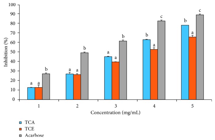
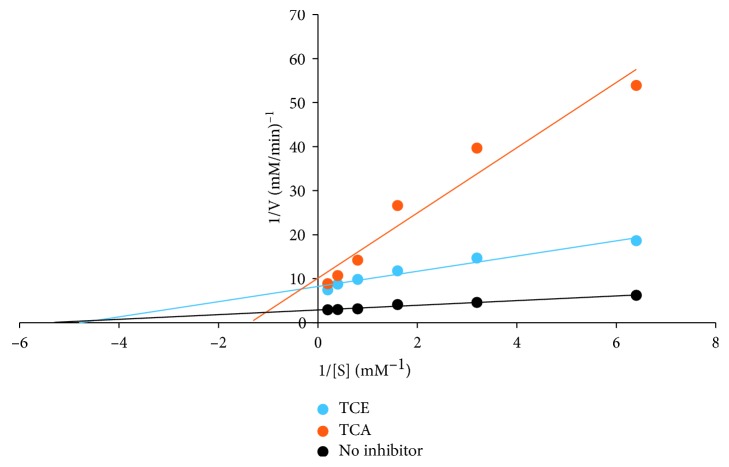
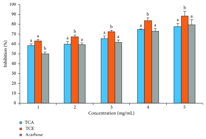
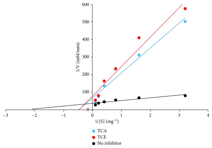
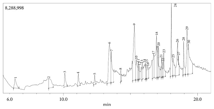
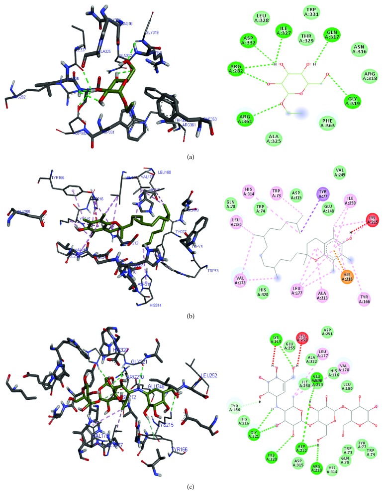
Terminalia catappa leaves are used in managing both diabetes mellitus and its complications in Southwest Nigeria. However, its inhibitory activity on enzymes implicated in diabetes is not very clear. This study investigated the in vitro inhibitory properties and mode of inhibition of T. catappa leaf extracts on enzymes associated with diabetes. The study also identified some bioactive compounds as well as their molecular interaction in the binding pocket of these enzymes. Standard enzyme inhibition and kinetics assays were performed to determine the inhibitory effects of aqueous extract (TCA) and ethanol extract (TCE) of T. catappa leaves on α-glucosidase and α-amylase activities. The phytoconstituents of TCA and TCE were determined using GC-MS. Molecular docking of the phytocompounds was performed using Autodock Vina. TCA and TCE were the most potent inhibitors of α-glucosidase (IC50 = 3.28 ± 0.47 mg/mL) and α-amylase (IC50 = 0.24 ± 0.08 mg/mL), respectively. Both extracts displayed a mixed mode of inhibition on α-amylase activity, while mixed and noncompetitive modes of inhibition were demonstrated by TCA and TCE, respectively, on α-glucosidase activity. The GC-MS analytic chromatogram revealed the presence of 24 and 22 compounds in TCE and TCA, respectively, which were identified mainly as phenolic compounds, terpenes/terpenoids, fatty acids, and other phytochemicals. The selected compounds exhibited favourable interactions with the enzymes compared with acarbose. Overall, the inhibitory effect of T. catappa on α-amylase and α-glucosidase may be ascribed to the synergistic action of its rich phenolic and terpene composition giving credence to the hypoglycaemic nature of T. catappa leaves.


Conflict of interest statement

The authors declare that there are no conflicts of interest regarding the publication of this paper.

Figures

Figure 1
Figure 1
T. catappa leaf extract inhibitory effect on α-glucosidase activity. Bars are expressed as means ± SD of triplicate determinations. Bars with different superscripts on each concentration denote significant difference (p < 0.05).
Figure 2
Figure 2
T. catappa leaf extract mode of inhibition on α-glucosidase activity.
Figure 3
Figure 3
T. catappa leaf extract inhibitory effect on α-amylase activity. Bars are expressed as means ± SD of triplicate determinations. Bars with different superscripts on each concentration denote significant difference (p < 0.05).
Figure 4
Figure 4
T. catappa leaf extract mode of inhibition on α-amylase activity.
Figure 5
Figure 5
GC chromatogram of T. catappa ethanolic leaf extract.
Figure 6
Figure 6
GC chromatogram of T. catappa aqueous leaf extract.
Figure 7
Figure 7
Binding of ligands in the active and allosteric pockets of (a) α-glucosidase and (b) α-amylase. The ligands ethyl-α-D-glucopyranoside, vitamin E, n-hexadecanoic acid, phytol, and acarbose were colour coded as black, blue, purple, green, and red, respectively.
Figure 8
Figure 8
3D and 2D diagram of (a) ethyl-α-D-glucopyranoside, (b) vitamin E, and (c) acarbose in their α-amylase binding pocket using Autodock Vina. Green and blue broken lines represent conventional and carbon-hydrogen bonds, respectively; magenta, purple, and orange represent π bonds, while red broken lines represent unfavourable bonds.
Figure 9
Figure 9
3D and 2D diagram of (a) ethyl-α-D-glucopyranoside, (b) n-hexadecanoic acid, (c) phytol, (d) vitamin E, and (e) acarbose in their α-glucosidase binding pocket using Autodock Vina. Green and blue broken lines represent conventional and carbon-hydrogen bonds, respectively, while magenta and red broken lines represent p and unfavourable bonds.

Similar articles

Cited by

References

    1. International Diabetes Federation. IDF Diabetes Atlas. 8th. Brussels, Belgium: International Diabetes Federation; 2017. http://www.diabetesatlas.org. - PubMed
    1. Broadgate S., Kiire C., Halford S., Chong V. Diabetic macular oedema: under-represented in the genetic analysis of diabetic retinopathy. Acta Ophthalmologica. 2018;96:1–51. doi: 10.1111/aos.13678. - DOI - PubMed
    1. Holman N., Young B., Gadsby R. Current prevalence of type 1 and type 2 diabetes in adults and children in the UK. Diabetic Medicine. 2015;32(9):1119–1120. doi: 10.1111/dme.12791. - DOI - PubMed
    1. Akanji M. A., Olukolu S. O., Kazeem M. I. Leaf extracts of aerva lanata inhibit the activities of type 2 diabetes-related enzymes and possess antioxidant properties. Oxidative Medicine and Cellular Longevity. 2018;2018:7. doi: 10.1155/2018/3439048.3439048 - DOI - PMC - PubMed
    1. Katiki L. M., Gomes A. C. P., Barbieri A. M. E., et al. Terminalia catappa: chemical composition, in vitro and in vivo effects on Haemonchus contortus. Veterinary Parasitology. 2017;246:118–123. doi: 10.1016/j.vetpar.2017.09.006. - DOI - PubMed

LinkOut - more resources