

Endoscopic Mucosal Resection of a Proximal Esophageal Pyogenic Granuloma
- PMID: 31662914
- PMCID: PMC6791219
- DOI: 10.1155/2019/9869274
Endoscopic Mucosal Resection of a Proximal Esophageal Pyogenic Granuloma
Abstract
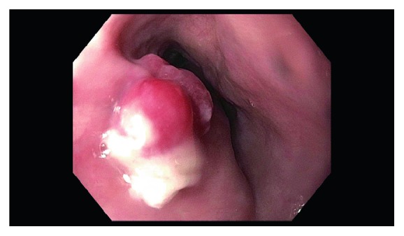
Pyogenic Granuloma (PG), also known as lobular capillary hemangioma, is usually seen as a polypoid red lesion found on the skin or the mucosal surface of the oral cavity. PG of the gastrointestinal tract is rare, in particular involving the esophagus, only 14 cases have been reported in the English literature. We present an 80-year-old male who underwent endoscopy for evaluation of dysphagia and was found to have a single, red, bilobed 10 mm polyp with adherent white exudate approximately 19 cm from the incisors. Endoscopic ultrasound was performed with a 20 mHz miniprobe which showed the lesion contained to the mucosal layer with no muscularis propria invasion. A decision was made to perform endoscopic mucosal resection (EMR). A mixture of saline and methylene blue was injected into the submucosal plane to raise the lesion with subsequent successful mucosal hot snare resection. The resection defect was then approximated and closed with a hemostatic clip to prevent bleeding. Pathology of the specimen revealed small capillary vessels growing in a lobular architecture with an edematous stroma and a florid inflammatory infiltrate representing a pyogenic granuloma. EMR allows for an en bloc resection of mucosal lesions with tumor-free margins, thereby providing both diagnostic and prognostic information. Comparing EMR with the novel technique of endoscopic submucosal dissection (ESD), the incidence of bleeding and perforation is much lower; making EMR the best and safest resection option for this rare hemangioma. In this case, we demonstrate that EMR is a safe technique in removing a pyogenic granuloma in the esophagus.
Copyright © 2019 Elias Estifan et al.
Conflict of interest statement
The authors declare that they have no conflicts of interest.
Figures


References
-
- Suarez-Zamora D. A., Rodriguez-Urrego P. A., Solano-Mariño J., Sierra-Arango F., Palau-Lazaro M. A. Esophageal pyogenic granuloma: a case report and review of the literature. International Journal of Surgical Pathology. 2018;26(8):735–738. - PubMed
-
- Poncet A. D. L. Botryomycose humaine. Rev Chir. 1897;18:996–1003.
-
- Hartzell M. B. Granuloma pyogenicus. Journal of Cutaneous Diseases. 1904;22:520–523.
-
- Bhaskar S. N., Jacoway J. R. Pyogenic granuloma–clinical features, incidence, histology, and result of treatment: report of 242 cases. Journal Oral Surgery. 1966;24(5):391–398. - PubMed
Publication types
LinkOut - more resources
Full Text Sources
Research Materials
Miscellaneous
