. 2019 Sep 22:2019:5423703.
doi: 10.1155/2019/5423703. eCollection 2019.

Chlorogenic Acid Attenuates Kidney Ischemic/Reperfusion Injury via Reducing Inflammation, Tubular Injury, and Myofibroblast Formation

Affiliations

Chlorogenic Acid Attenuates Kidney Ischemic/Reperfusion Injury via Reducing Inflammation, Tubular Injury, and Myofibroblast Formation

Nur Arfian et al. Biomed Res Int. .

Abstract

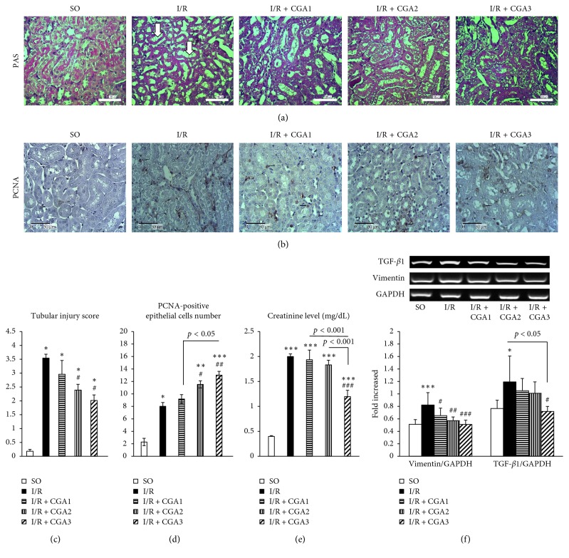
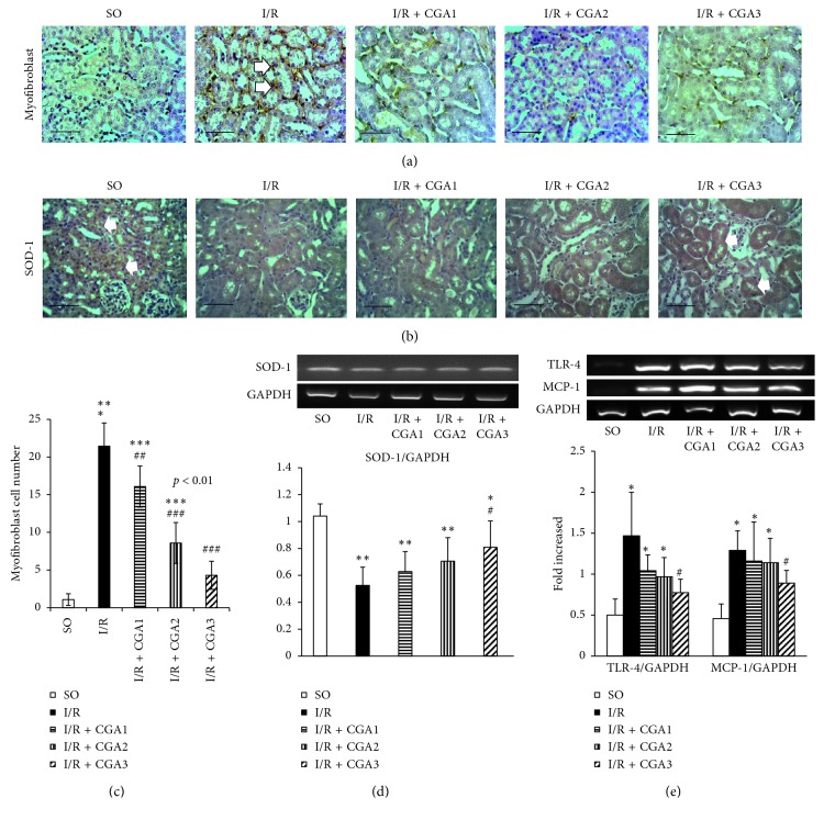
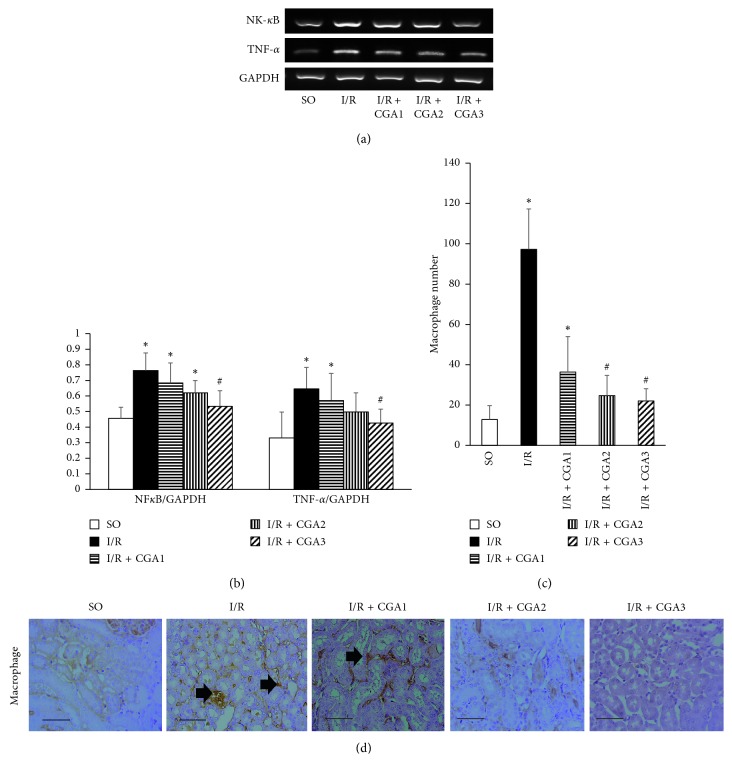
Kidney ischemic/reperfusion (I/R) injury is the main cause of acute kidney injury (AKI) involving renal function deterioration, renal architecture damage, and inflammation. This condition may lead to kidney fibrosis with epithelial to mesenchymal transition (EMT) and myofibroblast formation. Inhibition of chronic effects of kidney I/R injury may provide effective strategies for treating AKI and chronic kidney diseases (CKDs). Chlorogenic acid (CGA) is recognized as a powerful antioxidant, with anti-inflammatory and antifibrotic properties in many conditions. However, the effect of CGA on kidney I/R injury has not been elucidated yet. Kidney I/R injury was performed on male Swiss background mice (I/R group, n = 5, 3-4 months, 30-40 g) which underwent bilateral renal pedicles clamping for 30 minutes and then were euthanized on day three after operation. Three groups of I/R were treated with 3 different doses of CGA intraperitoneally for 2 days: 3.5 (I/R + CGA1 group), 7 (I/R + CGA2 group), and 14 (I/R + CGA3 group) mg/kg of body weight. Tubular injury was quantified based on Periodic Acid-Schiff staining, while reverse transcriptase PCR (RT-PCR) was performed to quantify mRNA expression of TGF-β1, vimentin, SOD-1, TLR-4, TNF-α, NF-κB and MCP-1. Immunohistochemical staining was done to quantify proliferating cell nuclear antigen (PCNA), myofibroblast (α-SMA), SOD-1 and macrophage (CD68) number. Kidney I/R demonstrated tubular injury and increased inflammatory mediator expression, macrophage number, and myofibroblast expansion. Meanwhile, histological analysis showed lower tubular injury with higher epithelial cell proliferation in CGA-treated groups compared to the I/R group. RT-PCR also revealed significantly lower TGF-β1 and vimentin mRNA expressions with higher SOD-1 mRNA expression. CGA-treated groups also demonstrated a significantly lower macrophage and myofibroblast number compared to the I/R group. These findings associated with lower mRNA expression of TLR-4, TNF-α, NF-κB, and MCP-1 as inflammatory mediators in CGA groups. I/R + CGA3 represented the highest amelioration effect among other CGA-treated groups. CGA treatment attenuates kidney I/R injury through reducing inflammation, decreasing myofibroblast expansion, and inducing epithelial cells proliferation.


Conflict of interest statement

The authors declare that they have no conflicts of interest.

Figures

Figure 1
Figure 1
(a) Representative pictures of PAS staining in all groups. The SO group had normal histology of kidney tissue with brush border. The IR group had tubular injury characteristics with dilatation and brush-border loss (white arrow). (b) Immunostaining of PCNA showed positive staining in epithelial cells' nuclei (black arrows). (c, d). Quantification of tubular injury score and PCNA-positive cell number showed attenuation of tubular injury which associated with the increased number of proliferating epithelial cells in CGA-treated groups (especially IR + CGA3 group). (e) Creatinine level measurement showed reduction in the IR + CGA3 group. (f) Reverse transcriptase-PCR (RT-PCR) analysis of vimentin (mesenchymal marker) and TGF-β1 (profibrotic factor). The IR + CGA3 group demonstrated significantly lower vimentin and TGF-β1 mRNA expressions with the IR group. p < 0.05 vs. SO group; ∗∗p < 0.01 vs. IR group; ∗∗∗p < 0.001 vs. SO group. #p < 0.05 vs. IR group; ##p < 0.01 vs. IR group; ###p < 0.001 vs. IR group.
Figure 2
Figure 2
(a) Representative pictures of α-SMA immunostaining for myofibroblast marker. Positive staining is shown in interstitial areas (white arrow). (b) Representative pictures of SOD-1 immunostaining. Positive staining is shown in epithelial cells in SO, meanwhile reduction in the IR group. The IR + CGA3 group demonstrated positive staining in epithelial cells. (c) Quantification of the myofibroblast number showed reduction of the numbers in CGA-treated groups, especially the IR + CGA3 group. (d) Reverse transcriptase-PCR (RT-PCR) analysis of SOD-1. The IR + CGA3 group demonstrated significantly higher SOD-1 mRNA expression compared to the IR group. (e) Reverse transcriptase-PCR (RT-PCR) analysis of inflammatory markers (TLR-4 and MCP-1). The IR + CGA3 group demonstrated significantly lower MCP-1 and TLR-4 mRNA expressions with the IR group. p < 0.05 vs. SO group; ∗∗p < 0.01 vs. SO group; ∗∗∗p < 0.001 vs. SO group. #p < 0.05 vs. IR group; ##p < 0.01 vs. IR group; ###p < 0.001 vs. IR group.
Figure 3
Figure 3
(a, b) Reverse transcriptase-PCR (RT-PCR) analysis of NF-κB and TNF-α mRNA expressions. The IR + CGA3 group demonstrated significantly lower NF-κB and TNF-α mRNA expressions compared to the IR group. (b) Representative pictures of CD68 immunostaining for macrophage marker. Positive staining is shown in interstitial areas (black arrow). (c) Quantification of the macrophage number showed reduction of the numbers in CGA-treated groups, especially the IR + CGA3 group. (d) Representative pictures of CD68 immunostaining for demonstrating macrophage infiltration (black arrows). p < 0.05 vs. SO group. #p < 0.05 vs. IR group.

References

    1. Hsu C.-Y., McCulloch C. E., Fan D., Ordoñez J. D., Chertow G. M., Go A. S. Community-based incidence of acute renal failure. Kidney International. 2007;72(2):208–212. doi: 10.1038/sj.ki.5002297. - DOI - PMC - PubMed
    1. Waikar S. S., Liu K. D., Chertow G. M. Diagnosis, epidemiology and outcomes of acute kidney injury. Clinical Journal of the American Society of Nephrology. 2008;3(3):844–861. doi: 10.2215/cjn.05191107. - DOI - PubMed
    1. Cerdá J., Lameire N., Eggers P., et al. Epidemiology of acute kidney injury. Clinical Journal of the American Society of Nephrology. 2008;3(3):881–886. doi: 10.2215/cjn.04961107. - DOI - PubMed
    1. Kezić A., Stajic N., Thaiss F. Innate immune response in kidney ischemia/reperfusion injury: potential target for therapy. Journal of Immunology Research. 2017;2017:10. doi: 10.1155/2017/6305439.6305439 - DOI - PMC - PubMed
    1. Bonventre J. V., Yang L. Cellular pathophysiology of ischemic acute kidney injury. Journal of Clinical Investigation. 2011;121(11):4210–4221. doi: 10.1172/jci45161. - DOI - PMC - PubMed

MeSH terms