. 2019 Oct 16;5(10):eaax8855.
doi: 10.1126/sciadv.aax8855. eCollection 2019 Oct.

Cryo-electron microscopy structure and analysis of the P-Rex1-Gβγ signaling scaffold

Affiliations

Cryo-electron microscopy structure and analysis of the P-Rex1-Gβγ signaling scaffold

Jennifer N Cash et al. Sci Adv. .

Abstract

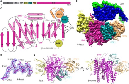
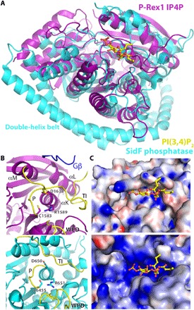
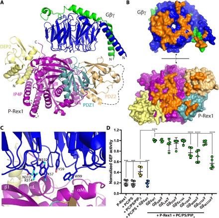
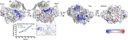
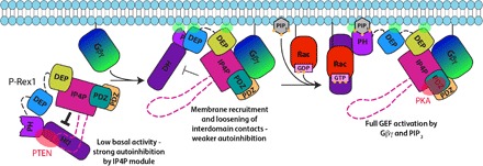
PIP3-dependent Rac exchanger 1 (P-Rex1) is activated downstream of G protein-coupled receptors to promote neutrophil migration and metastasis. The structure of more than half of the enzyme and its regulatory G protein binding site are unknown. Our 3.2 Å cryo-EM structure of the P-Rex1-Gβγ complex reveals that the carboxyl-terminal half of P-Rex1 adopts a complex fold most similar to those of Legionella phosphoinositide phosphatases. Although catalytically inert, the domain coalesces with a DEP domain and two PDZ domains to form an extensive docking site for Gβγ. Hydrogen-deuterium exchange mass spectrometry suggests that Gβγ binding induces allosteric changes in P-Rex1, but functional assays indicate that membrane localization is also required for full activation. Thus, a multidomain assembly is key to the regulation of P-Rex1 by Gβγ and the formation of a membrane-localized scaffold optimized for recruitment of other signaling proteins such as PKA and PTEN.


Figures

Fig. 1
Fig. 1. Cryo-EM structure of the P-Rex1–Gβγ complex reveals a complex Gβγ-binding module comprising four P-Rex1 domains.
(A) Domain layout of P-Rex1. The domains shown in shades of gray were conformationally heterogeneous with respect to the Gβγ-binding module. (B) Cryo-EM map of the C-terminal 1153 residues of P-Rex1 in complex with Gβγ. (C) Topology diagram of the P-Rex1 IP4P domain. The DEP2 and PDZ domains are shown as circles and adopt canonical folds. The PDZ2 domain has two extra helices (α2´ and α3) and an additional β strand (β4′, not shown). Gβγ interaction sites are indicated with black lines. Parentheses indicate the number of unmodeled residues in loops. Green circles correspond to canonical phosphatase catalytic residues. (D) Example map density and fitted model from the IP4P domain. (E) “Top” and “bottom” views of P-Rex1 relative to (B) with Gβγ removed for clarity. Dashed lines represent disordered loops.
Fig. 2
Fig. 2. Sequence alignment of P-Rex1 with its close homologs along with other annotated characteristics.
Although residues 38 to 505 of P-Rex1 were present in the protein used for cryo-EM analysis, they were not observed in the high-resolution reconstruction and are not shown here. Clustal Omega was used to align the sequences of human P-Rex1 (UniProtKB ID Q8TCU6), P-Rex2 (ID Q70Z35), and inositol 3,4-bisphosphate 4-phosphatase (INPP4A; ID Q96PE3). Residues 1 to 227 of INPP4A, corresponding to its N-terminal C2 domain, were excluded. The dots above the alignment correspond to every 10th amino acid in the P-Rex1 sequence. The secondary structure elements observed in P-Rex1 are shown above the alignment, with α helices depicted as rounded rectangles, β strands as arrows, and coil as a black line. They are colored by their corresponding domain as defined in Fig. 1. The absence of indicated secondary structure indicates that these residues were not observed in the structure. Thick red bars above the sequence correspond to P-Rex1 regions that are >90% exchanged with deuterium after 1000 s (see fig. S9 and data file S1). Thick blue bars indicate regions that are significantly stabilized (4% or greater protection at 1000 s) during HDX-MS in the presence of Gβγ (see Fig. 5 and data files S1 and S2). Residues highlighted in blue correspond to those that bury ≥5 Å2 of accessible surface area in the Gβγ complex (out of a total of 1000 Å2 buried accessible surface area on P-Rex1). Most also correspond to regions that are stabilized in HDX-MS upon complex formation. Residues highlighted in yellow correspond to canonical cysteine and arginine active site residues found in PTEN and Legionella phosphoinositide phosphatases SidF and SidP. Residues in P-Rex1 reported to be phosphorylated are highlighted in orange and are found in the more dynamic loops of the structure where protein kinases would have easier access. P-Rex2 residues that are associated with mutation in cancer patients are highlighted in green. Gβγ-binding residues are not conserved in INPP4A, and its phosphatase active site is much more basic than that of P-Rex1 in part due to the presence of two lysines in its P loop, analogous to those conserved in PTEN, SidF, and SidP, consistent with their robust phosphatase activity.
Fig. 3
Fig. 3. The P-Rex1 IP4P domain is a structural homolog of the Legionella phosphatase SidF.
(A) IP4P domain superimposed on SidF (residues 183 to 743) in complex with PI(3,4)P2. For the figure, residues 1579 to 1589 of P-Rex1 were aligned with residues 641 to 651 of SidF. (B) Residues in P-Rex1 (top) compared with catalytic residues in SidF (bottom). A likely counterpart of Asp650 in SidF is Asp1638 on the TI loop of P-Rex1. Canonical phosphatase loops are indicated in yellow. A loop from Gβ is shown to indicate its proximity. (C) Surface representation colored by electrostatic potential of the structures shown in (B).
Fig. 4
Fig. 4. The P-Rex1 IP4P domain forms an extensive docking site for Gβγ.
(A) Overview of Gβγ in complex with the P-Rex1 C-terminal Gβγ-binding module. (B) Surface representation of the complex in an “open book” view, with Gβγ and P-Rex1 peeled away from each other to visualize surfaces buried on each during complex formation (orange). (C) Close-up view of key interactions between Gβγ and the IP4P domain. (D) Liposome-based GEF assay demonstrating dependence of P-Rex1 activation on PIP3 and on Gβγ residues that interact with the IP4P domain. GEF activity in each experiment was normalized to that of WT Gβγ + PC/PS/PIP3.
Fig. 5
Fig. 5. HDX-MS suggests allosteric changes in P-Rex1 upon Gβγ binding.
Differences in HDX upon complex formation with Gβγ (at 1000 s) were plotted onto the cryo-EM structure of the P-Rex1 Gβγ-binding module. Red regions, more dynamic behavior upon Gβγ binding; blue regions, less dynamic behavior upon Gβγ binding. Graph shows a comparison of the exchange over time for the indicated structural features. Changes occur distal from the Gβγ-binding site (dashed ovals), suggesting that binding may cause allosteric changes in P-Rex1. These experiments were performed twice, and the data shown represent the average of two experiments. See also data files S1 and S2.
Fig. 6
Fig. 6. Allosteric activation model for P-Rex1.
Our cryo-EM data (fig. S4B) and HDX-MS data (Fig. 5) suggest that the DH/PH/DEP1 domains could interact with the C-terminal Gβγ-binding module, perhaps through the unanticipated domain found within the loops of the IP4P domain (pink dashed line). This low-activity, autoinhibited form is predicted to have weak affinity for the plasma membrane. Gβγ localizes P-Rex1 to the cell membrane and allosterically loosens the autoinhibitory interdomain contacts. Binding to PIP3 results in complete activation and provides full substrate access to the RhoGEF active site through an undetermined mechanism. There are multiple points of contact of this complex with the cell membrane, through either lipid modifications (solid black lines) or basic patches on P-Rex1 domains (green transparent ovals). The quaternary arrangement of domains in P-Rex1 is thought to be important for scaffolding interactions with other signaling proteins such as PTEN (specific for P-Rex2) and PKA (red transparent ovals), although these proteins may, in fact, bind P-Rex either at the cell membrane or in the cytoplasm. Dashed lines indicate domains (pink) or flexible linker regions (black) that have not been observed in this or previous structures.

References

    1. Wong C.-Y. A., Wuriyanghan H., Xie Y., Lin M.-F., Abel P. W., Tu Y., Epigenetic regulation of phosphatidylinositol 3,4,5-triphosphate-dependent Rac exchanger 1 gene expression in prostate cancer cells. J. Biol. Chem. 286, 25813–25822 (2011). - PMC - PubMed
    1. Montero J. C., Seoane S., Ocaña A., Pandiella A., P-Rex1 participates in Neuregulin-ErbB signal transduction and its expression correlates with patient outcome in breast cancer. Oncogene 30, 1059–1071 (2011). - PubMed
    1. Lindsay C. R., Lawn S., Campbell A. D., Faller W. J., Rambow F., Mort R. L., Timpson P., Li A., Cammareri P., Ridgway R. A., Morton J. P., Doyle B., Hegarty S., Rafferty M., Murphy I. G., McDermott E. W., Sheahan K., Pedone K., Finn A. J., Groben P. A., Thomas N. E., Hao H., Carson C., Norman J. C., Machesky L. M., Gallagher W. M., Jackson I. J., van Kempen L., Beermann F., der C., Larue L., Welch H. C., Ozanne B. W., Sansom O. J., P-Rex1 is required for efficient melanoblast migration and melanoma metastasis. Nat. Commun. 2, 555–559 (2011). - PMC - PubMed
    1. Sosa M. S., Lopez-Haber C., Yang C., Wang H. B., Lemmon M. A., Busillo J. M., Luo J., Benovic J. L., Klein-Szanto A., Yagi H., Gutkind J. S., Parsons R. E., Kazanietz M. G., Identification of the Rac-GEF P-Rex1 as an essential mediator of ErbB signaling in breast cancer. Mol. Cell 40, 877–892 (2010). - PMC - PubMed
    1. Qin J., Xie Y., Wang B., Hoshino M., Wolff D. W., Zhao J., Scofield M. A., Dowd F. J., Lin M. F., Tu Y., Upregulation of PIP3-dependent Rac exchanger 1 (P-Rex1) promotes prostate cancer metastasis. Oncogene 28, 1853–1863 (2009). - PMC - PubMed

Publication types

MeSH terms

Substances