. 2019 Jul 23;5(3):186-204.
doi: 10.3934/microbiol.2019.3.186. eCollection 2019.

Purification, kinetic characterization, and site-directed mutagenesis of Methanothermobacter thermautotrophicus RFAP Synthase Produced in Escherichia coli

Affiliations

Purification, kinetic characterization, and site-directed mutagenesis of Methanothermobacter thermautotrophicus RFAP Synthase Produced in Escherichia coli

Matthew E Bechard et al. AIMS Microbiol. .

Abstract

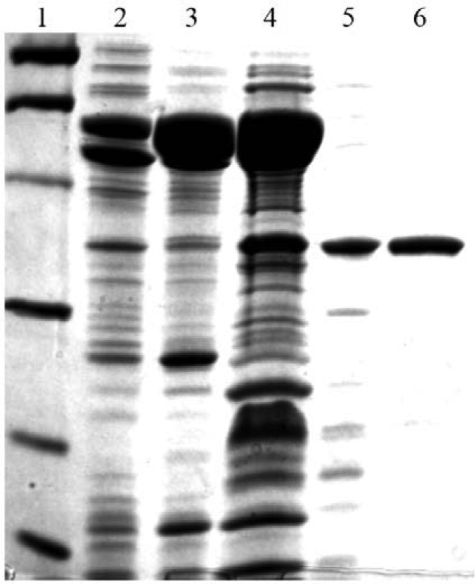
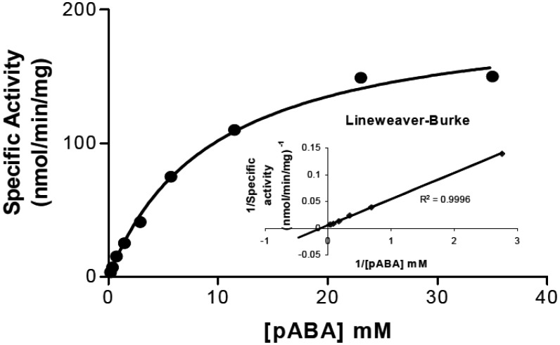
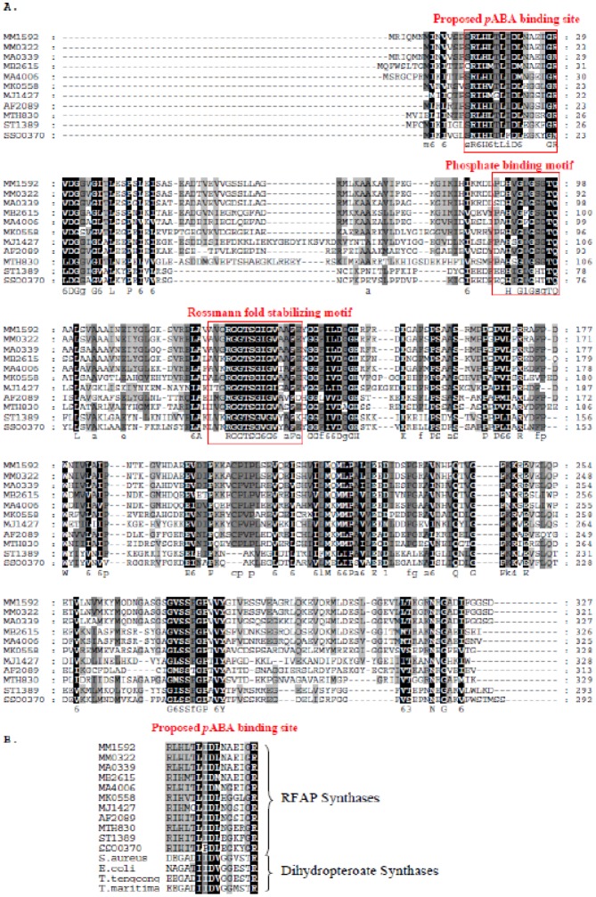
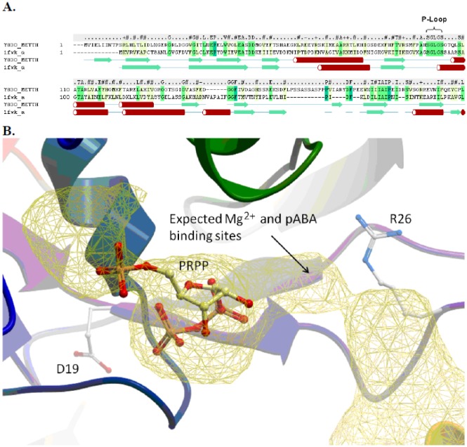
Methane-producing archaea are among a select group of microorganisms that utilize tetrahydromethanopterin (H4MPT) as a one-carbon carrier instead of tetrahydrofolate. In H4MPT biosynthesis, β-ribofuranosylaminobenzene 5'-phosphate (RFAP) synthase catalyzes the production of RFAP, CO2, and pyrophosphate from p-aminobenzoic acid (pABA) and phosphoribosyl-pyrophosphate (PRPP). In this work, to gain insight into amino acid residues required for substrate binding, RFAP synthase from Methanothermobacter thermautotrophicus was produced in Escherichia coli, and site-directed mutagenesis was used to alter arginine 26 (R26) and aspartic acid 19 (D19), located in a conserved sequence of amino acids resembling the pABA binding site of dihydropteroate synthase. Replacement of R26 with lysine increased the KM for pABA by an order of magnitude relative to wild-type enzyme without substantially altering the KM for PRPP. Although replacement of D19 with alanine produced inactive enzyme, asparagine substitution allowed retention of some activity, and the K M for pABA increased about threefold relative to wild-type enzyme. A molecular model developed by threading RFAP synthase onto the crystal structure of homoserine kinase places R26 in the proposed active site. In the static model, D19 is located close to the active site, yet appears too far away to influence ligand binding directly. This may be indicative of the protein conformational change predicted previously in the Bi-Ter kinetic mechanism and/or formation of the active site at the interface of two subunits. Due to the vital role of RFAP synthase in H4MPT biosynthesis, insights into the mode of substrate binding and mechanism could be beneficial for developing RFAP synthase inhibitors designed to reduce the production of methane as a greenhouse gas.

Keywords: RFAP synthase; methanogenesis; methanopterin; site-directed mutagenesis; substrate binding.


Conflict of interest statement

Conflict of interest: All authors declare no conflicts of interest in this paper.

Figures

Figure 1.
Figure 1.. Reaction catalyzed by RFAP synthase.
Figure 2.
Figure 2.. SDS-PAGE gel of RFAP synthase purification. Protein samples were boiled in the presence of 7.5% 2-mercaptoethanol in SDS-PAGE sample buffer and loaded onto a 12% polyacrylamide gel. The gel was stained with Coomassie Brilliant Blue R-250 (Bio-Rad). Lane 1, Molecular mass markers. Lane 2, CFE (8 µg). Lane 3, heated CFE (10.2 µg). Lane 4, hydroxyapatite fraction (17 µg). Lane 5, Mono Q fraction (2 µg). Lane 6, Superdex 75 fraction (2 µg). The bands in lane 1 indicate the positions of the following molecular mass markers: phosphorylase b (97.4 kDa), bovine serum albumin (66.2 kDa), ovalbumin (45 kDa), carbonic anhydrase (31 kDa), soybean trypsin inhibitor (21.5 kDa), and lysozyme (14.4 kDa).
Figure 3.
Figure 3.. Determination of pABA KM for recombinant RFAP synthase. RFAP synthase activity was measured at a constant PRPP concentration of 8.8 mM while varying the pABA concentration from 181 µM to 36 mM. The inset shows the Lineweaver-Burke plot derived from the Michaelis-Menton plot. The estimated KM for pABA was 4.5 mM and the Vmax at 70 °C was approximately 190 nmol/min/mg protein.
Figure 4.
Figure 4.. Bioinformatic analysis of group I RFAP synthase genes. A. Multiple amino acid sequence alignment of Group I RFAP synthases using the alignment program GeneDoc . Possible phosphate binding motifs are indicated by brackets. The protein abbreviations are as follows: Methanosarcina mazei, MM1592 and MM0322; Methanosarcina acetivorans, MA4006 and MA0339; Methanosarcina barkeri, MB2615; Methanopyrus kandleri, MK0558; Methanocaldococcus jannaschii, MJ1427; Archaeoglobus fulgidus, AF2089; Methanothermobacter thermautotrophicus, MTH0830; Sulfolobus tokodaii, ST1329; Sulfolobus sulfataricus, SSO0370. B. Multiple amino acid sequence alignment showing homology between the conserved region of RFAP synthase and a predicted pABA-binding region of dihydropteroate synthase (DHPS) from Staphylococcus aureus, Escherichia coli; Thermoanaerobacter tengcongensis, and Thermotoga maritima.
Figure 5.
Figure 5.. Computational molecular model of MTH0830 RFAP synthase. A: Secondary structure and sequence alignment of the N-terminus of MTH0830 (Y830_METTH) (from residue 1 to 215) and homoserine kinase template structure (pdb code: 1fwk) . The secondary structure elements are colored green (beta Sheet) and red (alpha helix). The secondary structure of MTH0830 was calculated using the DSSP method , and the secondary structure of 1fwk is shown as defined in the Protein Data Bank file. B: The predicted PRPP and pABA binding pocket. The MTH0830 model is displayed in ribbon colored from the N to C terminus (blue – red), residues D19 and R26 are displayed as a stick representation. The docked PRPP ligand is displayed in stick representation with carbon atoms colored yellow. The binding pocket is displayed as a yellow mesh, and the predicted sites for pABA and Mg2+ are annotated.

References

    1. White RH. Biosynthesis of methanopterin. Biochemistry. 1996;35:3447–3456. - PubMed
    1. Rasche ME, White RH. Mechanism for the enzymatic formation of 4-(beta-D-ribofuranosyl) aminobenzene 5′-phosphate during the biosynthesis of methanopterin. Biochemistry. 1998;37:11343–11351. - PubMed
    1. Scott JW, Rasche ME. Purification, overproduction, and partial characterization of beta-RFAP synthase, a key enzyme in the methanopterin biosynthesis pathway. J Bacteriol. 2002;184:4442–4448. - PMC - PubMed
    1. Leigh JA. Levels of water-soluble vitamins in methanogenic and non-methanogenic bacteria. Appl Environ Microbiol. 1983;45:800–803. - PMC - PubMed
    1. Vanbeelen P, Labro JFA, Keltjens JT, et al. Derivatives of methanopterin, a coenzyme involved in methanogenesis. Eur J Bioche. 1984;139:359–365. - PubMed

LinkOut - more resources