Review
. 2019 Nov;13(11):1089-1105.
doi: 10.1080/17474124.2019.1685871. Epub 2019 Nov 8.

Identification and management of pancreas divisum

Affiliations
Review

Identification and management of pancreas divisum

Aditya Gutta et al. Expert Rev Gastroenterol Hepatol. 2019 Nov.

Abstract

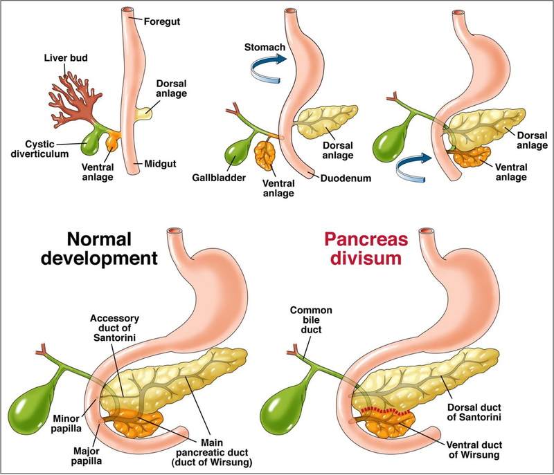
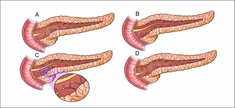
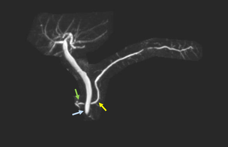
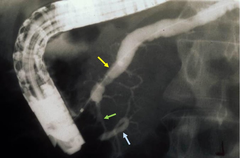
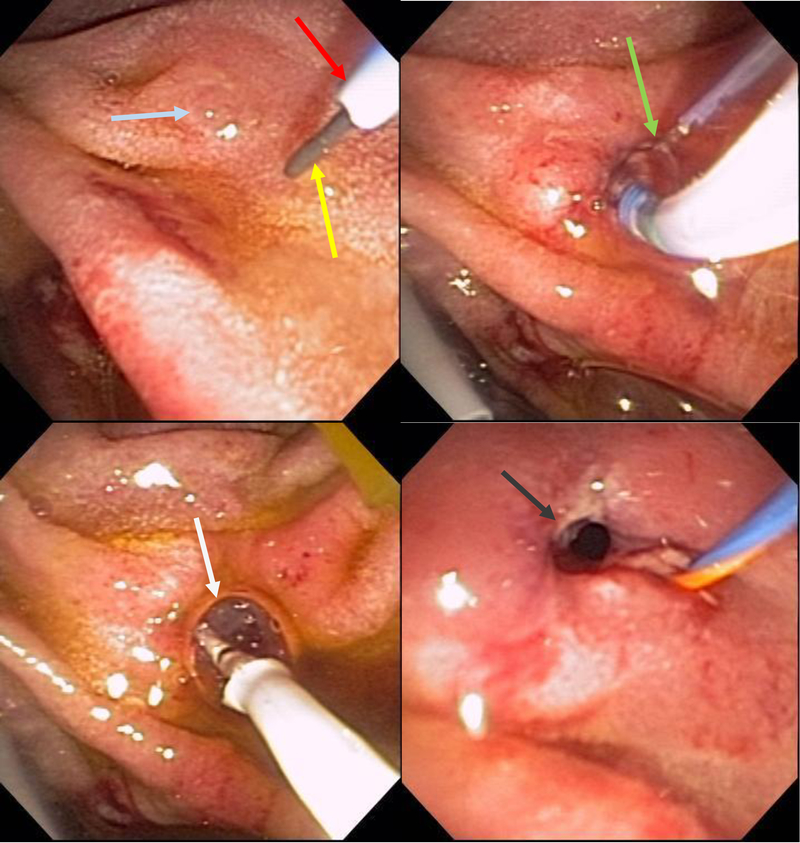
Introduction: Pancreas divisum is the most common congenital malformation of the pancreas with the majority asymptomatic. The etiological role, pathogenesis, clinical significance and management of pancreas divisum in pancreatic disease has not been clearly defined and our understanding is yet to be fully elucidated.Areas covered: This review describes the role of pancreas divisum in the development of pancreatic disease and the ambiguity related to it. In our attempt to offer clarity, a comprehensive search on PubMed, Ovid, Embase and Cochrane Library from inception to May 2019 was undertaken using key words "pancreas divisum", "idiopathic recurrent acute pancreatitis" and "chronic pancreatitis".Expert opinion: Current research fails to define a clear association between pancreas divisum and pancreatic disease. Though debatable, several studies do suggest a pathological role of pancreas divisum in pancreatic disease and a benefit of minor papilla therapy in the setting of acute recurrent pancreatitis. Surgical and endoscopic therapeutic modalities have not been directly compared. With the current data available, it would be imprudent to advise a definitive line of management for pancreatic disease associated with pancreas divisum and should involve a comprehensive discussion with the individual patient to define expectations before embarking on any medical and/or interventional therapy.

Keywords: Recurrent acute pancreatitis; chronic pancreatitis; dorsal duct syndrome; endoscopic retrograde cholangiopancreatography; idiopathic pancreatitis; magnetic resonance cholangiopancreatography; pancreas divisum.


Figures

Figure 1:
Figure 1:
Rotation of the ventral pancreatic bud/anlage (from which originates the hepatobiliary system) and fusion with the dorsal pancreatic bud/anlage in the foregut. The failure of fusion of the ductal systems of the dorsal and ventral anlage results in pancreas divisum with the majority of the pancreas draining via the dorsal duct of Santorini through the minor papilla.
Figure 2:
Figure 2:
A - Normal Pancreas; B - Classic Pancreas Divisum; C – Incomplete Pancreas Divisum (magnified image demonstrates the small communication between the dorsal and ventral pancreatic ductal systems); D – Reverse Pancreas Divisum
Figure 3:
Figure 3:
The dorsal pancreatic duct (yellow arrow) crosses the bile duct (blue arrow) and empties at a separate orifice/minor papilla (green arrow).
Figure 4:
Figure 4:
Incomplete Pancreas Divisum - Dorsal pancreatic duct (yellow arrow) is cannulated and opacified from the minor papilla with contrast passing into the ventral pancreatic duct (blue arrow) through a small communication between the two systems (green arrow).
Figure 5:
Figure 5:
This non-secretin stimulated MRCP shows an irregular dorsal main pancreatic duct (yellow arrow).
Figure 6:
Figure 6:
This secretin-stimulated MRCP shows a dilated and irregular dorsal pancreatic duct (yellow arrow) crossing the bile duct and opening separately at the minor papilla (blue arrow). Irregular side branches are also seen and are more prominent than the non-secretin enhanced image (Figure 5).
Figure 7:
Figure 7:
Placing the duodenoscope in the long position, along the greater curvature of the stomach (yellow arrow), provides the optimum angle for minor papilla cannulation and opacification of the dorsal pancreatic duct (blue arrow).
Figure 8:
Figure 8:
Duodenoscope can be reduced to a short position along the lesser curvature of the stomach (yellow arrow) once deep minor papilla cannulation is achieved to allow for improved stability to interrogate and instrument the dorsal pancreatic duct (blue arrow).
Figure 9:
Figure 9:
Minor papilla (yellow arrow) anterior and to the right of the major papilla (blue arrow)
Figure 10:
Figure 10:
Minor papilla (green arrow) identified with assistance of methylene blue and subsequently cannulated by a guide-wire (blue arrow) and a highly tapered catheter (yellow arrow). Deep cannulation of the minor papilla (grey arrow).
Figure 11:
Figure 11:
Minor papilla (green arrow) cannulated by a guide-wire (blue arrow) and a highly tapered catheter (yellow arrow).
Figure 12:
Figure 12:
Minor papilla (arrow) clearly identified after spraying duodenal mucosa with methylene blue and administration of secretin to enhance pancreatic secretion.
Figure 13:
Figure 13:
The minor papilla (yellow arrow) is easily identified after spraying the duodenal mucosa with methylene blue and administration of intravenous secretin. The pancreatic juice flow results in a zone of central clearing (white arrow).
Figure 14:
Figure 14:
Minor papilla pull-type sphincterotomy.
Figure 15:
Figure 15:
Free hand needle-knife (blue arrow) sphincterotomy (yellow arrow) of minor papilla (green arrow) followed by placement of a plastic stent (grey arrow) in the dorsal pancreatic duct.
Figure 16:
Figure 16:
Plastic stent (yellow arrow) placed in dorsal pancreatic duct after minor papilla endoscopic sphincterotomy (blue arrow). Stent in the dorsal pancreatic duct seen on fluoroscopy (green arrow)
Figure 17:
Figure 17:
Wire-guided (yellow arrow) cannulation of the minor papilla (blue arrow) with a highly tapered cannula (red arrow) followed by pull-type minor papilla sphincterotomy (green arrow) and a minor papilla endoscopic orifice dilation (white arrow). Widely open minor papilla orifice after endoscopic dilation (grey arrow).

References

    1. Kozu T, Suda K, Toki F. Pancreatic development and anatomical variation. Gastrointestinal endoscopy clinics of North America. 1995. January;5(1):1–30. - PubMed
    1. Smanio T Proposed nomenclature and classification of the human pancreatic ducts and duodenal papillae. Study based on 200 post mortems. International surgery. 1969. August;52(2):125–41. - PubMed
    1. Stimec B, Bulajic M, Korneti V, et al. Ductal morphometry of ventral pancreas in pancreas divisum. Comparison between clinical and anatomical results. The Italian journal of gastroenterology. 1996. Feb-Mar;28(2):76–80. - PubMed
    1. Bulow R, Simon P, Thiel R, et al. Anatomic variants of the pancreatic duct and their clinical relevance: an MR-guided study in the general population. European radiology. 2014. Dec;24(12):3142–9. doi: 10.1007/s00330-014-3359-7. - DOI - PubMed
    1. Bernard JP, Sahel J, Giovannini M, et al. Pancreas divisum is a probable cause of acute pancreatitis: a report of 137 cases. Pancreas. 1990. May;5(3):248–54. - PubMed

MeSH terms