. 2019 Nov 27;62(22):10391-10401.
doi: 10.1021/acs.jmedchem.9b01485. Epub 2019 Nov 11.

Validation of Human Sterol 14α-Demethylase (CYP51) Druggability: Structure-Guided Design, Synthesis, and Evaluation of Stoichiometric, Functionally Irreversible Inhibitors

Affiliations

Validation of Human Sterol 14α-Demethylase (CYP51) Druggability: Structure-Guided Design, Synthesis, and Evaluation of Stoichiometric, Functionally Irreversible Inhibitors

Laura Friggeri et al. J Med Chem. .

Abstract

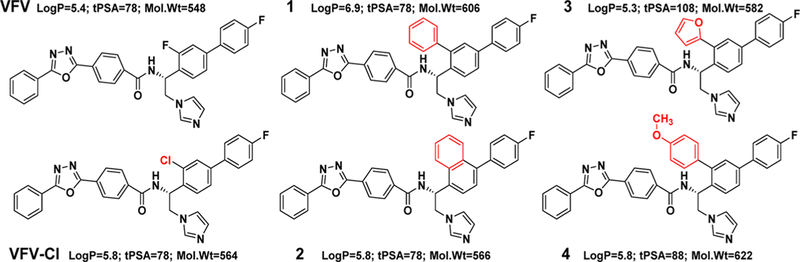
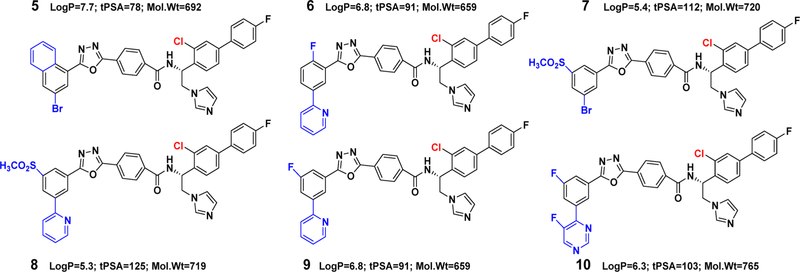
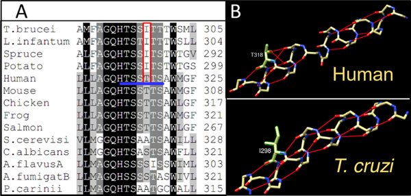
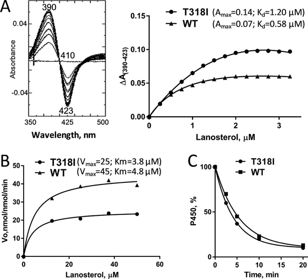
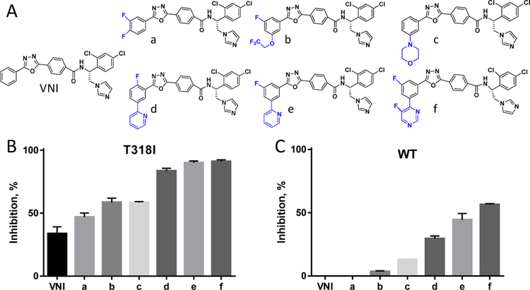
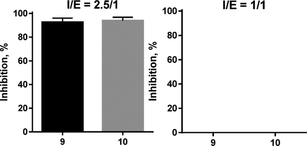
Sterol 14α-demethylases (CYP51) are the cytochrome P450 enzymes required for biosynthesis of sterols in eukaryotes, the major targets for antifungal agents and prospective targets for treatment of protozoan infections. Human CYP51 could be and, for a while, was considered as a potential target for cholesterol-lowering drugs (the role that is now played by statins, which are also in clinical trials for cancer) but revealed high intrinsic resistance to inhibition. While microbial CYP51 enzymes are often inhibited stoichiometrically and functionally irreversibly, no strong inhibitors have been identified for human CYP51. In this study, we used comparative structure/functional analysis of CYP51 orthologs from different biological kingdoms and employed site-directed mutagenesis to elucidate the molecular basis for the resistance of the human enzyme to inhibition and also designed, synthesized, and characterized new compounds. Two of them inhibit human CYP51 functionally irreversibly with their potency approaching the potencies of azole drugs currently used to inhibit microbial CYP51.


Conflict of interest statement

The authors declare no competing financial interest.

Figures

Figure 1.
Figure 1.
First generation of VFV derivatives. Although log P > 5 and mol wt > 500 are “violations of Lipinski’s rule of five”, there are numerous examples of successful drugs with larger values, especially those aimed at intracellular membrane-bound targets (e.g., cinacalcet/Sensipar at a log P of 6.5 and systemic clinical drug inhibitors of microbial CYP51 such as itraconazole (log P = 6.8, mol wt = 704) and posaconazole (log P = 5.5, mol wt = 701)).
Figure 2.
Figure 2.
Second generation of VFV derivatives.
Figure 3.
Figure 3.
Phylum-specific structural segments that might imply an evolutionary increase in the CYP51 flexibility. (A) Protozoan (3G1Q), (B) fungal (5TZ1), and (C) human (3I4K) CYP51. Distal P450 view.
Figure 4.
Figure 4.
VFV in the human CYP51 structure (4UHI). (A) “Butterfly-like” binding mode of the inhibitor: the imidazole ring of one VFV molecule is coordinated to the catalytic heme iron, and the biphenyl arm of the other VFV molecule is protruding from the substrate entry channel (A′, F″, and the β4 hairpin are marked). (B) Enlarged view. The biphenyl ortho-F atom of the heme-coordinated VFV molecule is located 3 Å from the I-helical backbone, pushing it downstream and thus shortening the loop-like region.
Figure 5.
Figure 5.
Middle portion of the CYP51 I-helix. (A) Fragment of sequence alignment; the six polar residues in human CYP51 are underlined in blue, and T318 and the corresponding protozoan/plant Ile are marked in red. (B) Backbone H-bonding in human vs Trypanosoma cruzi (T. cruzi) CYP51’s.
Figure 6.
Figure 6.
Functional characterization of the T318I mutant. (A) Spectral response to the substrate binding. Left: difference absorbance spectra upon T318I titration with lanosterol; right: comparative titration curves for the mutant and the WT (fit to one site–total, binding–saturation), ~2 μM P450. (B) Michaelis–Menten plots, 0.25 μM P450, 1 min reaction. (C) Stability at 42 °C monitored as the decrease in the P450 concentration; the initial P450 concentration was 2 μM.
Figure 7.
Figure 7.
Inhibition of human CYP51 with the VNI derivatives that are more potent than VNI as inhibitors of fungal CYP51. (A) Structural formulas. (B) The T318I mutation makes human CYP51 “more fungal-like”. (C) Three strongest T318I inhibitors display a clear inhibitory effect on the WT enzyme (1 h reaction, 0.5 μM P450, 50 μM lanosterol, and inhibitor/enzyme molar ratio = 50/1). Graphs represent mean ± SD of two independent experiments in duplicate.
Figure 8.
Figure 8.
Inhibition of human CYP51 at I/E molar ratios 2.5/1 vs 1/1 implied binding of two inhibitor molecules. 1 h reaction.
Figure 9.
Figure 9.
4UHI1 structures-based molecular model of compound 10 bound in the human CYP51 active site. (A) Position of the inhibitor molecules relative to the I-helix; the backbone helical H-bonds are red. The H-bonds predicted to have been formed by the inhibitor are presented as violet dashed lines. The participating atoms of the inhibitor molecules are shown in the corresponding colors in their structural formulas. (B) Inhibitor contacting residues. The complete list that relates the residues to the corresponding secondary structural elements (marked in Figure 10B) in the model and in the X-ray structures of human CYP51 is presented in Table S1.
Figure 10.
Figure 10.
Superimposed structures of VFV-bound (4UHI) and compound 10-bound (6Q2T) human CYP51, tan and blue ribbons, respectively. The carbon atoms of VFV, compound 10, and the detergent are orange, magenta, and yellow, respectively. (A) Overall (distal P450) view. The active site area is within the black square. A portion of the second molecule of compound 10 is seen at the top. (B) Enlarged view of the active site. The secondary structural elements that provide ligand-contacting residues (listed in Table S1) are marked.
Figure 11.
Figure 11.
Compound 10 in the human CYP51 active site. (A) Rearrangements of H236, W239, and M487 push the tail of the inhibitor (magenta) down relative to its position predicted by the VFV-structure-based model (gray). The arrows show the directions of the rearrangements. (B) Overall (distal P450) view of the compound 10-bound human CYP51 structure showing that the I-helix is straightened.
Scheme 1.
Scheme 1.. General Synthetic Method To Prepare the ortho-Cl-Substituted VFV Analogsa,b
a(R)-N-(1-(4-(4-fluorophenyl)naphthalen-1-yl)-2-(1H-imidazol-1-yl)ethyl)-4-(5-phenyl-1,3,4-oxadiazol-2-yl)benzamide (compound 2) was prepared using 1-(4-(4-fluorophenyl)naphthalen-1-yl)ethan-1-one as a starting material, and the synthesis was performed as previously reported. bRed box: Stille cross-coupling reaction. Blue box: enantioselective reduction of ketone to an (S)-alcohol.

Similar articles

Cited by

References

    1. Siegel RL; Miller KD; Jemal A Cancer statistics, 2018. CA: Cancer J. Clin 2018, 68, 7–30. - PubMed
    1. Hanahan D; Weinberg RA Hallmarks of cancer: the next generation. Cell 2011, 144, 646–674. - PubMed
    1. Chimento A; Casaburi I; Avena P; Trotta F; De Luca A; Rago V; Pezzi V; Sirianni R Cholesterol and its metabolites in tumor growth: therapeutic potential of statins in cancer treatment. Front. Endocrinol 2018, 9, 807. - PMC - PubMed
    1. Kuzu OF; Noory MA; Robertson GP The role of cholesterol in cancer. Cancer Res. 2016, 76, 2063–2070. - PMC - PubMed
    1. Murai T Cholesterol lowering: role in cancer prevention and treatment. Biol. Chem 2015, 396, 1–11. - PubMed

Publication types

MeSH terms

Substances

LinkOut - more resources