. 2019 Oct 30;11(20):8879-8891.
doi: 10.18632/aging.102308. Epub 2019 Oct 30.

CD8+CXCR5+T cells infiltrating hepatocellular carcinomas are activated and predictive of a better prognosis

Affiliations

CD8+CXCR5+T cells infiltrating hepatocellular carcinomas are activated and predictive of a better prognosis

Linsen Ye et al. Aging (Albany NY). .

Abstract

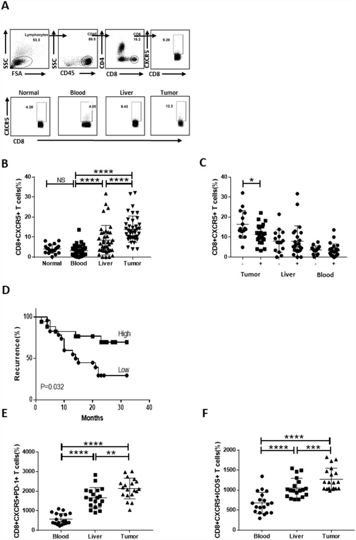
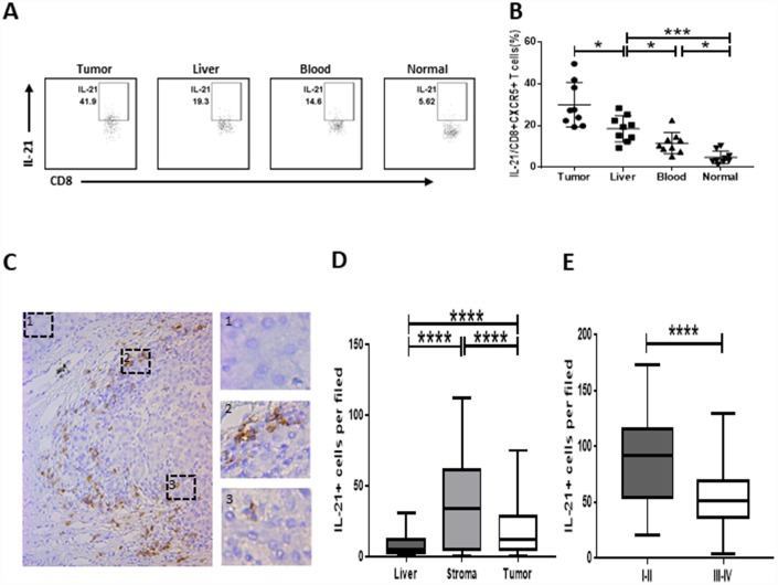
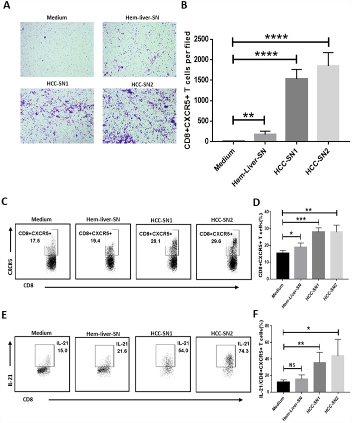
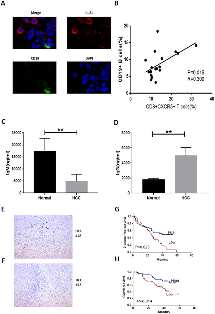
CD8+ T cells are thought to be the primary cytotoxic lymphocytes exerting antitumor effects. However, few studies have focused on the antitumor effects of CD8+ T cell-mediated humoral immunity or on interactions between CD8+ T cells and B cells in hepatocellular carcinoma (HCC). We found that the frequency of IL-21-producing CD8+CXCR5+ T cells was higher in HCC tumor tissue than in peritumoral tissue or peripheral blood from the same patients or in blood from healthy donors. Moreover, CD8+CXCR5+ T cells migrated in response to supernatants from primary HCC (HCC-SN) cells, and HCC-SN cells also powerfully induced CXCR5 expression in CD8+ T cells and IL-21 expression in CD8+CXCR5+ T cells. CD8+CXCR5+ T cells from HCC patients, but not those from healthy individuals, stimulated CD19+ B cells to differentiate into IgG-producing plasmablasts. These findings reveal that CD8+CXCR5+ T cells strongly infiltrate HCC tumors, and their infiltration is predictive of a better prognosis. Surprisingly, moreover, CD8+CXCR5+ T cells produced IL-21, which induced B cells to differentiate into IgG-producing plasmablasts and to play a key role in humoral immunity in HCC.

Keywords: cytotoxic T cells; hepatocellular carcinoma; humoral immunity; tumor microenvironment.


Conflict of interest statement

CONFLICTS OF INTEREST: The authors declare no potential conflicts of interest.

Figures

Figure 1
Figure 1
Strong infiltration of CD8+CXCR5+ T cells into HCC tumors predicts a better prognosis. Fresh samples were stained with anti-CD8, anti-CXCR5, anti-PD-1 and anti-ICOS antibodies. (AB) Following gating of CD8+ T cells, the frequencies of CD8+CXCR5+ T cells from healthy PBMCs (n=20), and matched HCC tumor tissue, peritumoral liver tissue and PBMC samples (n=40) were analyzed. (A) One representative experiment is shown. (C) Association of tumor-infiltrating CD8+CXCR5+ T cells with microvascular invasion (n=25 for positive, n=15 for negative) is shown. (BC) The data indicate the median with the interquartile range. (D) Patients were divided into two groups (Low/High) based on the median of the tumor-infiltrating CD8+CXCR5+ T cell percentages. The early recurrence rate was compared between the two groups using the log-rank test. (EF) PD-1 and ICOS expression by CD8+CXCR5+ T cells differed among tumor tissue and matched peritumoral tissues and peripheral blood (n=19). The data indicate the median with the interquartile range. *P<0.05, **P<0.01, ***P<0.001 and ****P<0.0001 determined using the Mann-Whitney U test (B, C, E and F).
Figure 2
Figure 2
Strong infiltration of IL-21-producing CD8+CXCR5+ T cells in HCC correlates with disease stage. (AB) Flow cytometric analysis of IL-21 production by CD8+CXCR5+ T cells (n=9). The cells were characterized using FACS with sequential gating of lymphocyte cells, CD45+ cells and then CD8+CXCR5+ cells. (A) One representative experiment is shown. (B) The data indicate the median with the interquartile range. (CE) Immunohistochemical staining of IL-21+ cells in paraffin-embedded HCC tissue (n=96). The distribution of IL-21+ cells is shown in (C and D). Micrographs at higher magnification show the stained peritumoral liver (1), peritumoral stromal region (2), and cancer nest (3). The association of the density of tumor-infiltrating IL-21+ cells with the TNM staging of patients is shown in (E). *P<0.05, **P<0.01, ***P<0.001 and ****P<0.0001 determined using Mann-Whitney U test (B and D) or student’s t test (E).
Figure 3
Figure 3
Differentiation and chemotaxis of CD8+CXCR5+ T cells is induced by HCC-SN cells. (AB) Culture supernatants from primary HCC cell cultures (HCC-SN1 and HCC-SN2 cells), but not normal liver cultures (Hem-liver-SN cells), induce chemotaxis of healthy blood CD8+CXCR5+ T cells sorted by FACS (n=3). (A) One representative experiment is shown. (B) The data indicate the mean±SD. (CE) HCC-SN1 and HCC-SN2 cells, but not Hem-liver-SN cells, are able to induce the CD8+CXCR5+ or IL-21+ CD8+CXCR5+ phenotype in healthy blood CD8+ T cells (n=3). The results shown represent four separate experiments. The data indicate the mean±SD. *P<0.05, **P<0.01, ***P<0.001 and ****P<0.0001 determined using student’s t test (B, D and F).
Figure 4
Figure 4
Tumor-infiltrating CD8+CXCR5+ T cells from HCC patients are potent inducers of plasmablasts differentiation in vitro. (A) Representative immunofluorescence images of CD19 (green), IL-21 (red) and nuclear staining with DAPI (blue) in HCC tissue. (B) Associations between tumor-infiltrating CD8+CXCR5+ T cells and tumor-infiltrating CD19+ B cells (n=19). (CD) Coculture of autologous CD19+ B cells with healthy blood CD8+CXCR5+ T cells or tumor-infiltrating CD8+CXCR5+ T cells. On day 5, the supernatants were harvested. IgG (C) and IgM levels (D) were determined using an ELISA (n=3). (EF) Immunohistochemical staining of CD138+ B cells in paraffin-embedded HCC tissue (n=96). (GH) Patients were divided into two groups (Low/High) based on the median of the tumor-infiltrating CD138+ B cell percentages. The DFS and OS curves between the two patient groups were compared using the log-rank test. **P<0.01.

References

    1. Shi L, Feng Y, Lin H, Ma R, Cai X. Role of estrogen in hepatocellular carcinoma: is inflammation the key? J Transl Med. 2014; 12:93. 10.1186/1479-5876-12-93 - DOI - PMC - PubMed
    1. Nordenstedt H, White DL, El-Serag HB. The changing pattern of epidemiology in hepatocellular carcinoma. Dig Liver Dis. 2010. (Suppl 3); 42:S206–14. 10.1016/S1590-8658(10)60507-5 - DOI - PMC - PubMed
    1. Mossanen JC, Tacke F. Role of lymphocytes in liver cancer. Oncoimmunology. 2013; 2:e26468. 10.4161/onci.26468 - DOI - PMC - PubMed
    1. Aravalli RN. Role of innate immunity in the development of hepatocellular carcinoma. World J Gastroenterol. 2013; 19:7500–14. 10.3748/wjg.v19.i43.7500 - DOI - PMC - PubMed
    1. Mellman I, Coukos G, Dranoff G. Cancer immunotherapy comes of age. Nature. 2011; 480:480–89. 10.1038/nature10673 - DOI - PMC - PubMed

Publication types