. 2019 Oct 29;10(1):4760.
doi: 10.1038/s41467-019-12683-8.

Cryo-EM structure and polymorphism of Aβ amyloid fibrils purified from Alzheimer's brain tissue

Affiliations

Cryo-EM structure and polymorphism of Aβ amyloid fibrils purified from Alzheimer's brain tissue

Marius Kollmer et al. Nat Commun. .

Abstract

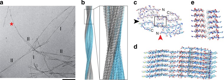
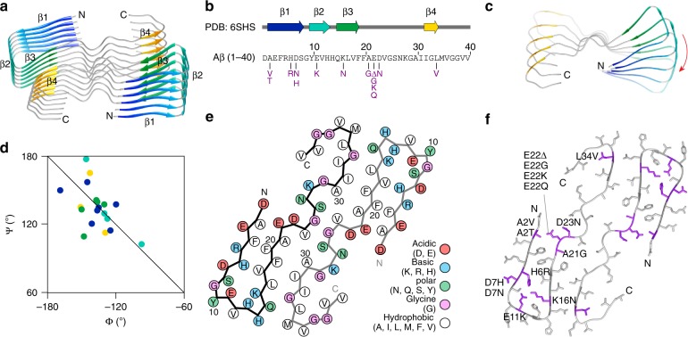
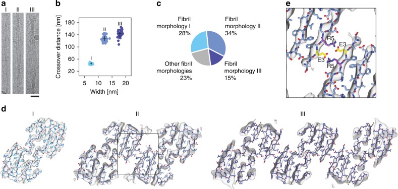
The formation of Aβ amyloid fibrils is a neuropathological hallmark of Alzheimer's disease and cerebral amyloid angiopathy. However, the structure of Aβ amyloid fibrils from brain tissue is poorly understood. Here we report the purification of Aβ amyloid fibrils from meningeal Alzheimer's brain tissue and their structural analysis with cryo-electron microscopy. We show that these fibrils are polymorphic but consist of similarly structured protofilaments. Brain derived Aβ amyloid fibrils are right-hand twisted and their peptide fold differs sharply from previously analyzed Aβ fibrils that were formed in vitro. These data underscore the importance to use patient-derived amyloid fibrils when investigating the structural basis of the disease.


Conflict of interest statement

The authors declare no competing interests.

Figures

Fig. 1
Fig. 1
Different structure of brain-derived and in vitro formed Aβ fibrils. a Negative stain TEM images of brain-derived amyloid fibrils or in vitro formed Aβ fibrils. Scale bar: 200 nm. b, c TEM (b) and SEM (c) images of brain-derived amyloid fibrils or in vitro formed Aβ fibrils after platinum side shadowing. Scale bars: 100 nm
Fig. 2
Fig. 2
Cryo-EM structure of fibril morphology I. a Representative cryo-EM micrograph indicating fibril morphologies I and II. Asterisk: morphology I-like fibril emanating from morphology II. Scale bar: 50 nm. b Side view of the reconstructed 3D map. c Cross-sectional view of one molecular layer of the fibril superimposed with the molecular model. d Side view of the 3D map of a six-layer peptide stack superimposed with the molecular model. The stack is viewed along the red arrow head in panel (c). e Side view of six molecular layers of the boxed region from panel (c), viewed along the black arrow head. The figure illustrates the staggering of the two peptide stacks. The two peptide stacks are colored gray and blue in panels (bd)
Fig. 3
Fig. 3
Peptide fold of fibril morphology I. a Ribbon diagram of six layers of the fibril. b Location of β-strands β1–β4 within the Aβ(1−40) sequence. Mutations are shown in magenta. c Top view of a peptide stack showing the right-hand β-sheet twist. Only every sixth molecule shown. d Section of the Ramachandran plot showing the Φ/Ψ-pairs of the residues within β-strands β1−β4. The β-strand color coding is kept consistent in panels (ad). e Packing scheme of one molecular layer of the fibril. f Cross-sectional view of the fibril with the known mutational variants highlighted in magenta (3)
Fig. 4
Fig. 4
Polymorphism of brain-derived Aβ fibrils. a 300 kV Cryo-EM images of fibril morphologies I−III. Scale bar: 20 nm. b Crossover distance and fibril width values of fibril morphologies I−III (n = 30). Black cross: average value and standard deviation. c Relative abundance of fibril morphologies I−III in our sample. d Cross-sectional densities of fibril morphologies I−III (gray) superimposed with the molecular model obtained with fibril morphology I. e Close-up of the PF−PF interface of fibril morphology II, indicated by a box in panel (d), showing the juxtaposed residues Glu3 and Arg5

References

    1. Chiti F, Dobson CM. Protein misfolding, amyloid formation, and human disease: a summary of progress over the last decade. Annu. Rev. Biochem. 2017;86:27–68. doi: 10.1146/annurev-biochem-061516-045115. - DOI - PubMed
    1. Selkoe DJ, Hardy J. The amyloid hypothesis of Alzheimer’s disease at 25 years. EMBO Mol. Med. 2016;8:595–608. doi: 10.15252/emmm.201606210. - DOI - PMC - PubMed
    1. Thal DR, Walter J, Saido TC, Fändrich M. Neuropathology and biochemistry of Aβ and its aggregates in Alzheimer’s disease. Acta Neuropathol. 2015;129:167–182. doi: 10.1007/s00401-014-1375-y. - DOI - PubMed
    1. Charidimou A, et al. Emerging concepts in sporadic cerebral amyloid angiopathy. Brain. 2017;140:1829–1850. doi: 10.1093/brain/awx047. - DOI - PMC - PubMed
    1. Glenner GG, Wong CW. Alzheimer’s disease: initial report of the purification and characterization of a novel cerebrovascular amyloid protein. Biochem. Biophys. Res. Commun. 1984;120:885–890. doi: 10.1016/S0006-291X(84)80190-4. - DOI - PubMed

Publication types