. 2019 Oct 29;10(1):4923.
doi: 10.1038/s41467-019-12895-y.

Hypothalamus-hippocampus circuitry regulates impulsivity via melanin-concentrating hormone

Affiliations

Hypothalamus-hippocampus circuitry regulates impulsivity via melanin-concentrating hormone

Emily E Noble et al. Nat Commun. .

Abstract

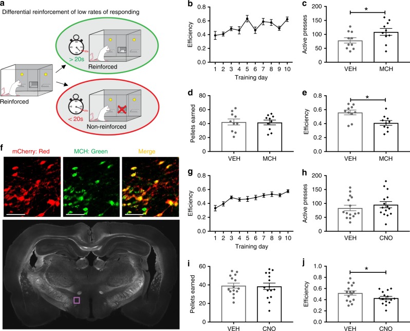
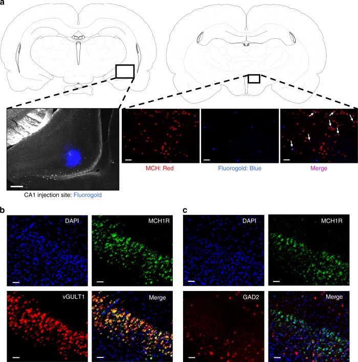
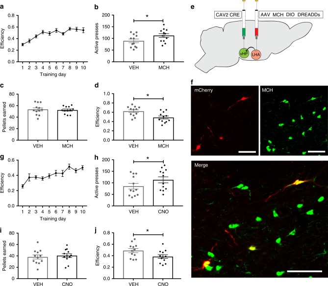
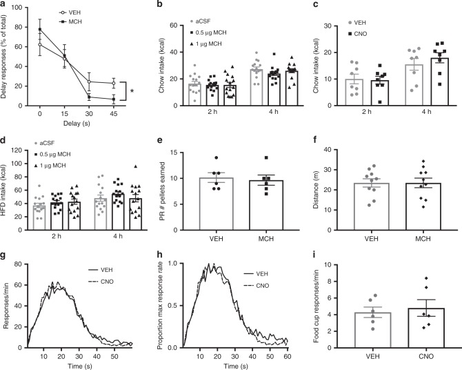
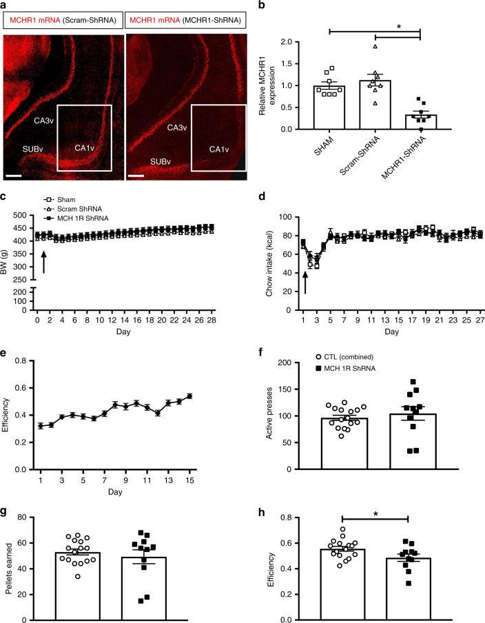
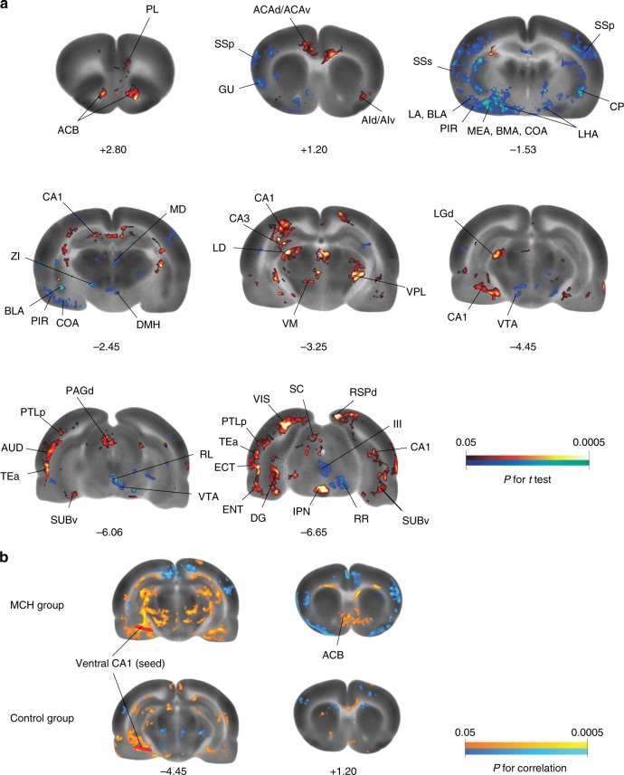
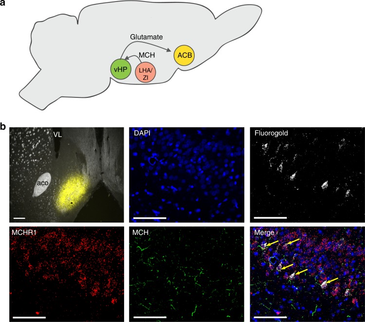
Behavioral impulsivity is common in various psychiatric and metabolic disorders. Here we identify a hypothalamus to telencephalon neural pathway for regulating impulsivity involving communication from melanin-concentrating hormone (MCH)-expressing lateral hypothalamic neurons to the ventral hippocampus subregion (vHP). Results show that both site-specific upregulation (pharmacological or chemogenetic) and chronic downregulation (RNA interference) of MCH communication to the vHP increases impulsive responding in rats, indicating that perturbing this system in either direction elevates impulsivity. Furthermore, these effects are not secondary to either impaired timing accuracy, altered activity, or increased food motivation, consistent with a specific role for vHP MCH signaling in the regulation of impulse control. Results from additional functional connectivity and neural pathway tracing analyses implicate the nucleus accumbens as a putative downstream target of vHP MCH1 receptor-expressing neurons. Collectively, these data reveal a specific neural circuit that regulates impulsivity and provide evidence of a novel function for MCH on behavior.


Conflict of interest statement

M.R.H. has received research support from investigator-initiated sponsored proposals from Novo Nordisk, Zealand Pharma, and Boehringer-Ingelheim. The other authors declare no competing interests.

Figures

Fig. 1
Fig. 1
MCH signaling increases impulsive responding for food. a A schematic diagram of the differential reinforcement of low rates of responding task (DRL). be Intracerebroventricular (ICV) injection of MCH (5 µg) effects on food impulsivity in the DRL task: b efficiency data during the training phase under the DRL 20 schedule (ce; data were analyzed using Student’s two-tailed, paired t tests; n = 10), c active lever presses in the DRL test session (P = 0.006), d number of pellets earned in the DRL test session, and e efficiency in the DRL test session (P = 0.0009). f Representative images showing the localization of the mCherry fluorescence reporter (red) in MCH neurons (green) following injections of AAV2-rMCHp-hM3D(Gq)-mCherry (colocalization in yellow). The region shown in the upper images is indicated by the purple box outlined in the coronal rat brain section below (~3.5 mm posterior to bregma). gj Chemogenetic activation of MCH DREADDs-containing neurons following ICV CNO injections effects on food impulsivity in the DRL task. g Efficiency data during the training phase in DRL 20 (hj; data were analyzed using Student’s two-tailed, paired t tests; n = 15), h number of active lever presses in the DRL test session, i number of pellets earned in the DRL test session, and j efficiency in the DRL test session (P = 0.02). Data shown as mean ± SEM; scale = 50 µm; *P < 0.05. Source data are provided as a source data file
Fig. 2
Fig. 2
MCH neurons communicate to glutamatergic neurons in the CA1v. a (left) A representative injection site for fluorogold in the CA1 region of the vHP; scale = 200 µm. (right) Fluorogold (pseudocolored blue) colocalizes with MCH immunofluorescence (red) in a region of the lateral hypothalamic area (white arrows indicate colocalization); scale = 100 µm. b Fluorescence in situ hybridization for MCHR1 mRNA (green) and the glutamatergic marker vGLUT1 (red), with DAPI counterstain (blue). c Fluorescence in situ hybridization for MCHR1 mRNA (green) and the GABAergic marker GAD2 (red), with DAPI counterstain (blue); scale = 50 µm
Fig. 3
Fig. 3
Activation of MCH-vHP signaling increases food impulsivity. ad Effects of vHP injection of MCH on impulsive responding for food in the DRL task: a efficiency data during the training phase in DRL 20. (bd; data were analyzed using Student’s two-tailed, paired t tests; n = 12). b Number of active lever presses during test phase (P = 0.02). c Number of pellets earned during test phase. d Efficiency in the DRL test phase (P = 0.008). e Schematic cartoon depicting methods for dual virus chemogenetic approach. The retroactively transported canine adenovirus 2 (CAV2 CRE) was used to deliver the transgene for cre recombinase to vHP projection neurons following direct injection into the vHP. A cre-dependent adeno-associated virus containing excitatory MCH DREADDs–mCherry transgene (AAV2 MCH DIO DREADDs) was injected into the LHA and ZI. f Representative images of fluorescent reporter localization in MCH neurons. g–j Effects of chemogenetic activation of MCH neurons that project to the vHP on impulsive responding for food in the DRL test session: g efficiency data during the training phase in DRL 20. (h–j; data were analyzed using Student’s two-tailed, paired t tests; n = 12), h number of active lever presses during DRL testing (P = 0.03), i number of pellets earned during DRL testing, and j efficiency during the DRL testing (P = 0.03). Data shown as mean ± SEM; scale = 50 µm; *P < 0.05. Source data are provided as a source data file
Fig. 4
Fig. 4
vHP MCH signaling increases impulsive choice but not food motivation or timing accuracy. a MCH (1 µg in the vHP) effects on performance in the delay discounting task (two-way repeated measures ANOVA with delay × drug treatment; n = 9). Results show a significant delay × drug interaction (F(3,24) = 5.53; P = 0.005), with main effect of delay time (F(3,24) = 18.21; P < 0.0001). (bd; Two-way repeated measures ANOVA (time × drug)). b Effect of vHP MCH injection (n = 15) and c chemogenetic activation of vHP projecting MCH neurons (n = 8) on standard chow intake in the home cage. (d Effect of vHP MCH injection on high fat/high sugar (HFD) diet intake (n = 15). e MCH (1 µg in the vHP) effect on the number of pellets earned in the progressive ratio task (Student’s two-tailed, paired t test; n = 6). f Effect of 1 µg of MCH (0.5 µg/side) in the vHP on distance traveled in the open field test (Student’s two-tailed, paired t test; n = 10). gi Effect of chemogenetic activation of MCH neurons on peak interval timing (n = 6). g Peak interval response rates during probe trials, with increased response rates that peaks near the 20 s criterion duration; h normalized function for peak rate, where the peak function for each animal was converted to a percentage of maximum response rate. This analysis revealed no horizontal shift in the peak interval timing function (two-way repeated measures ANOVA; F(1,5) = 1.63, 0.25) following activation of MCH neurons by lateral ventricle CNO injections. i MCH neuronal activation also had no influence on food cup approach behavior during the peak interval test (Student’s two-tailed, paired t test). Data shown as mean ± SEM; (*P < 0.05). Source data are provided as a source data file
Fig. 5
Fig. 5
MCHR1 knockdown increases food impulsivity. a Representative images showing MCHR1 gene expression via fluorescence in situ hybridization and b relative MCHR1 gene expression (relative to Sham controls) via qPCR in CA1v in scrambled control shRNA (Scram-shRNA) or MCHR1 shRNA injected animals (one-way ANOVA with Tukey’s multiple comparisons test; n = 8/group; P < 0.05); scale = 200 µm. c Body weight and d chow intake before (day 0) and after surgery in animals injected with either Scram-shRNA (n = 8), MCHR1 shRNA (n = 11), or Sham injected (n = 8); arrow indicates surgery date. e Efficiency data during the training phase in DRL 20 (before RNAi AAV injections). fh; Student’s two-tailed, unpaired t test; n = 11, 16 (combined controls) f Number of active lever presses, g number of pellets earned and h efficiency in the DRL task following RNAi AAV incubation (P = 0.04). Data shown as mean ± SEM; *P < 0.05. Source data are provided as a source data file
Fig. 6
Fig. 6
Neural responses following vHP MCHR1 activation. a Color-coded overlays over a selection of representative coronal sections of the template brain show significant MCH-induced changes in regional cerebral glucose uptake (rCGU) as an indirect measurement of brain activation (Student’s t test, red/blue: increase/decrease in rCGU in the MCH group compared to the control group; n = 9 and 8, respectively). Numbers under images are approximate level along the anterior–posterior axis relative to the bregma (in mm). b Seed correlation analysis revealed statistically significant positive correlation between the seed in the ventral CA1 and ACB shell area in the MCH but not in the control group (Pearson’s correlation, orange/blue: positive/negative correlation with the seed). ACB nucleus accumbens, ACAd/ACAv anterior cingulate area, dorsal/ventral part, AId/AIv agranular insular area, dorsal/ventral part, AUD auditory area, TEa temporal association areas, BLA basolateral amygdalar nucleus, BMA basomedial amygdalar nucleus, CA1/CA3 CA1/CA3 area of the HP, COA cortical amygdalar nucleus, CP caudoputamen, DG dentate gyrus, DMH dorsomedial hypothalamic nucleus, ECT ectorhinal area, ENT entorhinal area, GU gustatory area, III oculomotor nucleus, IPN interpeduncular nucleus, LA lateral amygdalar nucleus, LD lateral dorsal nucleus thalamus, LGd lateral geniculate complex, dorsal part, LHA lateral hypothalamic area, MD mediodorsal nucleus thalamus, MEA medial amygdalar nucleus, PAGd periaqueductal gray, dorsal division, PIR piriform area, PL prelimbic area, PTLp parietal region, posterior association areas, RL rostral linear nucleus raphe, RR midbrain reticular nucleus, retrorubral area, RSPd retrosplenial area, dorsal part, SC superior colliculus, SSp primary somatosensory area, SSs supplemental somatosensory area, SUBv subiculum, ventral part, VIS visual areas, VM ventral medial nucleus thalamus, VPL ventral posterolateral nucleus thalamus, VTA ventral tegmental area, ZI zona incerta). Abbreviations are taken from ref.
Fig. 7
Fig. 7
The ACB is a target for vHP-projecting MCH neurons. a A cartoon schematic showing the projection pathway from MCH neurons in the lateral hypothalamus/zona incerta (LHA/ZI) to the vHP and then to the ACB. b (top left) Injection site for fluorogold in the ACB in a 30-µm coronal section taken from a rat brain at 1.1 mm anterior to bregma; scale = 200 µm. Retrogradely labeled cells are shown in the CA1 subregion of the vHP in a 30-µm coronal section taken from a rat brain at 4.7 mm posterior to bregma. DAPI nuclear counterstain is shown in blue, retrogradely labeled fluorogold-positive cells are shown in white. Fluorescence in situ hybridization for the MCHR1 is shown in red and immunofluorescence staining for MCH is in green. Arrows point to MCH fiber appositions to MCHR1-positive cells containing fluorogold, indicating that these vHP neurons both receive input from MCH neurons and project to the ACB. Images taken at ×20 magnification; scale = 50 µm. ACB nucleus accumbens, aco anterior commissure, LHA lateral hypothalamus, vHP ventral hippocampus, VL lateral ventricle, ZI zona incerta

References

    1. Dalley JW, Robbins TW. Fractionating impulsivity: neuropsychiatric implications. Nat. Rev. Neurosci. 2017;18:158–171. doi: 10.1038/nrn.2017.8. - DOI - PubMed
    1. Guerrieri R, Nederkoorn C, Jansen A. How impulsiveness and variety influence food intake in a sample of healthy women. Appetite. 2007;48:119–122. doi: 10.1016/j.appet.2006.06.004. - DOI - PubMed
    1. Guerrieri R, Nederkoorn C, Jansen A. The interaction between impulsivity and a varied food environment: its influence on food intake and overweight. Int. J. Obes. (Lond.) 2008;32:708–714. doi: 10.1038/sj.ijo.0803770. - DOI - PubMed
    1. Giel, K. E., Teufel, M., Junne, F., Zipfel, S. & Schag, K. Food-related impulsivity in obesity and binge eating disorder-a systematic update of the evidence. Nutrients9, E1170 (2017). - PMC - PubMed
    1. Nederkoorn C, Houben K, Hofmann W, Roefs A, Jansen A. Control yourself or just eat what you like? Weight gain over a year is predicted by an interactive effect of response inhibition and implicit preference for snack foods. Health Psychol. 2010;29:389–393. doi: 10.1037/a0019921. - DOI - PubMed

Publication types

LinkOut - more resources