. 2019 Oct 29;9(1):15559.
doi: 10.1038/s41598-019-52085-w.

Chloroquine modulates inflammatory autoimmune responses through Nurr1 in autoimmune diseases

Affiliations

Chloroquine modulates inflammatory autoimmune responses through Nurr1 in autoimmune diseases

Tae-Yoon Park et al. Sci Rep. .

Abstract

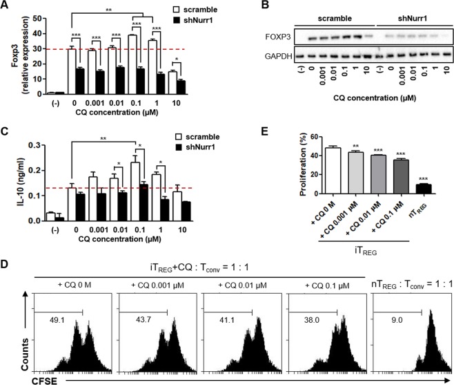
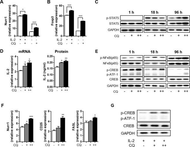
For over a half-century the anti-malarial drug chloroquine (CQ) has been used as a therapeutic agent, alone or in combination, to treat autoimmune diseases. However, neither the underlying mechanism(s) of action nor their molecular target(s) are well defined. The orphan nuclear receptor Nurr1 (also known as NR4A2) is an essential transcription factor affecting the development and maintenance of midbrain dopaminergic neurons. In this study, using in vitro T cell differentiation models, we demonstrate that CQ activates TREG cell differentiation and induces Foxp3 gene expression in a Nurr1-dependent manner. Remarkably, CQ appears to induce Nurr1 function by two distinct mechanisms: firstly, by direct binding to Nurr1's ligand-binding domain and promoting its transcriptional activity and secondly by upregulation of Nurr1 expression through the CREB signaling pathway. In contrast, CQ suppressed gene expression and differentiation of pathogenic TH17 cells. Importantly, using a valid animal model of inflammatory bowel disease (IBD), we demonstrated that CQ promotes Foxp3 expression and differentiation of TREG cells in a Nurr1-dependent manner, leading to significant improvement of IBD-related symptoms. Taken together, these data suggest that CQ ameliorates autoimmune diseases via regulating Nurr1 function/expression and that Nurr1 is a promising target for developing effective therapeutics of human inflammatory autoimmune diseases.


Conflict of interest statement

The authors declare no competing interests.

Figures

Figure 1
Figure 1
Nurr1-dependent regulation of iTREG differentiation by CQ. Mouse primary naïve CD4+CD25CD62Lhigh T cells were transfected with lenti-scramble- or lenti-shNurr1-plasmid. Cells were treated with CQ (0.001~10 μM) and stimulated with plate-bound anti-CD3 and soluble anti-CD28 antibodies for 96 h under iTREG-polarizing conditions. (A,B) The level of Foxp3 mRNA (A) or protein (B) expression was determined by quantitative real-time PCR or western blot, normalized with GAPDH. (C) The level of IL-10 in the culture media was analyzed by ELISA. (D) In vitro TREG suppression assays based on CFSE dilution by Tconv cells proliferating in the presence of CQ treated iTREG cells for 48 h and analyzed with flow cytometry. (E) Quantitation of the percentage ± SEM of proliferation of Tconv in the presence of iTREG with or without CQ treatment. These experiments were repeated three times in triplicate using independently prepared samples. Each error bar represents means ± s.e.m. *P < 0.05, **P < 0.01, ***P < 0.001.
Figure 2
Figure 2
Nurr1-independent regulation of pTH17 differentiation by CQ. Mouse primary naïve CD4+CD25CD62Lhigh T cells were transfected with lenti-scramble- or lenti-shNurr1-plasmid. Cells were treated with CQ (0.001~10 μM) and stimulated with plate-bound anti-CD3 and soluble anti-CD28 antibodies for 72 h under pTH17-polarizing conditions. (AC) The levels of RORγt (A), IL-23R (B) or IL-17A (C) mRNA expression were determined by quantitative real-time PCR and normalized with GAPDH. (D) The levels of Foxp3 mRNA expression were determined by quantitative real-time PCR and normalized with GAPDH. These experiments were repeated three times in triplicate using independently prepared samples. Each error bar represents means ± s.e.m. *P < 0.05, **P < 0.01, ***P < 0.001.
Figure 3
Figure 3
CQ interacts with Nurr1-LBD and directly regulates Nurr1 transcriptional activity. NMR titration experiments of Nurr1-LBD with CQ. (A) Overlay of 1H-15N HSQC spectra of uniformly 15N labeled Nurr1-LBD (red) and Nurr1-LBD in the presence of CQ at molar ratio of 1 to 2.5 (green) and 1 to 5 (blue). (B) Expanded views of chemical shift perturbations upon CQ binding. The perturbations of chemical shifts from free Nurr1-LBD (red) to CQ bound forms (green and blue) are indicated by arrows. (C) Chemical Shift Perturbation Plot of Nurr1-LBD upon CQ binding at molar ratio of 1 (Nurr1-LBD) to 5 (CQ). The difference in chemical shifts was calculated using the following formula, Δδ = [(1Hfree − 1Hbound)2 + (15Nfree − 15Nbound)2)]1/2. Interaction site mapping of Nurr1-LBD and CQ based on the NMR data. (D) Surface mapping of CQ binding site and interaction residues on Nurr1-LBD based on 1H-15N HSQC titration data. Perturbed amino acid residues are displayed according to their chemical shift perturbation: red (Δδ > 0.1), blue (0.8 < Δδ < 0.1). (E) Ribbon representation of mapping data in Fig. 3D. Perturbed amino acid residues were displayed by stick representation and chemical shift values were used in the same manner as in Fig. 3D. (F) Impact of Nurr1-LBD point mutation on CQ’s effect on Nurr1 transcriptional activity. Luciferase activities were diminished in full length mutant Nurr1 transfected HEK293 cell lines compared to wild-type Nurr1 transfected control. Each error bar represents means ± s.e.m. ***P < 0.001. NT, non-treated condition.
Figure 4
Figure 4
Regulation of Nurr1 expression by CQ. Mouse primary naïve CD4+CD25CD62Lhigh T cells were treated with 100 nM (+) or 1 μM (++) CQ and stimulated with plate-bound anti-CD3 and soluble anti-CD28 antibodies for 1–96 h under iTREG-polarizing conditions (with or without IL-2). (A,B) The level of Nurr1 (A) and Foxp3 (B) mRNA expression were analyzed by quantitative real-time PCR and normalized with GAPDH. (C) The expression of p-STAT5, STAT5, and GAPDH proteins were confirmed by western blot after 1, 18, and 96 h treatment with CQ under iTREG-polarizing condition without IL-2 treatment. (D) The level of IL-2 mRNA and protein expression were analyzed by quantitative real-time PCR and ELISA, respectively. (E) The expression of p-NFkB(p65), NFkB(p65), p-CREB, CREB, and GAPDH proteins were confirmed by western blot after 1, 18, and 96 h treatment with CQ under iTREG-polarizing condition without IL-2 treatment. (F) The level of Nurr1, CD25, and FASL mRNA expression were determined by quantitative real-time PCR and normalized with GAPDH. (G) Mouse primary naïve CD4+CD25CD62Lhigh T cells were treated with 100 nM (+) or 1 μM (++) CQ and stimulated with plate-bound anti-CD3 and soluble anti-CD28 antibodies for 96 h under iTREG-polarizing conditions (with IL-2). The expression of p-CREB, CREB, and GAPDH proteins were confirmed by western blot. These experiments were repeated more than twice in triplicate using independently prepared samples. Each error bar represents means ± s.e.m. *P < 0.05, **P < 0.01, ***P < 0.001.
Figure 5
Figure 5
Nurr1-dependent attenuation of DSS-induced colitis by CQ. Male mice were infected with none, scrambled-lentivirus (Scr) or shNurr1-lentivirus (shNurr1) 7 days prior to treatment with water (No DSS) or 3% DSS for 8 days. During the 8 days, each group, Scr + CQ and shNurr1 + CQ, received 50 mg/kg/day of CQ intraperitoneally. Mice were sacrificed at day 8. (A) Body weight change after DSS induction of colitis was evaluated and expressed as a percentage of the initial weight. (B) Colon length was measured. (C) Histological colitis scores were recorded. (D) Representative histologic images of H&E-stained colon sections. (E) MLN CD4+ T cells’ Foxp3 levels were analyzed by flow cytometry. (F) Quantification of results in E. (G) Representative histologic images of Foxp3/Hoechst-stained colon sections. Data are representative of two experiments with ten mice per group. Each error bar represents means ± s.e.m. *P < 0.05.

References

    1. O’Dell JR. Therapeutic strategies for rheumatoid arthritis. N. Engl. J. Med. 2004;350:2591–2602. doi: 10.1056/NEJMra040226. - DOI - PubMed
    1. Lee SJ, Silverman E, Bargman JM. The role of antimalarial agents in the treatment of SLE and lupus nephritis. Nat. Rev. Nephrol. 2011;7:718–729. doi: 10.1038/nrneph.2011.150. - DOI - PubMed
    1. O’Dell JR, et al. Therapies for active rheumatoid arthritis after methotrexate failure. N. Engl. J. Med. 2013;369:307–318. doi: 10.1056/NEJMoa1303006. - DOI - PubMed
    1. Haladyj E, Sikora M, Felis-Giemza A, Olesinska M. Antimalarials – are they effective and safe in rheumatic diseases? Reumatologia. 2018;56:164–173. doi: 10.5114/reum.2018.76904. - DOI - PMC - PubMed
    1. Olsen NJ, Schleich MA, Karp DR. Multifaceted effects of hydroxychloroquine in human disease. Semin. Arthritis. Rheum. 2013;43:264–272. doi: 10.1016/j.semarthrit.2013.01.001. - DOI - PubMed

Publication types

MeSH terms

Substances