. 2020 Jun;11(3):541-551.
doi: 10.1007/s12975-019-00745-4. Epub 2019 Oct 30.

CD47 Blocking Antibody Accelerates Hematoma Clearance After Intracerebral Hemorrhage in Aged Rats

Affiliations

CD47 Blocking Antibody Accelerates Hematoma Clearance After Intracerebral Hemorrhage in Aged Rats

Chuanyuan Tao et al. Transl Stroke Res. 2020 Jun.

Abstract

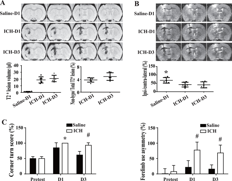
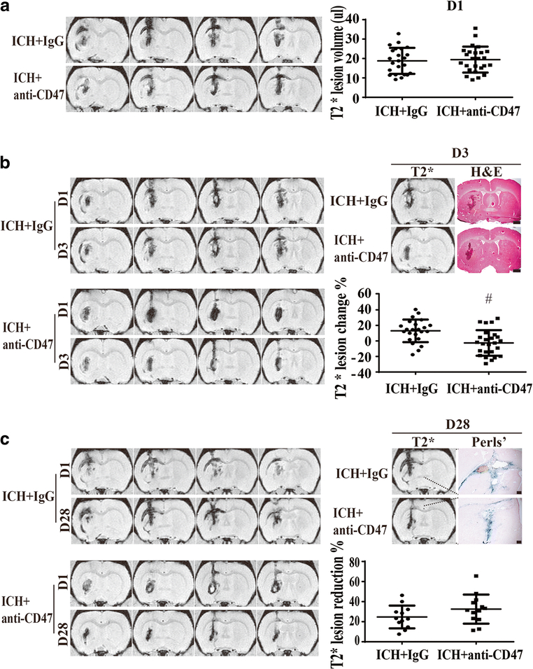
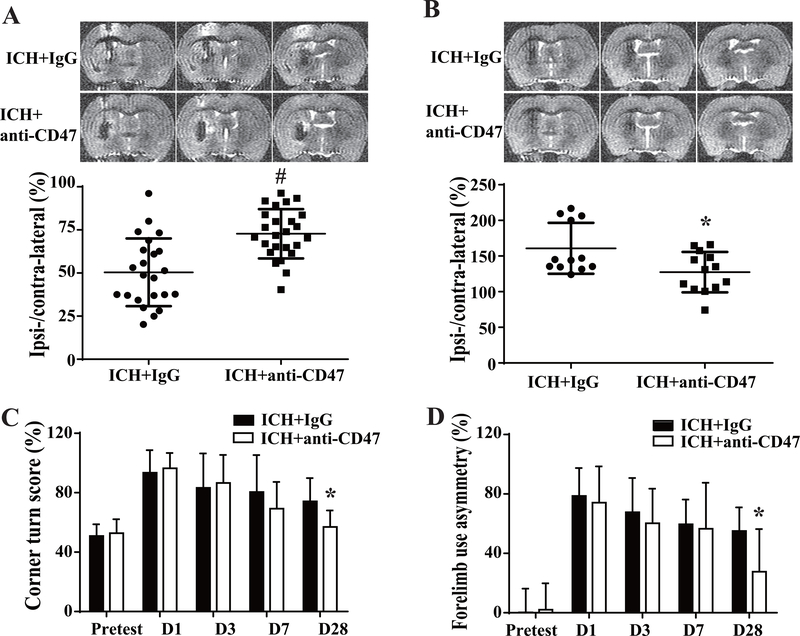
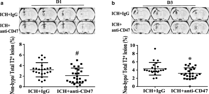
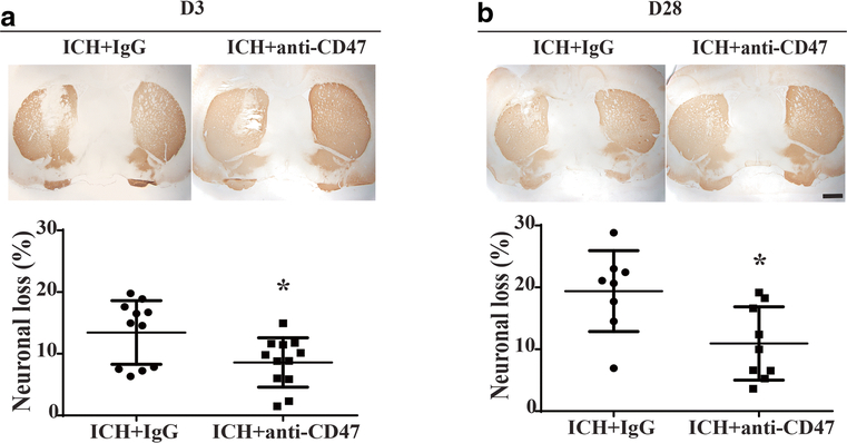
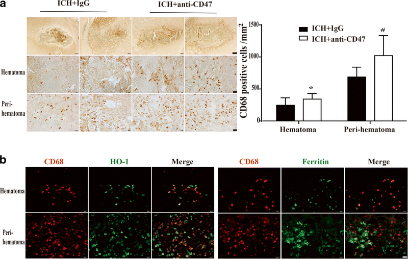
Both experimental studies and surgical clinical trials suggest that hematoma clearance is a therapeutic target in intracerebral hemorrhage (ICH). We have investigated effects of CD47, a "don't eat me" signal expressed on erythrocytes, on hematoma resolution after ICH in young mice. This study expands those findings by examining the effects on a CD47 blocking antibody in aged rats. First, male Fischer 344 rats (18 months old) received an intracaudate injection of 50 μL autologous whole blood or saline. Hematoma features of magnetic resonance imaging (MRI) and neurological deficits were evaluated within 3 days. Second, rats had an intracaudate co-injection of 50 μL autologous blood with either CD47 blocking antibody or IgG. MRI was used to quantify hematoma/iron volume, hemolysis, brain swelling, and atrophy at different time points, behavioral tests to assess neurological deficits, and immunohistochemistry to assess brain injury and neuroinflammation. The CD47 blocking antibody significantly promoted hematoma clearance, attenuated brain swelling, hemolysis, and neuronal loss and increased the number of phagocytic macrophages in and around hematoma 3 days after ICH. Moreover, CD47 blockade reduced neuronal loss, brain atrophy, and neurobehavioral deficits at day 28. These results indicate that a CD47 blocking antibody can accelerate hematoma clearance and alleviate short- and long-term brain injury after ICH in aged rats and that it might be a therapeutic strategy for ICH.

Keywords: Aged rats; CD47 blocking antibody; Cerebral hemorrhage; Macrophage activation.


Conflict of interest statement

Compliance with Ethical Standards

Conflict of Interests: Chuanyuan Tao, Richard F. Keep, Guohua Xi, and Ya Hua declare no conflict of interests.

Figures

Figure 1.
Figure 1.
The characteristics of ICH modeled by intracaudate injection of 50μl blood in aged rats. (A) Representative T2*-weighted MRI showing the hematoma volume and early hemolysis at day 1 and day 3 after ICH. Hemolysis is indicated by the non-hypointense area at the center of the hematoma. A saline-injected control is shown for comparison. (B) Brain swelling was evaluated by measuring ventricular compression on T2-weighted imaging. Ipsi/Contra = ipsilateral/contralateral ventricular volume. (C) Corner turn and forelimb use asymmetry were tested before and after ICH or saline injection. Values are means±SD; n=6 in each group, * P< 0.05 vs. other groups by one-way ANOVA; *P<0.05 and # P< 0.01 vs. saline group by two-way ANOVA.
Figure 2.
Figure 2.
Effect of CD47 blocking antibody on hematoma resolution as assessed by T2* MRI. (A) Examples of T2* MRIs at day 1 after injection of blood with anti-CD47 antibody or IgG controls. T2* lesion volumes (hematoma volumes) were calculated for the two groups. Values are mean ±SD, anti-CD47 antibody (n=25) or IgG (n=23). There was no significant difference in hematoma volume at day 1. (B) Representative examples of T2* MRIs at day 3 and a comparison between T2* lesions and H&E stain showing the T2* lesion corresponded to the hematoma at day 3. The change in T2* lesion volume (day 3 – day 1) was calculated and normalized by dividing by the day 1 volume. Values are mean±SD, scale bar= 1mm; # P< 0.01 vs. IgG group by unpaired t test; n=23 in IgG group, n=25 in anti-CD47 antibody group. (C) Representative examples of T2* MRIs at day 28 and a comparison between T2* lesions and Perls’ staining showing T2* lesion corresponded to the area of iron deposition at day 28. The change in T2* lesion volume (day 28 – day 1) was calculated and normalized by dividing by the day 1 volume. Values are mean ±SD, scale bar= 200μm; n=12 in IgG group, n=13 in anti-CD47 antibody group.
Figure 3.
Figure 3.
Brain swelling and atrophy, neurological deficits assessment at different time points after ICH in IgG and anti-CD47 treated rats. (A) Ipsilateral ventricular compression was measured on T2 MRIs (representative images shown) to assess brain swelling at day 3 after ICH. Ipsilateral ventricular volume was expressed as a % of contralateral ventricular volume, n=23 in IgG group, n=25 in anti-CD47 antibody group. (B) Ipsilateral ventricular dilation at day 28 was measured on T2 MRIs (representative images shown) to assess brain atrophy. Ipsilateral ventricular volume was expressed as a % of contralateral ventricular volume, n=12 in IgG group and n=13 in anti-CD47 antibody group. For (A) and (B) values are means±SD; * P< 0.05, # P< 0.01 vs. IgG group by unpaired t test. (C) Corner turn and (D) forelimb use asymmetry evaluation before and after blood+IgG or blood+anti-CD47 antibody injection, n=23 in IgG group, n=25 in anti-CD47 antibody group at pretest, days 1 and 3; n=12 in IgG group, n=13 in anti-CD47 antibody group at days 7 and 28. Values are means±SD; * P< 0.05 vs. IgG group by two-way ANOVA.
Figure 4.
Figure 4.
Representative hemolysis in hematoma center as hyper- or iso-intense on T2* imaging at day 1 (A) and day 3 (B). Values are means ± SD; n=23 in IgG group, n=25 in anti-CD47 antibody group; * P< 0.05, # P< 0.01 vs. IgG group by unpaired t test.
Figure 5.
Figure 5.
Neuronal loss after ICH with and without CD47 blocking antibody treatment. (A) and (B) Examples of DARPP-32 staining in aged rats treated with IgG or anti-CD47 blocking antibody at day 3 and 28 after ICH, scale bars=1mm. Loss of DARPP-32 staining was quantified as (contralateral − ipsilateral) / contralateral DARPP-32-positive area. Values are means ± SD; n=11 in IgG group, n=12 in anti-CD47 antibody group at day 3; n=8 in IgG group, n=9 in anti-CD47 antibody group at day 28, * P< 0.05 vs. IgG group by unpaired t test.
Figure 6.
Figure 6.
Effect of CD47 blocking antibody on CD68 expression in microglia/macrophages at day 3 after ICH and co-localization of CD68 with HO-1 and ferritin. (A) Examples of CD68 immunohistochemistry from animals treated with IgG or anti-CD47 antibody; upper scale bar=200 μm and lower scale bar=20 μm. Quantification of CD68 positive cells in and around hematoma. Values are means ± SD; n=11 in IgG group, n=12 in anti-CD47 antibody group; * P< 0.05, # P< 0.01 vs. IgG group by unpaired t test. (B) Immunofluorescent double labeling of CD68 positive cells with HO-1 and ferritin positive cells in an anti-CD47 treated animal, scale bar=20 μm.

References

    1. Broderick JP, Brott TG, Duldner JE, Tomsick T, Huster G. Volume of intracerebral hemorrhage. A powerful and easy-to-use predictor of 30-day mortality. Stroke. 1993;24:987–993 - PubMed
    1. Zhao X, Sun G, Ting SM, Song S, Zhang J, Edwards NJ, et al. Cleaning up after ich: The role of nrf2 in modulating microglia function and hematoma clearance. J Neurochem. 2015;133:144–152 - PMC - PubMed
    1. Wilkinson DA, Keep RF, Hua Y, Xi G. Hematoma clearance as a therapeutic target in intracerebral hemorrhage: From macro to micro. J Cereb Blood Flow Metab. 2018;38:741–745 - PMC - PubMed
    1. Hanley DF, Thompson RE, Rosenblum M, Yenokyan G, Lane K, McBee N, et al. Efficacy and safety of minimally invasive surgery with thrombolysis in intracerebral haemorrhage evacuation (mistie iii): A randomised, controlled, open-label, blinded endpoint phase 3 trial. Lancet. 2019;393:1021–1032 - PMC - PubMed
    1. Zhao X, Sun G, Zhang J, Strong R, Song W, Gonzales N, et al. Hematoma resolution as a target for intracerebral hemorrhage treatment: Role for peroxisome proliferator-activated receptor gamma in microglia/macrophages. Ann Neurol. 2007;61:352–362 - PubMed

Publication types