. 2020 Jan;30(1):126-133.
doi: 10.1111/jon.12673. Epub 2019 Oct 30.

Standardized Brain MRI Acquisition Protocols Improve Statistical Power in Multicenter Quantitative Morphometry Studies

Affiliations

Standardized Brain MRI Acquisition Protocols Improve Statistical Power in Multicenter Quantitative Morphometry Studies

Allan George et al. J Neuroimaging. 2020 Jan.

Abstract

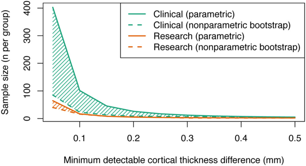
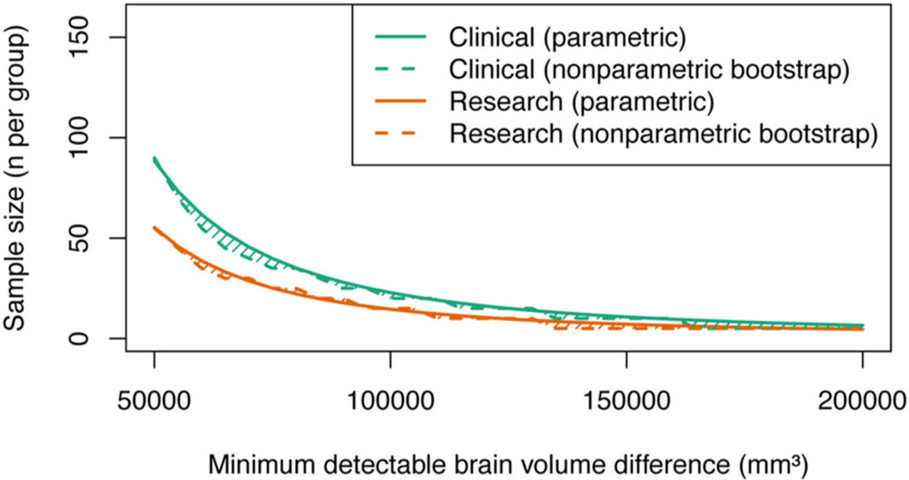
Background and purpose: In this study, we used power analysis to calculate required sample sizes to detect group-level changes in quantitative neuroanatomical estimates derived from MRI scans obtained from multiple imaging centers. Sample size estimates were derived from (i) standardized 3T image acquisition protocols and (ii) nonstandardized clinically acquired images obtained at both 1.5 and 3T as part of the multicenter Human Epilepsy Project. Sample size estimates were compared to assess the benefit of standardizing acquisition protocols.

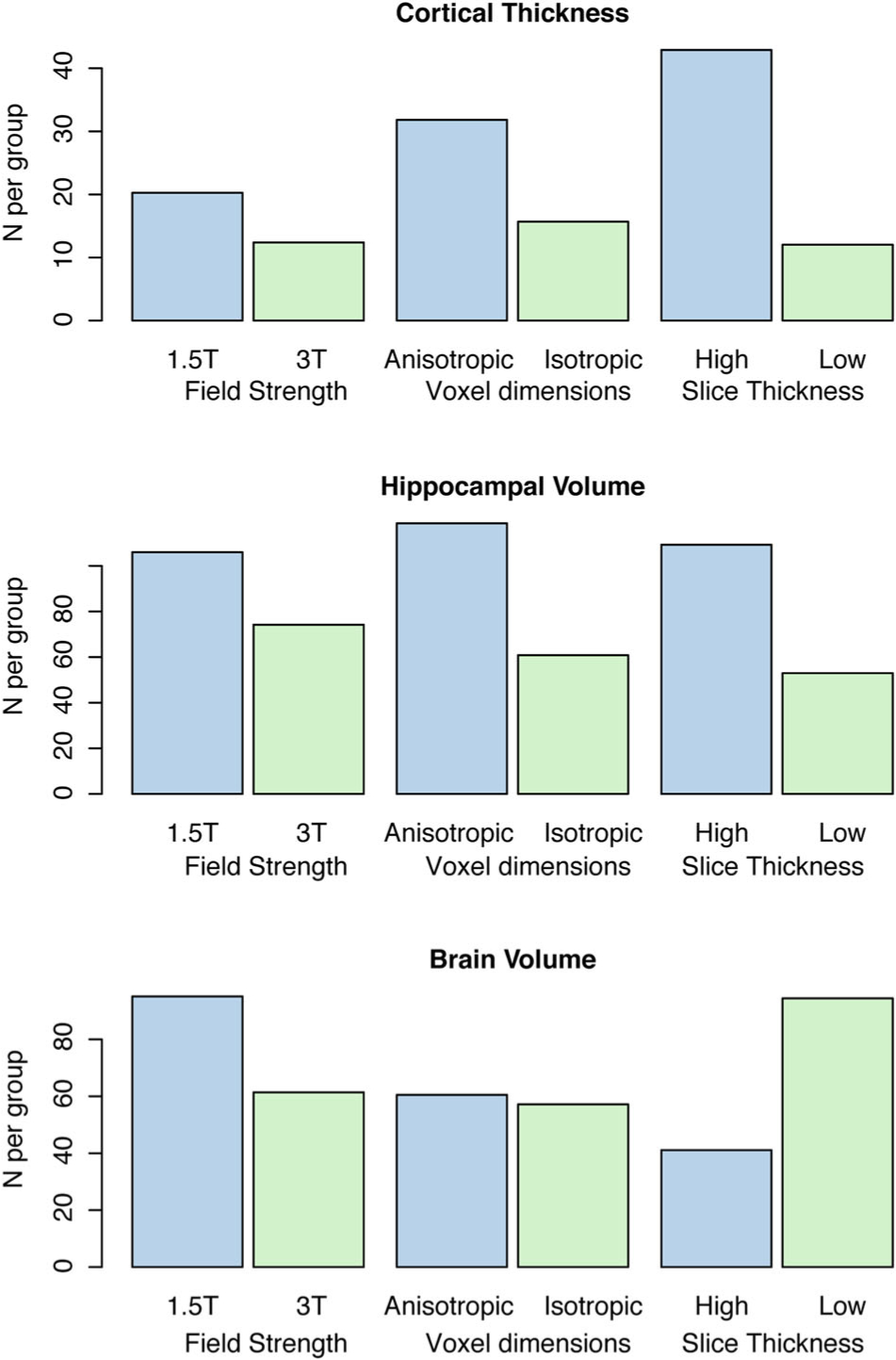
Methods: Cortical thickness, hippocampal volume, and whole brain volume were estimated from whole brain T1-weighted MRI scans processed using Freesurfer v6.0. Sample sizes required to detect a range of effect sizes were calculated using (i) standard t-test based power analysis methods and (ii) a nonparametric bootstrap approach.

Results: A total of 32 participants were included in our analyses, aged 29.9 ± 12.62 years. Standard deviation estimates were lower for all quantitative neuroanatomical metrics when assessed using standardized protocols. Required sample sizes per group to detect a given effect size were markedly reduced when using standardized protocols, particularly for cortical thickness changes <.2 mm and hippocampal volume changes <10%.

Conclusions: The use of standardized protocols yielded up to a five-fold reduction in required sample sizes to detect disease-related neuroanatomical changes, and is particularly beneficial for detecting subtle effects. Standardizing image acquisition protocols across scanners prior to commencing a study is a valuable approach to increase the statistical power of multicenter MRI studies.

Keywords: Brain morphometry; multisite studies; power analysis; quantitative neuroanatomy.


Conflict of interest statement

Conflict of Interest: The authors have no conflicts of interest to disclose.

Figures

Fig 1.
Fig 1.
Standardization of image acquisition protocols improves statistical power for multicenter cortical thickness studies. The figure demonstrates a substantive reduction in required sample size when using a standardized image acquisition protocol (orange lines) compared with a nonstandardized protocol (green lines). The solid lines show sample size estimates obtained from conventional power analysis techniques that assume values are sampled from a normal distribution, dashed lines indicate sample size estimates obtained using a nonparametric bootstrap approach.
Fig 2.
Fig 2.
The use of a standardized image acquisition protocol improves statistical power for detection of hippocampal volume changes in multicenter imaging studies. The plot shows that the number of subjects required per group is less for a given effect size (hippocampal volume difference) when using a standardized protocol (orange lines) compared to a nonstandardized protocol (green lines). As an example, to detect a 200 mm3 volume change (5%) requires approximately 110 subjects for a standardized protocol and approximately 220 subjects for a nonstandardized protocol.
Fig 3.
Fig 3.
The use of a standardized image acquisition protocol improves statistical power for detection of brain volume changes in multicenter imaging studies. The plot shows that the number of subjects required per group is less for a given effect size when using a standardized protocol (orange lines) compared to a nonstandardized protocol (green lines). As an example, to detect a 50,000 mm3 volume change (~5%) requires approximately 60 subjects for a standardized protocol and approximately 90 subjects for a nonstandardized protocol.
Fig 4.
Fig 4.
The relationship between image acquisition parameters and sample size estimates obtained using nonstandardized clinical imaging. The figure shows that 3T imaging with isotropic voxel size and low slice thickness allows lower sample sizes and, therefore, higher power for detection of changes in cortical thickness and hippocampal volume.

References

    1. Cannon TD, Sun F, McEwen SJ, et al. Reliability of neuroanatomical measurements in a multisite longitudinal study of youth at risk for psychosis. Hum Brain Mapp 2014;35:2424–34. - PMC - PubMed
    1. Jovicich J, Marizzoni M, Sala-Llonch R, et al. Brain morphometry reproducibility in multi-center 3t MRI studies: a comparison of cross-sectional and longitudinal segmentations. Neuroimage 2013;83:472–84. - PubMed
    1. Keshavan A, Paul F, Beyer MK, et al. Power estimation for nonstandardized multisite studies. Neuroimage 2016;134:281–94. - PMC - PubMed
    1. Friedman L, Stern H, Brown GG, et al. Test-retest and between-site reliability in a multicenter fMRI study. Hum Brain Mapp 2008;29:958–72. - PMC - PubMed
    1. Gouttard S, Styner M, Prastawa M, Piven J, Gerig G. Assessment of reliability of multi-site neuroimaging via traveling phantom study. Med Image Comput Comput Assist Interv 2008;11:263–70. - PMC - PubMed

Publication types