Review
. 2019 Nov 26;13(11):12301-12321.
doi: 10.1021/acsnano.9b06522. Epub 2019 Nov 5.

Aptamers as Modular Components of Therapeutic Nucleic Acid Nanotechnology

Affiliations
Review

Aptamers as Modular Components of Therapeutic Nucleic Acid Nanotechnology

Martin Panigaj et al. ACS Nano. .

Abstract

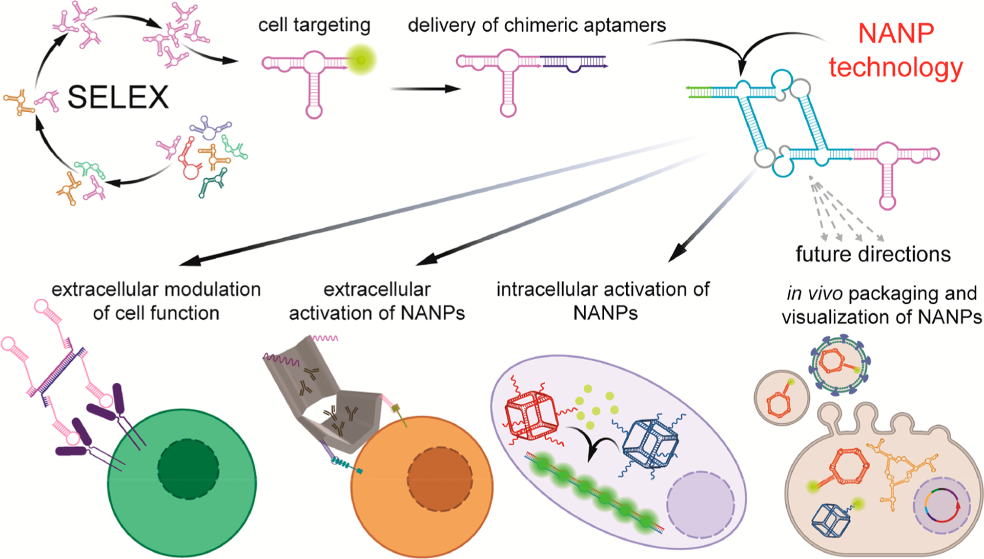
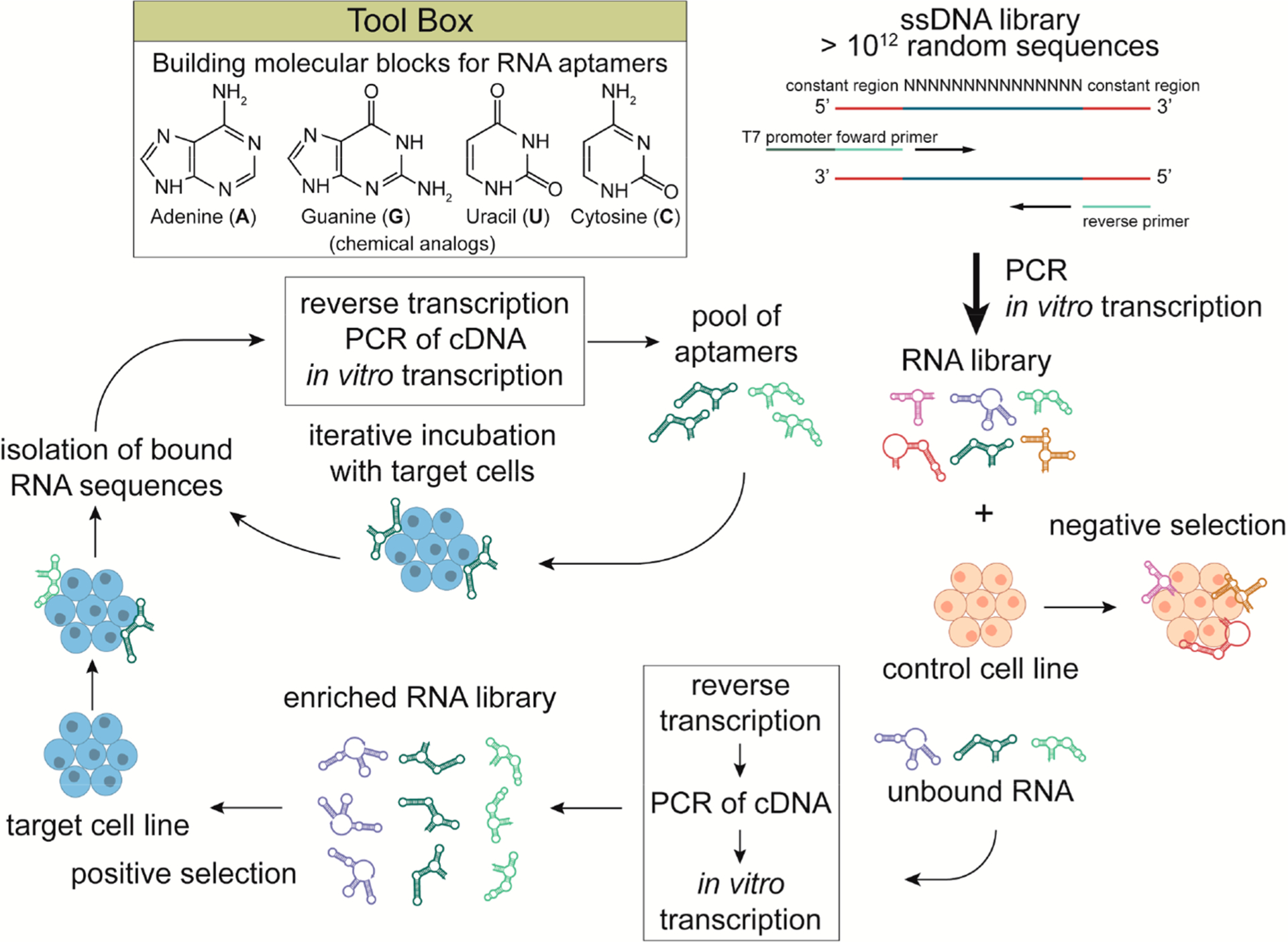
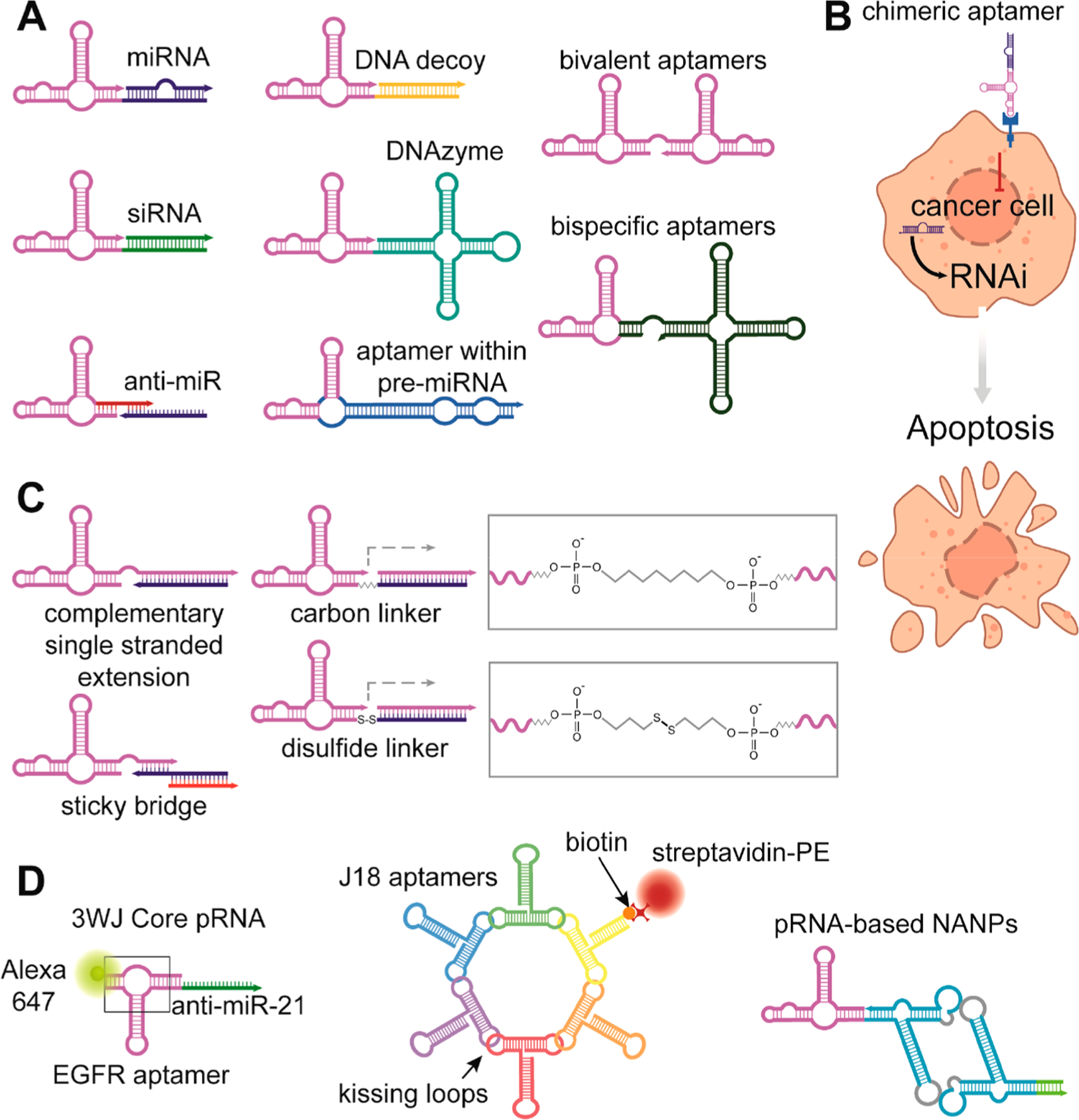
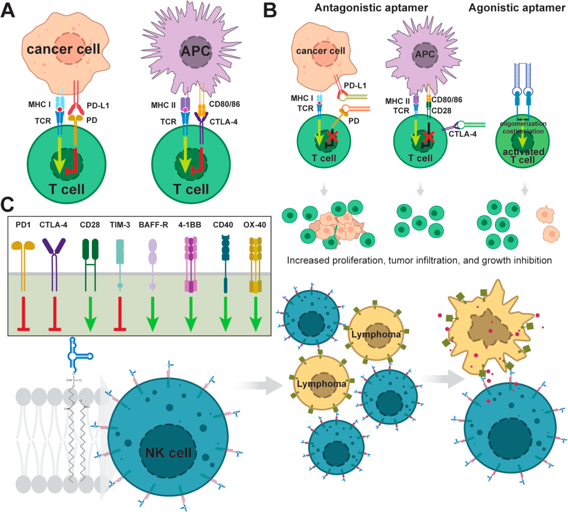
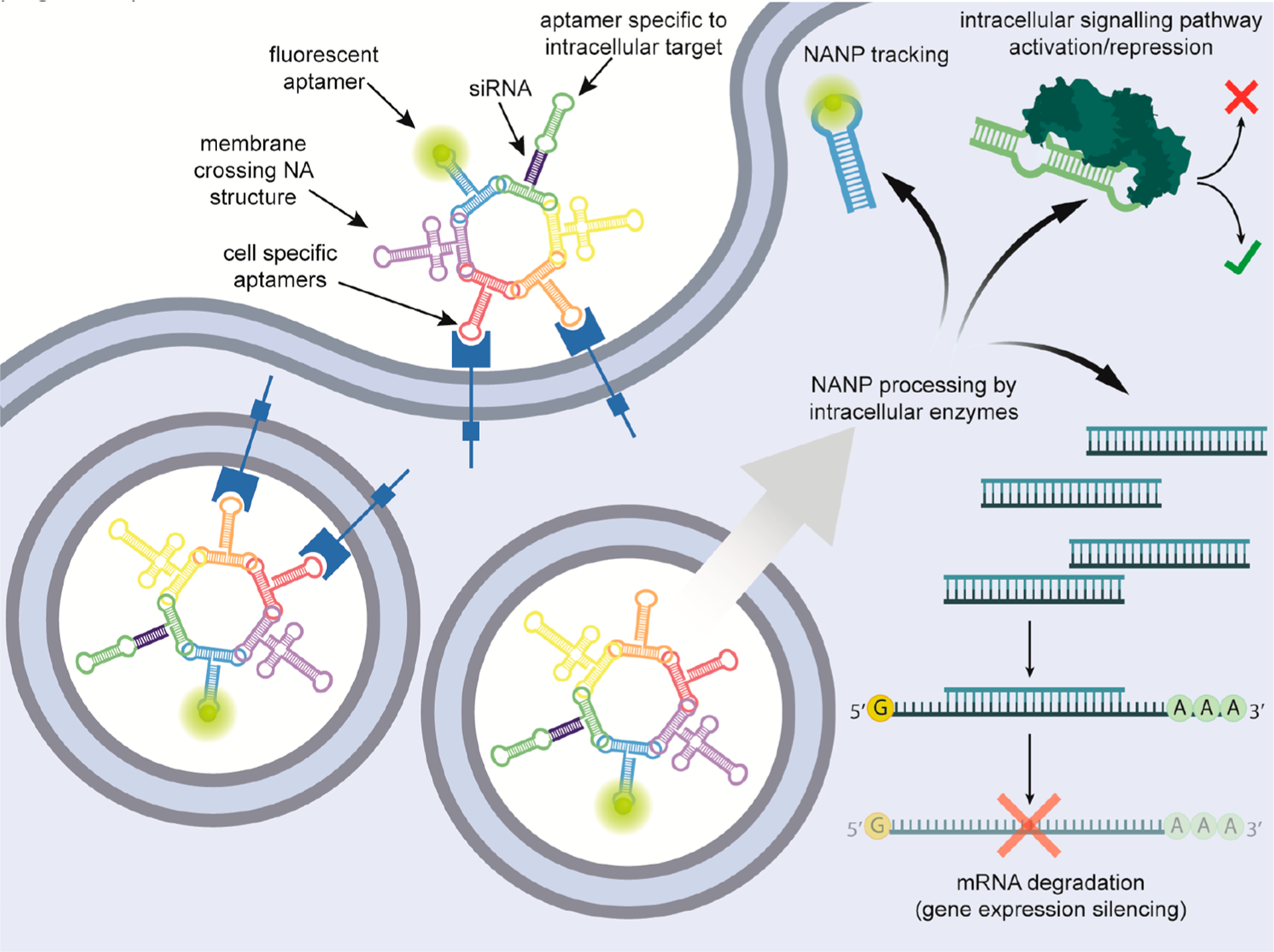
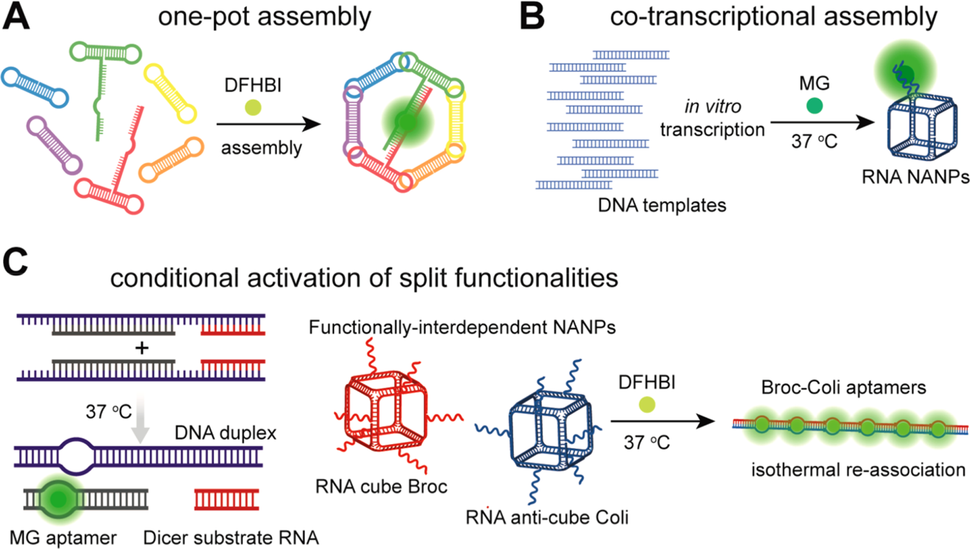
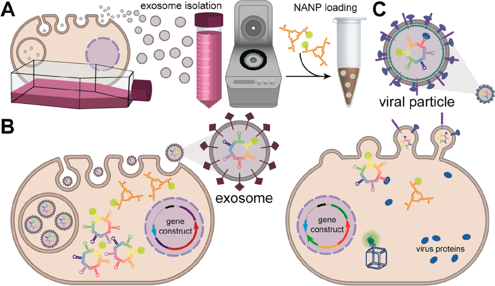
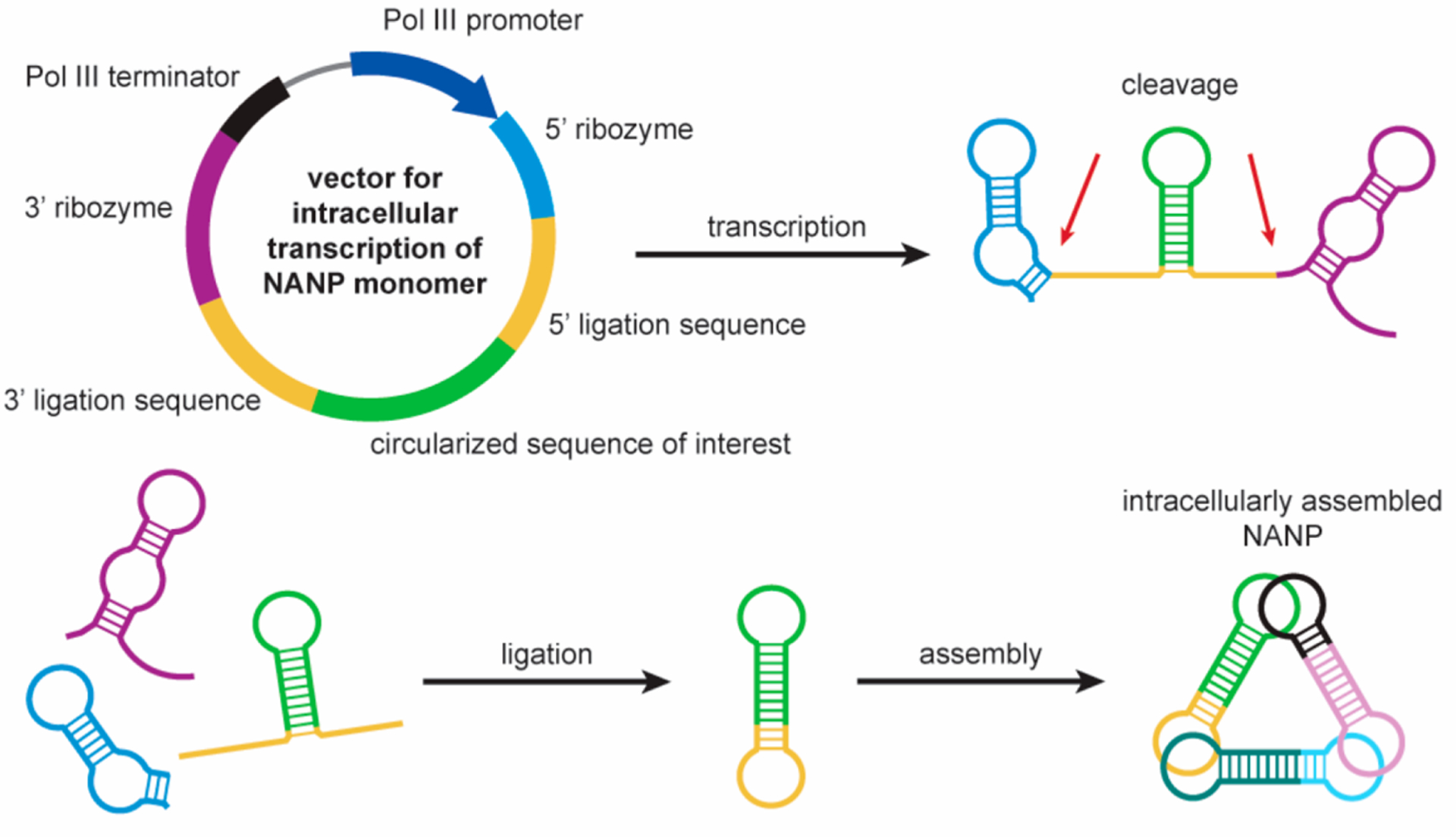
Nucleic acids play a central role in all domains of life, either as genetic blueprints or as regulators of various biochemical pathways. The chemical makeup of ribonucleic acid (RNA) or deoxyribonucleic acid (DNA), generally represented by a sequence of four monomers, also provides precise instructions for folding and higher-order assembly of these biopolymers that, in turn, dictate biological functions. The sequence-based specific 3D structures of nucleic acids led to the development of the directed evolution of oligonucleotides, SELEX (systematic evolution of ligands by exponential enrichment), against a chosen target molecule. Among the variety of functions, selected oligonucleotides named aptamers also allow targeting of cell-specific receptors with antibody-like precision and can deliver functional RNAs without a transfection agent. The advancements in the field of customizable nucleic acid nanoparticles (NANPs) opened avenues for the design of nanoassemblies utilizing aptamers for triggering or blocking cell signaling pathways or using aptamer-receptor combinations to activate therapeutic functionalities. A recent selection of fluorescent aptamers enables real-time tracking of NANP formation and interactions. The aptamers are anticipated to contribute to the future development of technologies, enabling an efficient assembly of functional NANPs in mammalian cells or in vivo. These research topics are of top importance for the field of therapeutic nucleic acid nanotechnology with the promises to scale up mass production of NANPs suitable for biomedical applications, to control the intracellular organization of biological materials to enhance the efficiency of biochemical pathways, and to enhance the therapeutic potential of NANP-based therapeutics while minimizing undesired side effects and toxicities.

Keywords: NANPs; RNA nanotechnology; SELEX; aptamers; exosomes; immunotherapy; nucleic acid delivery; therapeutic nucleic acids.


Conflict of interest statement

The authors declare no competing financial interest.

Figures

Figure 1.
Figure 1.
Schematic description of growing structural and functional complexity of aptamer involvements into nucleic acid nanotechnology. Fluorescently labeled aptamers that are specific to cell receptors can be used for cell detection. Their interactions with receptors often result in modulation of the receptor signaling. Later development led to the design of aptamer chimeras, where aptamers deliver the functional RNA or DNA moieties to target cells. Inclusion of aptamers to NANPs enhances the combinatorial applications of aptamers in changing cellular pathways and allowing for NANPs to logically respond to the presence of key triggers. In addition, light-up aptamers are potentially suitable reporters of NANP assembly or real-time monitoring of mutual interactions of NANPs in vivo. One of the future applications of aptamers in NANP technology could be a transport of NANPs to cell vesicles or viral vectors that would be mediated by aptamers targeted to vesicle- or virus-specific proteins.
Figure 2.
Figure 2.
Schematic illustration of whole-cell SELEX. A library of commercially available ssDNAs is amplified by PCR and subsequently in vitro transcribed to an RNA library. This is possible due to the constant 5′ and 3′ sequences that are the same for each ssDNA and contain complementary sites for PCR as well as a T7 promoter for transcription. The variable body of aptamers that is unique for each strand is located between common 5′ and 3′ sequences required for PCR amplification. In the first step, the RNA library is incubated with the control cell population that does not express target receptors. In the next step, the unbound sequences are recovered and reverse transcribed to cDNA that is amplified by PCR. The subsequent in vitro transcribed RNA library is enriched with sequences with low or no affinity to the control cell line. The library is then incubated with target cells, unbound RNA is washed out, and bound strands are isolated and again reamplified. In vitro transcribed RNA in this step is used for the next round of selection. With each cycle, specific aptamers should prevail in the reamplified population. The final sequence(s) with the highest affinity is identified by sequencing analysis. RNA structures can be visualized using the NUPACK Web Application.
Figure 3.
Figure 3.
Schematic depiction of various chimeric aptamers. Multiple nucleic-acid-based functionalities can be linked to cell-specific aptamers with many different approaches that are beyond the scope of the article. (A) Post-transcriptional silencing of gene expression is achieved by delivery of miRNA or siRNA. Transcription of aptamers within pre-miRNA from gene constructs offers prolonged production of chimeric RNA. Downregulation of some genes by endogenous miRNAs during tumorigenesis can be reverted by the delivery of anti-miRs that block binding of miRNAs to target mRNAs. Delivery of a DNAzyme that cleaves specific mRNA is another way to repress gene expression. Conjugation of two identical aptamers is used for oligomerization of receptors in comparison with two different aptamers that may attach to cell surface proteins from intercellular space or the bloodstream as well as interconnect two different cells. (B) Schematic illustration of cell targeting by chimeric aptamer with a synergistic effect. An aptamer blocks signaling, while the therapeutic payload silences genes crucial for cell survival. Thus, simultaneously both functional parts promote apoptosis. (C) Most utilized ways of chimeric TNA conjugations. (D) From left to right: 3WJ-EGFR aptamer/anti-miR-21 nanoparticles harboring three functional modules: EGFR RNA aptamer for targeted delivery, anti-miR-21 LNA for therapy, and Alexa-647 dye for imaging. The RNA nanoring carrying five J18 aptamers for cell targeting, connected to the RNA ring. One biotinylated oligonucleotide provides fluorescent readout after coupling to a streptavidin–phycoerythrin conjugate. The 3WJ pRNA motif can be used to multiply assemble scaffolds.
Figure 4.
Figure 4.
Illustration of aptamer involvement in cancer immunotherapy. (A) Two most crucial (most known) immune checkpoints. (B) Antagonistic aptamers, selected against either receptor or ligand, inhibit their interaction. Therefore, the downstream pathway is not triggered. Agonistic aptamers dimerize receptor dimers that result in switching on respective signaling. (C) Selected immune system receptors that have already been targeted by aptamers. (D) Whereas standard approaches use aptamers as targeting agents to deliver therapeutic cargo to cells, aptamer-engineered NK cells are directed by cell-membrane-embedded aptamers to lymphoma cells.
Figure 5.
Figure 5.
Vision of uptake of multifunctional NANPs composed purely from nucleic acids (NA). Cell-specific aptamers facilitate targeted delivery of NANPs. Fluorescent aptamers would provide tracking of NANPs from the cell surface, through the endosome compartment, to cytoplasm. Membrane crossing NAs, currently hypothetical in structure, promote escape of NANPs to the cytoplasm. Subsequently, intracellular enzyme machineries would “liberate” functional parts—siRNAs and aptamers against intracellular targets—resulting in a synergistic effect.
Figure 6.
Figure 6.
Fluorescent aptamers as a tool in NANP assembly. (A) Signal from individually reassembled nanoparticles’ parts forming complete NANPs correlates with full ring assembly. (B) Assembly of 3D nanostructures is possible during the parallel transcription of all templates.(C) Embedding split light-up aptamers into conditionally pairwise reshaping NANPs enables the monitoring of their interactions in real time.
Figure 7.
Figure 7.
Schematic description proposing NANP loading into exosomes or lentiviruses. (A) In vitro loading of NANPs to isolated exosomes.(B) Genetically encoded NANPs are loaded to exosomes. (C) NANPs are loaded into viral particles via the aptamers binding to virus proteins.
Figure 8.
Figure 8.
Intracellular assembly of NANPs can be designed on the basis of the Tornado system. NANP building blocks transcribed from one or several vectors are flanked by ribozymes that, after self-cleavage, create termini—a 5′ hydroxyl and a 2′,3′-cyclic phosphate at the 3′ end that are recognized by endogenous RtcB ligase. The resulting circularized sequence is chemically stable and, in addition to the Broccoli aptamer, can also code for another functional aptamer (e.g., NF-κB). This system is inspiring for the creation of intracellular structures that can assemble into NANPs.

References

    1. Cech TR; Steitz JA The Noncoding RNA Revolution-Trashing Old Rules to Forge New Ones. Cell 2014, 157, 77–94. - PubMed
    1. Lieberman J Tapping the RNA World for Therapeutics. Nat. Struct. Mol. Biol 2018, 25, 357–364. - PMC - PubMed
    1. Chakraborty C; Sharma AR; Sharma G; Doss CGP; Lee S-S Therapeutic miRNA and siRNA: Moving from Bench to Clinic as Next Generation Medicine. Mol. Ther.–Nucleic Acids 2017, 8, 132–143. - PMC - PubMed
    1. Zhou J; Neff CP; Swiderski P; Li H; Smith DD; Aboellail T; Remling-Mulder L; Akkina R; Rossi JJ Functional In Vivo Delivery of Multiplexed Anti-HIV-1 siRNAs via a Chemically Synthesized Aptamer with a Sticky Bridge. Mol. Ther 2013, 21, 192–200. - PMC - PubMed
    1. Zhou J; Satheesan S; Li H; Weinberg MS; Morris KV; Burnett J; Rossi J Cell-Specific RNA Aptamer Against Human CCR5 Specifically Targets HIV-1 Susceptible and Inhibits HIV-1 Infectivity. Chem. Biol 2015, 22, 379–390. - PMC - PubMed

Publication types