. 2019 Dec 1;142(12):3834-3851.
doi: 10.1093/brain/awz331.

Tumour-associated macrophage-derived interleukin-1 mediates glioblastoma-associated cerebral oedema

Affiliations

Tumour-associated macrophage-derived interleukin-1 mediates glioblastoma-associated cerebral oedema

Cameron J Herting et al. Brain. .

Abstract

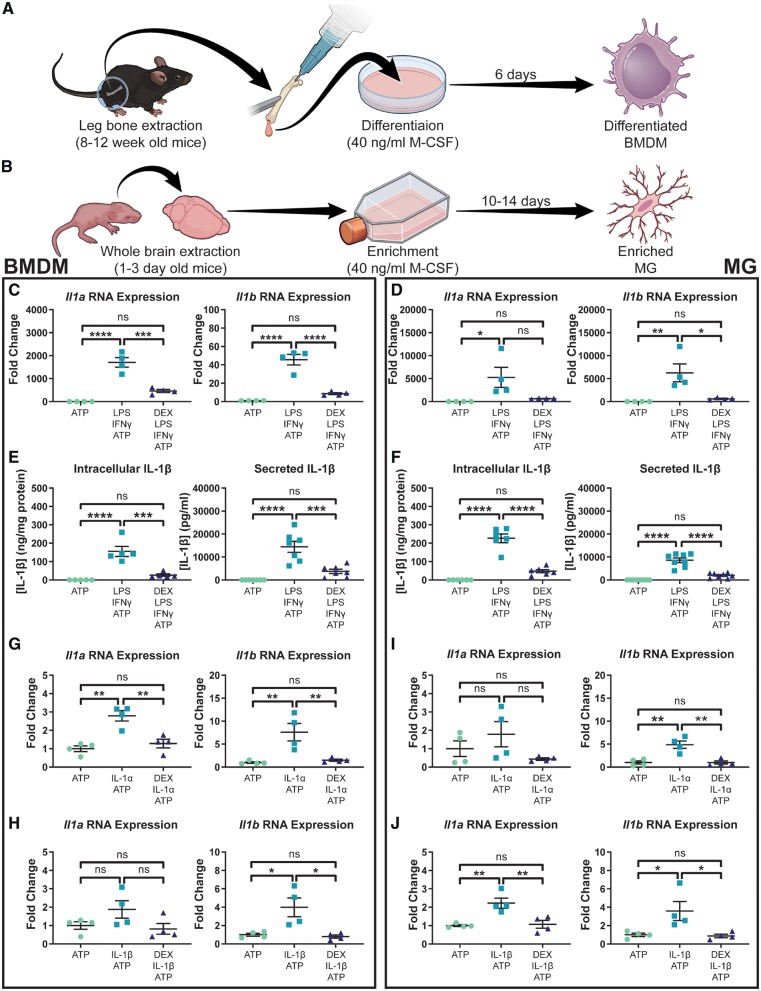
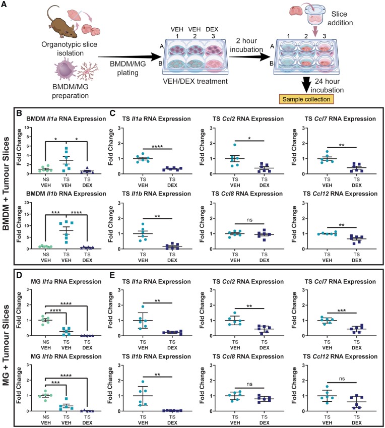
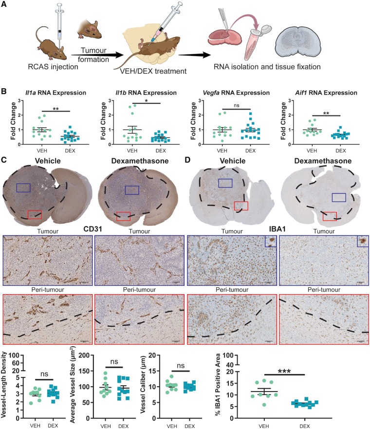
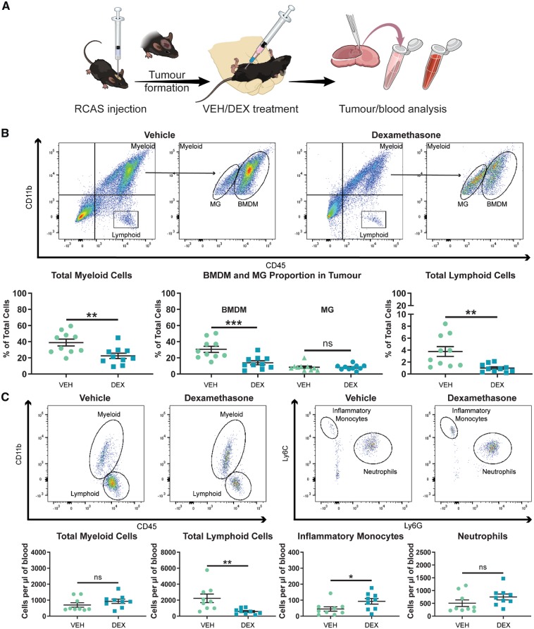
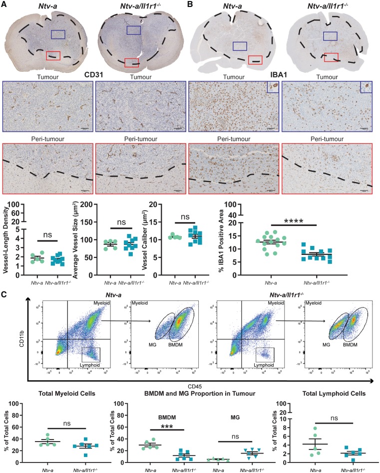
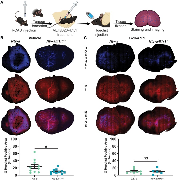
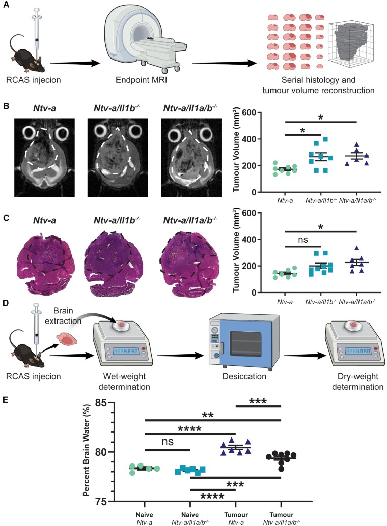
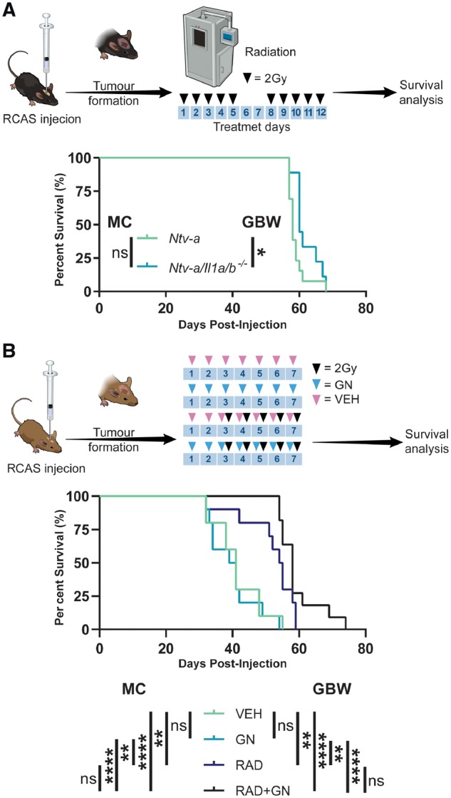
Glioblastoma is the most common and uncompromising primary brain tumour and is characterized by a dismal prognosis despite aggressive treatment regimens. At the cellular level, these tumours are composed of a mixture of neoplastic cells and non-neoplastic cells, including tumour-associated macrophages and endothelial cells. Cerebral oedema is a near-universal occurrence in patients afflicted with glioblastoma and it is almost exclusively managed with the corticosteroid dexamethasone despite significant drawbacks associated with its use. Here, we demonstrate that dexamethasone blocks interleukin-1 production in both bone marrow-derived and brain resident macrophage populations following stimulation with lipopolysaccharide and interferon gamma. Additionally, dexamethasone is shown to inhibit downstream effectors of interleukin-1 signalling in both macrophage populations. Co-culture of bone marrow-derived macrophages with organotypic tumour slices results in an upregulation of interleukin-1 cytokines, an effect that is absent in co-cultured microglia. Genetic ablation of interleukin-1 ligands or receptor in mice bearing RCAS/tv-a-induced platelet-derived growth factor B-overexpressing glioblastoma results in reduced oedema and partial restoration of the integrity of the blood-brain barrier, respectively; similar to results obtained with vascular endothelial growth factor neutralization. We establish that tumours from dexamethasone-treated mice exhibit reduced infiltration of cells of the myeloid and lymphoid compartments, an effect that should be considered during clinical trials for immunotherapy in glioblastoma patients. Additionally, we emphasize that caution should be used when immune profiling and single-cell RNA sequencing data are interpreted from fresh glioblastoma patient samples, as nearly all patients receive dexamethasone after diagnosis. Collectively, this evidence suggests that interleukin-1 signalling inhibition and dexamethasone treatment share therapeutic efficacies and establishes interleukin-1 signalling as an attractive and specific therapeutic target for the management of glioblastoma-associated cerebral oedema.

Keywords: cerebral oedema; glioblastoma; interleukin-1; macrophage; microglia.


Figures

Figure 1
Figure 1
Dexamethasone blocks LPS/IFNγ and IL-1-induced IL-1 expression in primary BMDMs and microglia. Primary murine BMDMs (A) and microglia (B) were cultured using modifications of previously established protocols. Il1 expression in BMDMs (n = 4) (C) and microglia (n = 4) (D) following stimulation with LPS and IFNγ. Intracellular (n = 5) and secreted (n = 7) IL-1β protein levels in BMDMs (E) as well as intracellular (n = 6) and secreted (n = 8) IL-1β protein levels in microglia (F) following stimulation with LPS and IFNγ. IL-1α (G) and IL-1β (H) stimulation of BMDMs (n = 4). IL-1α (I) and IL-1β (J) stimulation of microglia (n = 4). One-way ANOVA, ns = not significant. *P < 0.05, **P < 0.01, ***P < 0.001, ****P < 0.0001. MG = microglia.
Figure 2
Figure 2
Dexamethasone treatment ablates Il1 upregulation by BMDMs exposed to organotypic tumour slices and reduces expression of MCP cytokines by tumour tissue. (A) An experimental outline of the co-culture of BMDMs and microglia (MG) with organotypic tumour slices (TS). (B) Expression of Il1a and Il1b in BMDMs co-cultured with tumour slices (n = 6). (C) Il1a, Il1b, Ccl2, Ccl7, Ccl8, and Ccl12 expression in tumour slices co-cultured with BMDMs (n = 6). (D) Expression of Il1a and Il1b in microglia co-cultured with tumour slices (n = 5). (E) Il1a, Il1b, Ccl2, Ccl7, Ccl8, and Ccl12 expression in tumour slices co-cultured with microglia (n = 6). In all experiments, dexamethasone (DEX) was added at a dose of 5 µM where indicated. One-way ANOVA and two-tailed Student’s t-test. ns = not significant. *P < 0.05, **P < 0.01, ***P < 0.001, ****P < 0.0001. DEX = dexamethasone; VEH = vehicle.
Figure 3
Figure 3
Dexamethasone treatment has no effect on tumour angiogenesis, but significantly reduces the amount of tumour-associated macrophages. (A) An experimental outline of the generation and treatment of PDGFB-overexpressing tumours. (B) Quantitative-PCR of tumour samples and interrogation of Il1a, Il1b, Vegfa, and Aif1 expression in tumour tissue samples from vehicle (VEH, n = 13) and dexamethasone (DEX)-treated mice (n = 15). Immunohistochemical analysis of CD31 (C) and IBA1 (D) expression in formalin-fixed, paraffin-embedded tumour slices from vehicle- (n = 9) and dexamethasone-treated mice (n = 11). Scale bars = 100 µm. Two-tailed Student’s t-test, ns = not significant. *P < 0.05, **P < 0.01, ***P < 0.001. RCAS = replication-competent avian sarcoma-leukosis virus long terminal repeat with splice acceptor.
Figure 4
Figure 4
Dexamethasone treatment significantly impairs the ability of immune cells to infiltrate the tumour from the circulation. (A) An experimental outline of the treatment of mice bearing PDGFB-overexpressing tumours with vehicle (VEH) or dexamethasone (DEX). (B) Representative flow cytometry plots and quantification of total myeloid cells, BMDMs, microglia (MG), and lymphoid cells in PDGFB-overexpressing tumours from mice treated with vehicle (n = 10) or dexamethasone (n = 10). (C) Representative flow cytometry plots and quantification of circulating myeloid cells, lymphoid cells, inflammatory monocytes, and neutrophils in the blood of tumour-bearing mice treated with vehicle (n = 9) or dexamethasone (n = 9) at endpoint. Two-tailed Student’s t-test, ns = not significant. *P < 0.05, **P < 0.01, ***P < 0.001. RCAS = replication-competent avian sarcoma-leukosis virus long terminal repeat with splice acceptor.
Figure 5
Figure 5
IL-1R1 ablation reduces the influx of circulating myeloid cells in the tumours of mice bearing PDGFB-overexpressing glioblastoma. Immunohistochemical analysis of CD31 (A) in Ntv-a (n = 5) and Ntv-a/Il1r1−/− (n = 9) mice. Immunohistochemical analysis of IBA1 (B) in Ntv-a (n = 14) and Ntv-a/Il1r1−/− (n = 11) mice. (C) Representative flow cytometry plots and quantification of total myeloid cells, BMDMs, microglia (MG), and lymphoid cells in tumours of Ntv-a (n = 5) and Ntv-a/Il1r1−/− (n = 6) mice bearing PDGFB-overexpressing glioblastoma. Two-tailed Student’s t-test, ns = not significant. ***P < 0.001, ****P < 0.0001.
Figure 6
Figure 6
Ablation of IL-1 signalling reduces blood–brain barrier permeability in mice bearing PDGFB-overexpressing tumours. (A) A schematic outlining tumour generation, treatment, and tissue processing. (B) Comparison of Hoechst dye leakage in tumours from Ntv-a (n = 10) and Ntv-a/Il1r1−/− (n = 11) mice treated with vehicle solution. (C) Comparison of Hoechst dye leakage in tumours from Ntv-a (n = 8) and Ntv-a/Il1r1−/− (n = 5) mice treated with the VEGF-neutralizing antibody B20-4.1.1. Two-tailed Student’s t-test, ns = not significant. *P < 0.05.
Figure 7
Figure 7
IL-1 ligand ablation reduces oedema formation in tumour-bearing mice. (A) A schematic illustration of the MRI and serial histology experimental workflow. (B) Comparison of endpoint tumour volumes between Ntv-a (n = 8), Ntv-a/Il1b−/− (n = 8), and Ntv-a/Il1a/b−/− (n = 6) mice with T2-weighted MRI. (C) Comparison of endpoint tumour volumes between Ntv-a (n = 7), Ntv-a/Il1b−/− (n = 9), and Ntv-a/Il1a/b−/− (n = 7) mice with serial sectioning followed by haematoxylin and eosin staining. (D) A schematic illustration of the wet/dry assay for oedema measurement. (E) Comparison of per cent brain water in naïve Ntv-a (n = 5), naïve Ntv-a/Il1a/b−/− (n = 5), tumour-bearing Ntv-a (n = 7), and tumour-bearing Ntv-a/Il1a/b−/− (n = 8) mice. One-way ANOVA, ns = not significant. *P < 0.05, **P < 0.01, ***P < 0.001, ****P < 0.0001.
Figure 8
Figure 8
Genetic and pharmacological inhibition of IL-1 signalling have no impact on radiotherapy efficacy. (A) A schematic illustration of the irradiation of tumour-bearing Ntv-a (n = 13) and Ntv-a/Il1a/b−/− (n = 9) mice with the associated survival curves. (B) A schematic illustration of the treatment of tumour-bearing mice with vehicle (n = 10), gallium nitrate (n = 10), vehicle plus radiation (n = 10), or gallium nitrate plus radiation (n = 11) with the associated survival curves. Mantel-Cox (MC) and Gehan-Breslow-Wilcoxon (GBW) tests, ns = not significant. *P < 0.05, **P < 0.01, ****P < 0.0001. RCAS = replication-competent avian sarcoma-leukosis virus long terminal repeat with splice acceptor.

References

    1. Albrecht U, Yang X, Asselta R, Keitel V, Tenchini ML, Ludwig S, et al. Activation of NF-kappaB by IL-1beta blocks IL-6-induced sustained STAT3 activation and STAT3-dependent gene expression of the human gamma-fibrinogen gene. Cell Signal 2007; 19(9): 1866–78. - PubMed
    1. Aranda PS, LaJoie DM, Jorcyk CL. Bleach gel: a simple agarose gel for analyzing RNA quality. Electrophoresis 2012; 33: 366–9. - PMC - PubMed
    1. Argaw AT, Zhang Y, Snyder BJ, Zhao ML, Kopp N, Lee SC, et al. IL-1beta regulates blood-brain barrier permeability via reactivation of the hypoxia-angiogenesis program. J Immunol 2006; 177: 5574–84. - PubMed
    1. Auphan N, DiDonato JA, Rosette C, Helmberg A, Karin M. Immunosuppression by glucocorticoids: inhibition of NF-kappa B activity through induction of I kappa B synthesis. Science 1995; 270: 286–90. - PubMed
    1. Badie B, Schartner JM, Paul J, Bartley BA, Vorpahl J, Preston JK. Dexamethasone-induced abolition of the inflammatory response in an experimental glioma model: a flow cytometry study. J Neurosurg 2000; 93: 634–9. - PubMed

Publication types

MeSH terms